Introduction
Small bowel obstructions are common, being secondary
in most cases to adhesions from previous surgeries, followed by
abdominal hernias and intestinal tumors. Intestinal intussusception
is one of the most common causes of intestinal obstruction in
children; however, the incidence in adults decreases, caused by
tumors in most cases. Small bowel tumors involve only 5% of all
gastrointestinal malignancies, of which 44% are carcinoid tumors,
33% are adenocarcinomas, 17% are stromal tumors and 8% lymphomas.
Melanoma of the gastrointestinal tract is relatively rare, with
only a few cases having been reported (1).
The majority of cases occur as metastasis from
cutaneous primary lesions, and the small bowel is the most common
location of melanoma metastases in the gastrointestinal tract
(2). The majority of melanomas are
cutaneous in origin; however, ~3% of cases occur as metastases with
an unknown primary source even following thorough investigations
(3). In some patients, it is
possible to describe a previous suspicious naevus, which
spontaneously regresses (4).
Patients with melanoma of unknown primary usually present with
locoregional melanoma metastases in the (sub)cutis, soft tissue,
and/or lymph nodes (i.e., stage III disease) or with distant
metastases including visceral metastases (i.e., stage IV disease)
(5). Metastases of malignant
melanoma also can lead to soft tissue sarcomas (6).
Case report
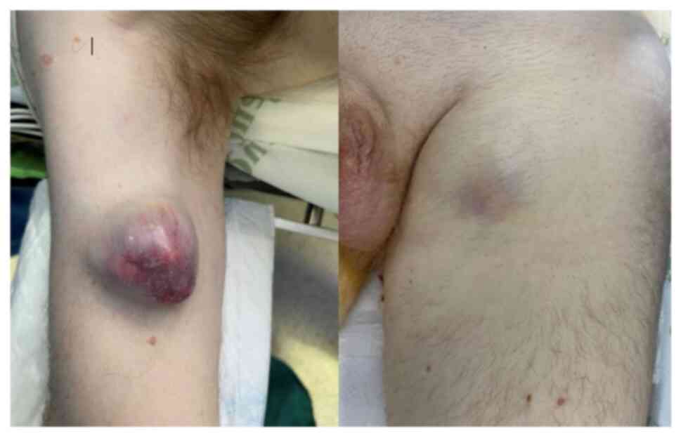
The present study describes the case of a
47-year-old male patient without any previous medical conditions,
who visited Príncipe de Asturias University Hospital (Alcalá de
Henares, Spain) for multiple soft tissue tumors, the largest one
being located in the left gluteal region, measuring 14x15x20 cm.
Other lesions were located in the left inguinal region, measuring
8x5x5 cm and in the right arm, measuring 5 cm, presenting with a
rapid growth pattern during the previous month (Fig. 1).
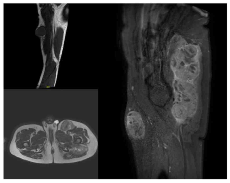
A magnetic resonance imaging scan was performed,
identifying the three masses (Fig.
2), and the gluteal tumor was biopsied reporting a differential
diagnosis between clear cell sarcoma and melanoma. Before other
tests could be performed to confirm the diagnosis, the patient was
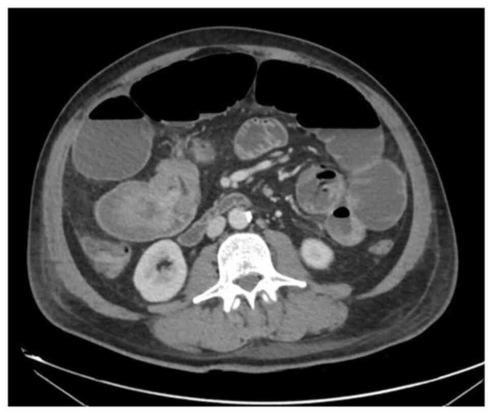
admitted to the emergency room for an intestinal obstruction. A
computed tomography scan was performed, which revealed an
ileo-ileal intussusception (Fig. 3).
Pulmonary nodules and bilateral liver lesions were also described
according to metastatic disease.
The patient was evaluated by a dermatologist,
without identifying any skin lesions compatible with cutaneous
melanomas and only a small nevus was signed to biopsy on the left
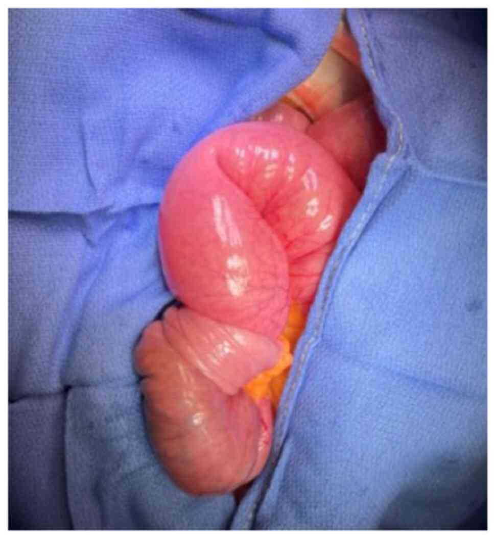
leg. An urgent laparotomy was performed, in which abdominal fluid
was observed (sent for cytological analysis), as well as an
invaginating tumor in the proximal ileum (Fig. 4). An ileal resection and a
side-to-side anastomosis were performed. Hepatic nodules, as well
as peritoneal implants in the greater omentum and meso-jejunum were
identified. The greater omentum and meso-jejunum implants were sent
for a pathological anatomical analysis, together with the tumor of
the right arm and the nevus signed by dermatology on the left
leg.
The results of the pathological analysis revealed
that the nevus in the left leg was a melanocytic nevus. The lesion
in the right arm, the cytology of the peritoneal fluid, the
peritoneal implants, the omentum, as well as the lesion in the
small intestine, corresponded to an undifferentiated malignant
neoplasm that, with the immunohistochemical analysis (performed by
another laboratory), exhibited positivity for vimentin, S100,
HMB-45, SOX 10, Melan-A, and negativity for CD 117, DOG 1, muscle
actin, CK7, CK20, synaptophysin, desmin and CD56.
The presence of the EWSRI I t(12;22)(ql3;ql2)] gene
rearrangement present in 90% of clear cell sarcoma cases was ruled
out, thus confirming the final diagnosis of melanoma.
The patient had a satisfactory immediate
post-operative period and was transferred to the oncological
department to commence treatment with chemotherapy. However, the
patient succumbed during the hospital stay due to a respiratory
infection on the 14th post-operative day.
Discussion
Intestinal intussusception is common among children,
but unusual in adults, where it is associated with an underlying
malignant process. Intussusception secondary to melanoma is
detected in 2-5% of patients with a history of melanoma, and it is
possible to find such intestinal metastases up to 10 years after
the skin lesion (7).
Small bowel melanoma is a rare entity, with the
majority of cases being secondary to metastasis of a primary tumor.
The small bowel is the location where a primary cutaneous melanoma
metastasizes most frequently to the gastrointestinal tract, due to
the rich vascular support of the splanchnic territory. There are a
limited number of published cases of primary melanoma in this
location, due to the absence of melanocytes (1). It is critical to make a differential
diagnosis between primary melanoma of the gastrointestinal tract
and metastasis as the prognosis is worse for primary intestinal
melanomas, which tend to grow at a more rapid rate and are more
aggressive (8).
When the skin melanoma is not identified, it is
mandatory to perform a colonoscopy (to rule out an anorectal
melanoma), gastroscopy (to rule out an oropharynx, esophagus or
stomach melanoma), as well as a fundoscopic examination (choroid
melanoma) (9). However, in up to 26%
of cases of intestinal melanomas, no extraintestinal primary lesion
can be identified. In such circumstances, the spontaneous regression
of the primary site may explain the lack of a primary melanoma. In
the case in the present study, it was not possible to perform these
tests as the patient suffered an intestinal obstruction and
ultimately succumbed during the hospitalization period due to a
respiratory infection on the 14th post-operative day; however, the
absence of any other lesion led to the conclusion that it was a
small bowel melanoma or soft tissue and visceral metastasis from an
unknown melanoma.
This type of metastasis is usually silent until the
moment of diagnosis. The majority of patients with metastatic
intestinal melanoma are asymptomatic and only 1-4% of metastases to
the gastrointestinal tract are detected before death (10). In the case in the present study, the
patient presented with multiple giant soft tissue tumors and an
ileal intussusception. There are previous reports of soft tissue
metastasis and ileal metastasis (6);
however, the patient in the present study suffered both site
metastasis.
The management of such cases consists of surgery to
solve the acute obstruction. In cases where metastatic disease is
limited and a resection is possible, surgery can be considered.
Even when a R0-status and a curative surgery cannot be achieved or
there is recurrent disease, tumor resection is recommended to
relieve symptoms or avoid future complications (11). Systemic treatment is not
standardized; however, systemic therapy with immunotherapy,
chemotherapy or molecular targeted therapy, such as vemurafenib,
ipilimumab, pembrolizumab or imatinib, are other options (12). They can also be useful as a
palliative treatment in metastatic intestinal melanoma; however,
role remains unclear (13).
In conclusion, gastrointestinal tract melanoma is a
rare entity, involving metastatic lesions in the majority of cases.
These lesions are usually diagnosed with the onset of symptoms,
presenting an ominous prognosis. In cases of bowel obstruction, a
surgical resection is required.
Acknowledgements
Not applicable.
Funding
Funding: No funding was received.
Availability of data and materials
The datasets used and/or analyzed during the current
study are available from the corresponding author on reasonable
request.
Authors' contributions
All authors (AV, ES, PB, MMD and AG) contributed to
the diagnosis and treatment of the patient, and to the design of
the study. JAVT was a major contributor to the writing of the
manuscript. AV and MMD confirm the authenticity of all the raw
data. All authors have read and approved the final manuscript.
Ethics approval and consent to
participate
The present study followed international and
national regulations and was in agreement with the Declaration of
Helsinki, and ethical principles. The patient signed an informed
consent form before the surgery was performed.
Patient consent for publication
The patient provided written informed consent for
the publication of any data and/or accompanying images, before the
surgery was performed. Patients have a right to anonymity and
privacy, and authors have a legal and ethical responsibility to
respect this right.
Competing interests
The authors declare that they have no competing
interests.
References
|
1
|
Li WX, Wei Y, Jiang Y, Liu YL, Ren L,
Zhong YS, Ye LC, Zhu DX, Niu WX, Qin XY and Xu JM: Primary colonic
melanoma presenting as ileocecal intussusception: Case report and
literature review. World J Gastroenterol. 20:9626–9630.
2014.PubMed/NCBI View Article : Google Scholar
|
|
2
|
Lens M, Bataille V and Krivokapic Z:
Melanoma of the small intestine. Lancet Oncol. 10:516–521.
2009.PubMed/NCBI View Article : Google Scholar
|
|
3
|
Katz KA, Jonasch E, Hodi FS, Soiffer R,
Kwitkiwski K, Sober AJ and Haluska FG: Melanoma of unknown primary:
Experience at Massachusetts general hospital and dana-farber cancer
institute. Melanoma Res. 15:77–82. 2005.PubMed/NCBI View Article : Google Scholar
|
|
4
|
Alvarez FA, Nicolás M, Goransky J, Vaccaro
CA, Beskow A and Cavadas D: Ileocolic intussusception due to
intestinal metastatic melanoma. Case report and review of the
literature. Int J Surg Case Rep. 2:118–121. 2011.PubMed/NCBI View Article : Google Scholar
|
|
5
|
Gershenwald JE, Scolyer RA, Hess KR,
Sondak VK, Long GV, Ross MI, Lazar AJ, Faries MB, Kirkwood JM,
McArthur GA, et al: Melanoma staging: Evidence-based changes in the
American joint committee on cancer eighth edition cancer staging
manual. CA Cancer J Clin. 67:472–492. 2017.PubMed/NCBI View Article : Google Scholar
|
|
6
|
Lodding P, Kindblom LG and Angervall L:
Metastases of malignant melanoma simulating soft tissue sarcoma. A
clinico-pathological, light- and electron microscopic and
immunohistochemical study of 21 cases. Virchows Arch A Pathol Anat
Histopathol. 417:377–388. 1990.PubMed/NCBI View Article : Google Scholar
|
|
7
|
Francken AB, Bastiaannet E and Hoekstra
HJ: Follow-up in patients with localised primary cutaneous
melanoma. Lancet Oncol. 6:608–621. 2005.PubMed/NCBI View Article : Google Scholar
|
|
8
|
Hadjinicolaou AV, Hadjittofi C,
Athanasopoulos PG, Shah R and Ala AA: Primary small bowel
melanomas: Fact or myth? Ann Transl Med. 4(113)2016.PubMed/NCBI View Article : Google Scholar
|
|
9
|
Cheung MC, Perez EA, Molina MA, Jin X,
Gutierrez JC, Franceschi D, Livingstone AS and Koniaris LG:
Defining the role of surgery for primary gastrointestinal tract
melanoma. J Gastrointest Surg. 12:731–738. 2008.PubMed/NCBI View Article : Google Scholar
|
|
10
|
Silva S, Tenreiro N, Melo A, Lage J,
Moreira H, Próspero F and Avelar P: Metastatic melanoma: An unusual
cause of gastrointestinal bleeding and intussusception-A case
report. Int J Surg Case Rep. 53:144–146. 2018.PubMed/NCBI View Article : Google Scholar
|
|
11
|
Aktas A, Hos G, Topaloglu S, Çalik A, Reis
A and Piskin B: Metastatic cutaneous melanoma presented with ileal
invagination: Report of a case. Turk J Trauma Emerg Surg.
16:469–472. 2010.PubMed/NCBI
|
|
12
|
Patti R, Cacciatori M, Guercio G, Territo
V and Di Vita G: Intestinal melanoma: A broad spectrum of clinical
presentation. Int J Surg Case Rep. 3:395–398. 2012.PubMed/NCBI View Article : Google Scholar
|
|
13
|
Albert JG, Gimm O, Stock K, Bilkenroth U,
Marsch WC and Helmbold P: Small-bowel endoscopy is crucial for
diagnosis of melanoma metastases to the small bowel: A case of
metachronous small-bowel metastases and review of the literature.
Melanoma Res. 17:335–338. 2007.PubMed/NCBI View Article : Google Scholar
|