Proteomic analysis of the cerebrospinal fluid in multiple sclerosis and neuromyelitis optica patients

  • Authors:
    • Shoufeng Jiang
    • Jiong Wu
    • Yi Yang
    • Jianren Liu
    • Yao Ding
    • Meiping Ding
  • View Affiliations

  • Published online on: August 8, 2012     https://doi.org/10.3892/mmr.2012.1025
  • Pages: 1081-1086
Metrics: Total Views: 0 (Spandidos Publications: | PMC Statistics: )
Total PDF Downloads: 0 (Spandidos Publications: | PMC Statistics: )


Abstract

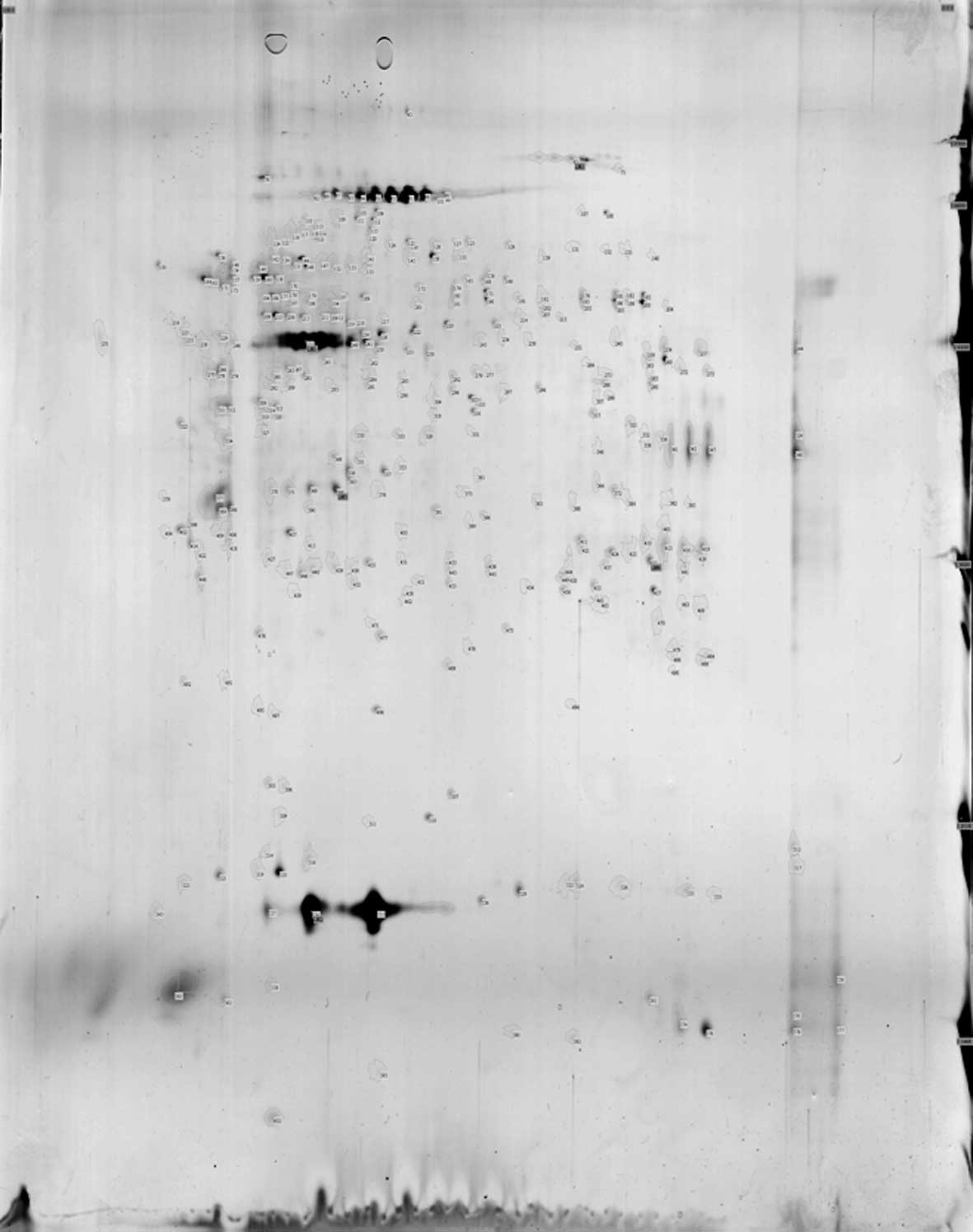
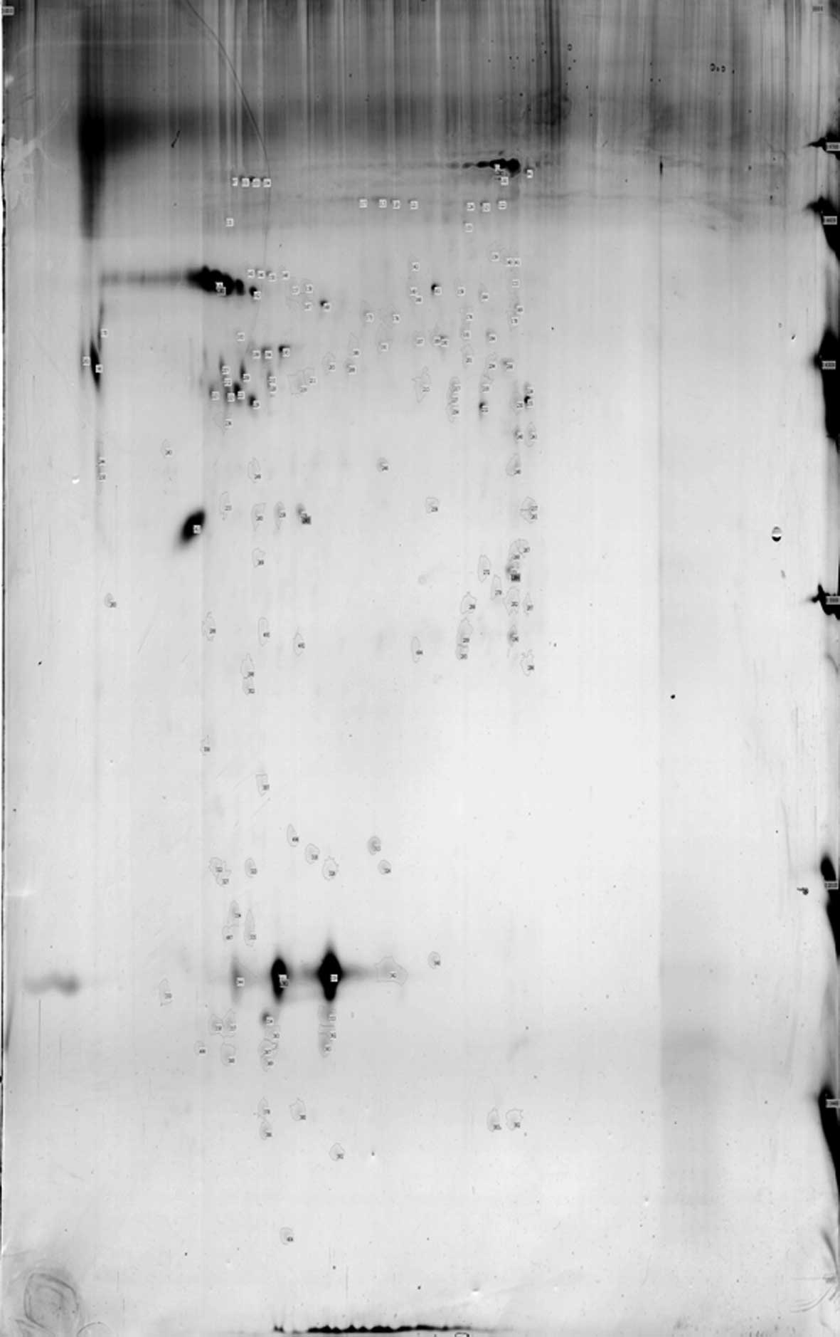
The present study aimed to compare the 2-dimensional (2D) electrophoresis pattern of the cerebrospinal fluid (CSF) in multiple sclerosis (MS), neuromyelitis optica (NMO) and control individuals, to identify the proteins with differential expression and to examine their significance. CSF samples from the three groups were collected and total protein was isolated and quantified using the Bradford method. 2D electrophoresis of the samples was conducted using equal amounts of CSF. In the CSF 2D gel electrophoresis map, 118 points were obtained from the MS group, 155 points from the NMO group and 350 points from the normal control group. Non-matching proteins appeared in 14 spots in the MS group and in 45 spots in the NMO group, and were also expressed in the 2D electrophoresis pattern of the normal control group. Four differential proteins were identified through the HD-MS/MS and MASCOT network search. Pre-albumin (PA) was found only in the CSF 2D gel electrophoresis map of the MS patients. Keratin 1 was expressed in the normal control group, but not in the MS group. The difference in the expression of keratin 9 in the MS group was twice that in the normal control group. The expression of keratin 1, keratin 9 and transferrin in the NMO group was twice that in the normal control group. The expression of PA was found only in the CSF 2D gel electrophoresis map of the MS patients, and not in the NMO group. Keratin 1 was expressed in the NMO group, but not in the MS group. The expression of a variety of proteins in the 2D electrophoresis pattern of CSF was significantly different in the MS, NMO and control groups. PA, keratin 1, transferrin and keratin 9 are identified as significantly differentially expressed proteins.
View Figures
View References

Related Articles

Journal Cover

November 2012
Volume 6 Issue 5

Print ISSN: 1791-2997
Online ISSN:1791-3004

Sign up for eToc alerts

Recommend to Library

Copy and paste a formatted citation
x
Spandidos Publications style
Jiang S, Wu J, Yang Y, Liu J, Ding Y and Ding M: Proteomic analysis of the cerebrospinal fluid in multiple sclerosis and neuromyelitis optica patients. Mol Med Rep 6: 1081-1086, 2012
APA
Jiang, S., Wu, J., Yang, Y., Liu, J., Ding, Y., & Ding, M. (2012). Proteomic analysis of the cerebrospinal fluid in multiple sclerosis and neuromyelitis optica patients. Molecular Medicine Reports, 6, 1081-1086. https://doi.org/10.3892/mmr.2012.1025
MLA
Jiang, S., Wu, J., Yang, Y., Liu, J., Ding, Y., Ding, M."Proteomic analysis of the cerebrospinal fluid in multiple sclerosis and neuromyelitis optica patients". Molecular Medicine Reports 6.5 (2012): 1081-1086.
Chicago
Jiang, S., Wu, J., Yang, Y., Liu, J., Ding, Y., Ding, M."Proteomic analysis of the cerebrospinal fluid in multiple sclerosis and neuromyelitis optica patients". Molecular Medicine Reports 6, no. 5 (2012): 1081-1086. https://doi.org/10.3892/mmr.2012.1025