Mouse forestomach carcinoma cells immunosuppress macrophages through transforming growth factor-β1
- Authors:
- Published online on: February 3, 2012 https://doi.org/10.3892/mmr.2012.777
- Pages: 988-992
Metrics: Total
Views: 0 (Spandidos Publications: | PMC Statistics: )
Total PDF Downloads: 0 (Spandidos Publications: | PMC Statistics: )
Abstract
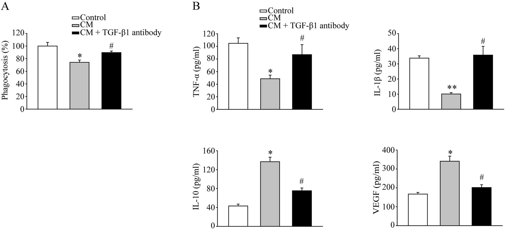
Peritoneal implantation metastasis of gastric cancer cells is associated with poor prognosis. Peritoneal macrophages are the most important immune cells in the abdominal cavity to control tumor metastasis. In the present study, the immunosuppressive effects of mouse forestomach cells on macrophages were examined. Conditioned medium from mouse forestomach cell cultures were used to treat isolated peritoneal macrophages. A colorimetry-based phagocytosis assay was performed to investigate the functional change of macrophages. The alteration of cytokine secretion by macrophages was measured by ELISA assay. Specific markers of macrophage polarization were analyzed by real-time RT-PCR. TGF-β1 signaling was evaluated by western blotting. Neutralization experiments were performed using an anti-TGF-β1 antibody. Conditioned medium reduced the phagocytotic capability of macrophages. Lower TNF-α and IL-1β levels and higher IL-10 and VEGF levels were observed. Real-time RT-PCR showed increased mRNA levels of M2 macrophage markers. Further study revealed that TGF-β1 was significantly elevated in the conditioned medium and TGF-β1 signaling was activated in the macrophages by the treatment of conditioned medium. Neutralization of TGF-β1 reversed the immunosuppressive effects on macrophages. Immunosuppressive macrophages can be induced by conditioned medium from mouse forestomach cell cultures. These effects appeared to occur through the production of TGF-β1 by the tumor cells. Targeted TGF-β1 intervention may help to control peritoneal metastasis of gastric cancers.