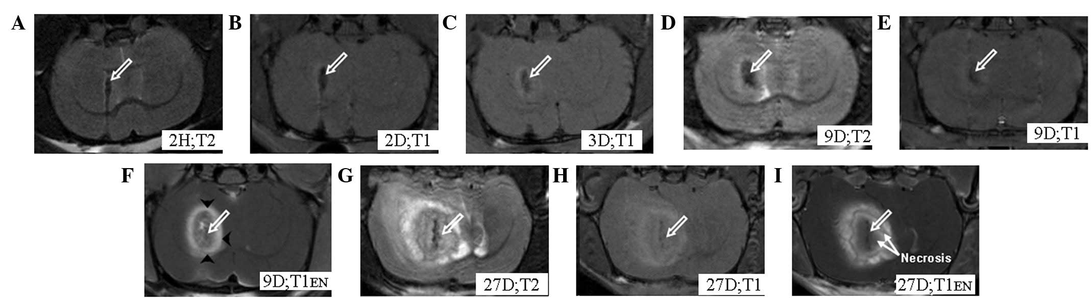
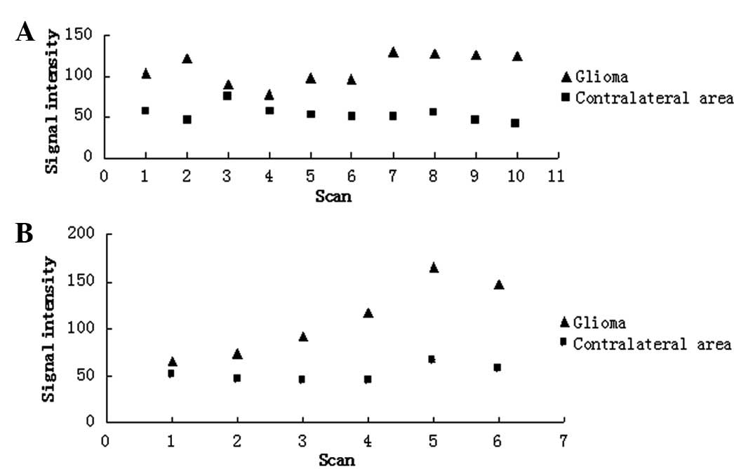
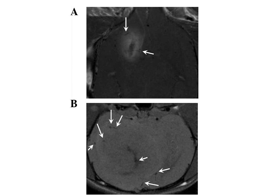
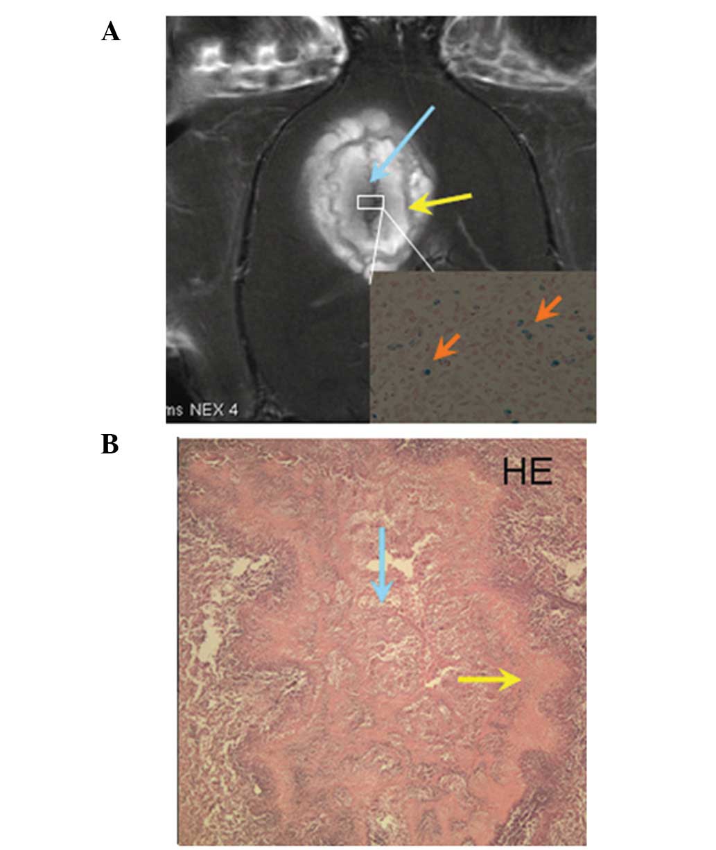
|
1
|
Wrensch M, Minn Y, Chew T, Bondy M and
Berger MS: Epidemiology of primary brain tumors: current concepts
and review of the literature. Neuro Oncol. 4:278–299.
2002.PubMed/NCBI
|
|
2
|
Matsukado Y, Maccarty CS and Kernohan JW:
The growth of glioblastoma multiforme (astrocytomas, grades 3 and
4) in neurosurgical practice. J Neurosurg. 18:636–644. 1961.
View Article : Google Scholar : PubMed/NCBI
|
|
3
|
Demuth T and Berens ME: Molecular
mechanisms of glioma cell migration and invasion. J Neurooncol.
70:217–228. 2004. View Article : Google Scholar : PubMed/NCBI
|
|
4
|
Weissleder R: Scaling down imaging:
molecular mapping of cancer in mice. Nat Rev Cancer. 2:11–18. 2002.
View Article : Google Scholar : PubMed/NCBI
|
|
5
|
Zhang F, Xie J, Liu G, He Y, Lu G and Chen
X: In vivo MRI tracking of cell invasion and migration in a rat
glioma model. Mol Imaging Biol. 13:695–701. 2011. View Article : Google Scholar : PubMed/NCBI
|
|
6
|
Shapiro EM, Skrtic S, Sharer K, Hill JM,
Dunbar CE and Koretsky AP: MRI detection of single particles for
cellular imaging. Proc Natl Acad Sci USA. 101:10901–10906. 2004.
View Article : Google Scholar : PubMed/NCBI
|
|
7
|
Walton RM, Magnitsky SG, Seiler GS,
Poptani H and Wolfe JH: Transplantation and magnetic resonance
imaging of canine neural progenitor cell grafts in the postnatal
dog brain. J Neuropathol Exp Neurol. 67:954–962. 2008. View Article : Google Scholar : PubMed/NCBI
|
|
8
|
Bulte JW and Kraitchman DL: Iron oxide MR
contrast agents for molecular and cellular imaging. NMR Biomed.
17:484–499. 2004. View
Article : Google Scholar : PubMed/NCBI
|
|
9
|
Valable S, Barbier EL, Bernaudin M,
Roussel S, Segebarth C, Petit E and Rémy C: In vivo MRI tracking of
exogenous monocytes/macrophages targeting brain tumors in a rat
model of glioma. Neuroimage. 40:973–983. 2008. View Article : Google Scholar
|
|
10
|
Reddy AM, Kwak BK, Shim HJ, Ahn C, Lee HS,
Suh YJ and Park ES: In vivo tracking of mesenchymal stem cells
labeled with a novel chitosan-coated superparamagnetic iron oxide
nanoparticles using 3.0T MRI. J Korean Med Sci. 25:211–219. 2010.
View Article : Google Scholar : PubMed/NCBI
|
|
11
|
Heyn C, Ronald JA, Ramadan SS, et al: In
vivo MRI of cancer cell fate at the single-cell level in a mouse
model of breast cancer metastasis to the brain. Magn Reson Med.
56:1001–1010. 2006. View Article : Google Scholar : PubMed/NCBI
|
|
12
|
Liu G, Xia C, Wang Z, Lv F, Gao F, Gong Q,
Song B, Ai H and Gu Z: Magnetic resonance imaging probes for
labeling of chondrocyte cells. J Mater Sci Mater Med. 22:601–606.
2011. View Article : Google Scholar : PubMed/NCBI
|
|
13
|
Benda P, Lightbody J, Sato G, Levine L and
Sweet W: Differentiated rat glial cell strain in tissue culture.
Science. 161:370–371. 1968. View Article : Google Scholar : PubMed/NCBI
|
|
14
|
Vince GH, Bendszus M, Schweitzer T,
Goldbrunner RH, Hildebrandt S, Tilgner J, Klein R, Solymosi L,
Christian Tonn J and Roosen K: Spontaneous regression of
experimental gliomas--an immunohistochemical and MRI study of the
C6 glioma spheroid implantation model. Exp Neurol. 190:478–485.
2004. View Article : Google Scholar : PubMed/NCBI
|
|
15
|
Sibenaller ZA, Etame AB, Ali MM, Barua M,
Braun TA, Casavant TL and Ryken TC: Genetic characterization of
commonly used glioma cell lines in the rat animal model system.
Neurosurg Focus. 19:E12005. View Article : Google Scholar : PubMed/NCBI
|
|
16
|
Doblas S, He T, Saunders D, Pearson J,
Hoyle J, Smith N, Lerner M and Towner RA: Glioma morphology and
tumor-induced vascular alterations revealed in seven rodent glioma
models by in vivo magnetic resonance imaging and angiography. J
Magn Reson Imaging. 32:267–275. 2010. View Article : Google Scholar
|
|
17
|
Corot C, Robert P, Idée JM and Port M:
Recent advances in iron oxide nanocrystal technology for medical
imaging. Adv Drug Deliv Rev. 58:1471–1504. 2006. View Article : Google Scholar : PubMed/NCBI
|
|
18
|
de Vries IJ, Lesterhuis WJ, Barentsz JO,
et al: Magnetic resonance tracking of dendritic cells in melanoma
patients for monitoring of cellular therapy. Nat Biotechnol.
23:1407–1413. 2005.PubMed/NCBI
|
|
19
|
Rice HE, Hsu EW, Sheng H, Evenson DA,
Freemerman AJ, et al: Superparamagnetic iron oxide labeling and
transplantation of adipose-derived stem cells in middle cerebral
artery occlusion-injured mice. AJR Am J Roentgenol. 188:1101–1108.
2007. View Article : Google Scholar : PubMed/NCBI
|
|
20
|
Fleige G, Nolte C, Synowitz M, Seeberger
F, Kettenmann H and Zimmer C: Magnetic labeling of activated
microglia in experimental gliomas. Neoplasia. 3:489–499. 2001.
View Article : Google Scholar : PubMed/NCBI
|
|
21
|
Meng XX, Wan JQ, Jing M, Zhao SG, Cai W
and Liu EZ: Specific targeting of gliomas with multifunctional
superparamagnetic iron oxide nanoparticle optical and magnetic
resonance imaging contrast agents. Acta Pharmacol Sin.
28:2019–2026. 2007. View Article : Google Scholar
|
|
22
|
Arbab AS, Bashaw LA, Miller BR, Jordan EK,
Lewis BK, Kalish H and Frank JA: Characterization of biophysical
and metabolic properties of cells labeled with superparamagnetic
iron oxide nanoparticles and transfection agent for cellular MR
imaging. Radiology. 229:838–846. 2003. View Article : Google Scholar : PubMed/NCBI
|
|
23
|
Schäfer R, Kehlbach R, Wiskirchen J,
Bantleon R, Pintaske J, Brehm BR, Gerber A, Wolburg H, Claussen CD
and Northoff H: Transferrin receptor upregulation: in vitro
labeling of rat mesenchymal stem cells with superparamagnetic iron
oxide. Radiology. 244:514–523. 2007.PubMed/NCBI
|
|
24
|
Walczak P, Kedziorek DA, Gilad AA, Barnett
BP and Bulte JW: Applicability and limitations of MR tracking of
neural stem cells with asymmetric cell division and rapid turnover:
the case of the shiverer dysmyelinated mouse brain. Magn Reson Med.
58:261–269. 2007. View Article : Google Scholar : PubMed/NCBI
|
|
25
|
Gilad AA, Winnard PT Jr, van Zijl PC and
Bulte JW: Developing MR reporter genes: promises and pitfalls. NMR
Biomed. 20:275–290. 2007. View
Article : Google Scholar : PubMed/NCBI
|
|
26
|
Gilad AA, McMahon MT, Walczak P, Winnard
PT Jr, Raman V, van Laarhoven HW, Skoglund CM, Bulte JW and van
Zijl PC: Artificial reporter gene providing MRI contrast based on
proton exchange. Nat Biotechnol. 25:217–219. 2007. View Article : Google Scholar : PubMed/NCBI
|