Phosphorylation of cMet tyrosine residues in murine ascitic hepatic cancer cell lines with different lymph node metastatic potentials

  • Authors:
    • Ying Li
    • Xiaohua Huang
    • Qiaoshu Zhang
    • Keli Ma
  • View Affiliations

  • Published online on: June 17, 2013     https://doi.org/10.3892/mmr.2013.1527
  • Pages: 655-661
Metrics: Total Views: 0 (Spandidos Publications: | PMC Statistics: )
Total PDF Downloads: 0 (Spandidos Publications: | PMC Statistics: )


Abstract

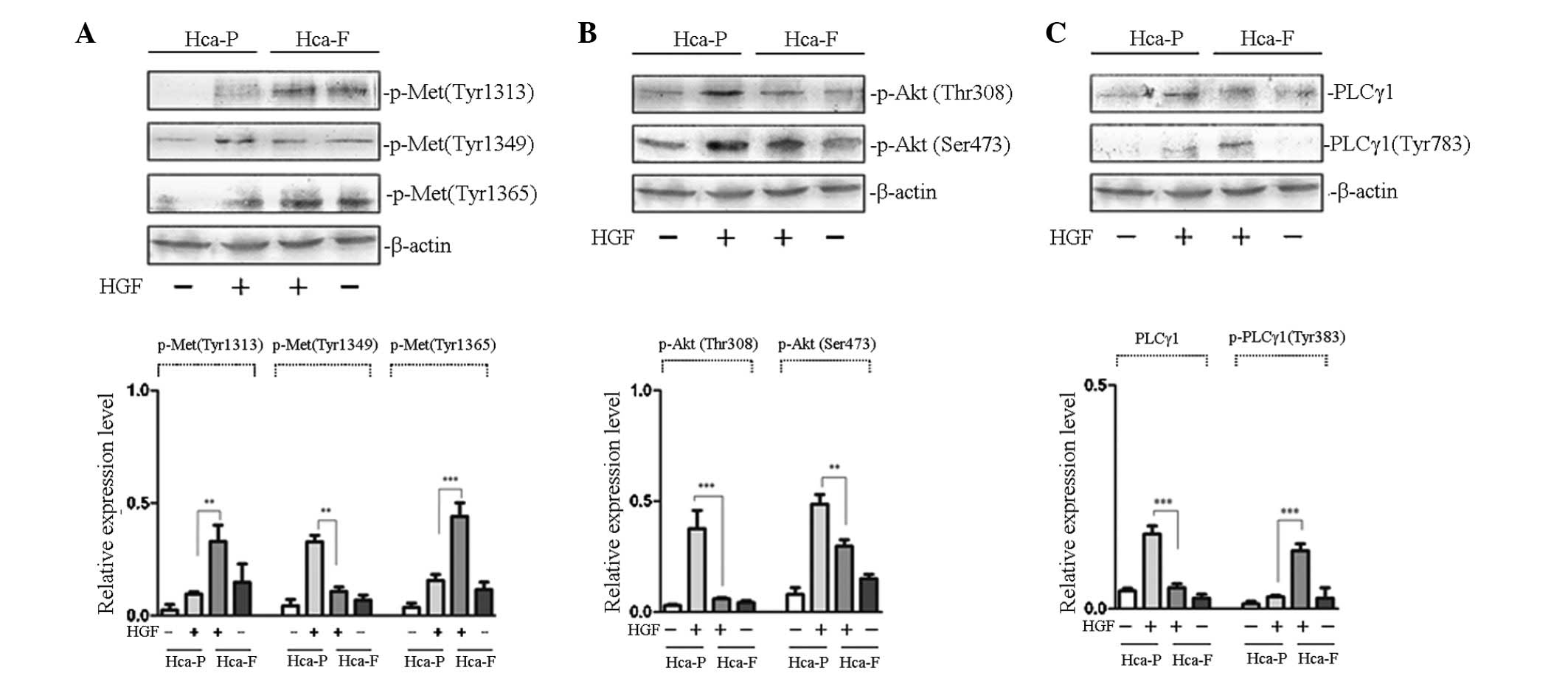
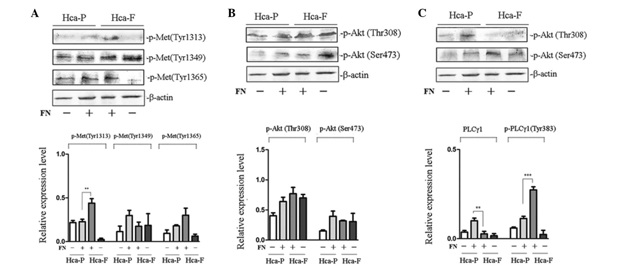
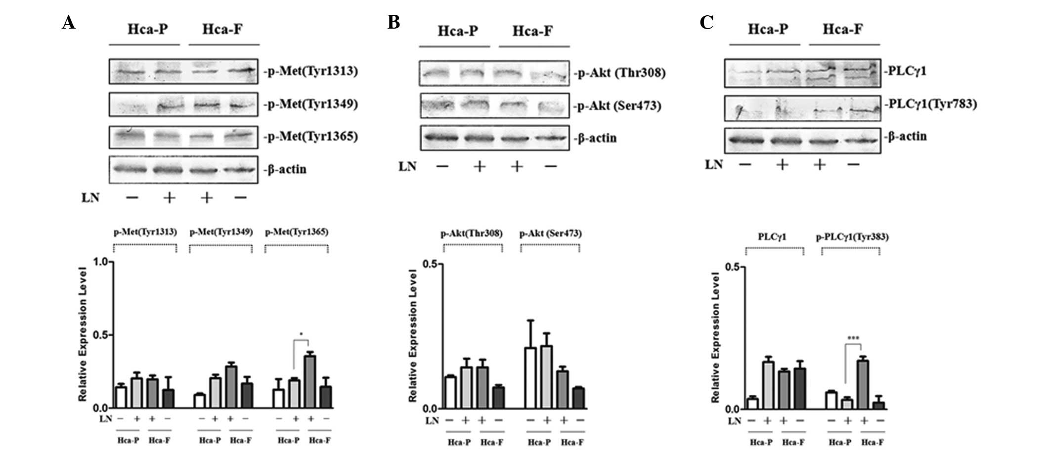
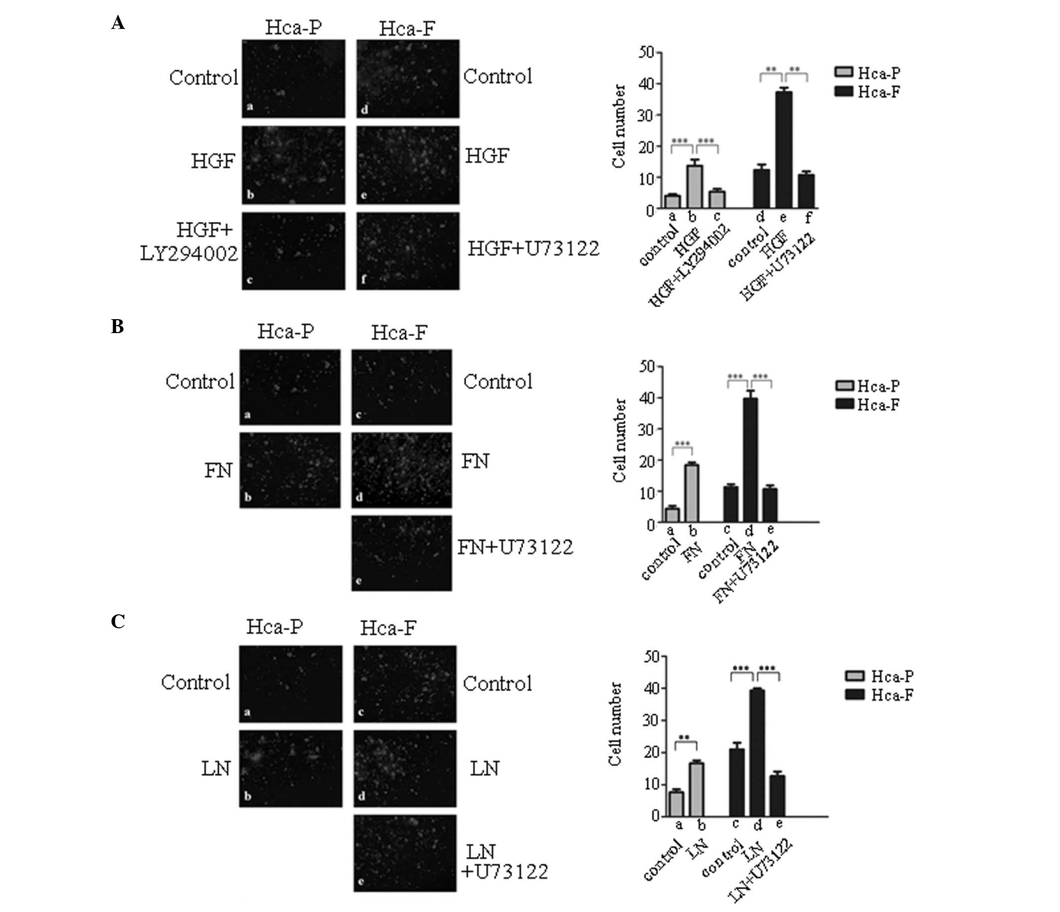
The aim of the present study was to determine the molecular mechanism by which the hepatocyte growth factor (HGF) receptor (cMet) regulates lymphatic metastasis in hepatocellular carcinoma. Mouse hepatoma ascites cell lines with different lymph node metastatic potentials, Hca‑F (high metastatic potential) and Hca‑P (low metastatic potential), were cultured in vitro. Cells were treated with HGF, fibronectin (FN) and laminin (LN), and the phosphorylated tyrosine residues of cMet and the activities of intracellular phospholipase Cγ/diacylglycerol/protein kinase C (PLCγ/DAG/PKC) and phosphoinositol‑3‑kinase/protein kinase B (PI3K/AKT) signaling pathways were analyzed comparatively in the two cell lines using western blot analysis and migration assays. Following HGF treatment, the phosphorylation of cMet at Tyr 1313 and 1365 in Hca‑F cells was higher, while the phosphorylation of cMet at Tyr 1349 was lower than that in Hca‑P. The activity of PLCγ/DAG/PKC was increased in Hca‑F cells compared with Hca‑P cells, whereas the activity of PI3K/AKT was reduced. After FN treatment, the phosphorylation of cMet at Tyr 1313 and the activity of the PLCγ/DAG/PKC signaling pathway was increased in Hca‑F cells compared with Hca‑P cells. Following LN treatment, the phosphorylation of cMet at Tyr 1365 and the activity of PLCγ/DAG/PKC was higher in Hca‑F cells than in Hca‑P cells. Results of the current study indicate that a number of ligands stimulate the phosphorylation of cMet at various tyrosine residues, activating different signaling transduction pathways. In addition, the same ligand was observed to phosphorylate different tyrosine residues on cMet in the two cell lines, as well as activate different intracellular signaling transduction pathways. After cMet is activated, various tyrosine residues are phosphorylated, leading to the activation of the PI3K/AKT and PLCγ/DAG/PKC signaling pathways to different extents in the two cells lines. These results may be important in determining the lymph node metastatic potentials of the two cell lines.
View Figures
View References

Related Articles

Journal Cover

August 2013
Volume 8 Issue 2

Print ISSN: 1791-2997
Online ISSN:1791-3004

Sign up for eToc alerts

Recommend to Library

Copy and paste a formatted citation
x
Spandidos Publications style
Li Y, Huang X, Zhang Q and Ma K: Phosphorylation of cMet tyrosine residues in murine ascitic hepatic cancer cell lines with different lymph node metastatic potentials. Mol Med Rep 8: 655-661, 2013.
APA
Li, Y., Huang, X., Zhang, Q., & Ma, K. (2013). Phosphorylation of cMet tyrosine residues in murine ascitic hepatic cancer cell lines with different lymph node metastatic potentials. Molecular Medicine Reports, 8, 655-661. https://doi.org/10.3892/mmr.2013.1527
MLA
Li, Y., Huang, X., Zhang, Q., Ma, K."Phosphorylation of cMet tyrosine residues in murine ascitic hepatic cancer cell lines with different lymph node metastatic potentials". Molecular Medicine Reports 8.2 (2013): 655-661.
Chicago
Li, Y., Huang, X., Zhang, Q., Ma, K."Phosphorylation of cMet tyrosine residues in murine ascitic hepatic cancer cell lines with different lymph node metastatic potentials". Molecular Medicine Reports 8, no. 2 (2013): 655-661. https://doi.org/10.3892/mmr.2013.1527