A pivotal role of the vascular endothelial growth factor signaling pathway in the formation of venous hypertension-induced dural arteriovenous fistulas
- Authors:
- Published online on: March 11, 2014 https://doi.org/10.3892/mmr.2014.2037
- Pages: 1551-1558
-
Copyright: © Li et al. This is an open access article distributed under the terms of Creative Commons Attribution License [CC BY_NC 3.0].
Metrics: Total
Views: 0 (Spandidos Publications: | PMC Statistics: )
Total PDF Downloads: 0 (Spandidos Publications: | PMC Statistics: )
Abstract
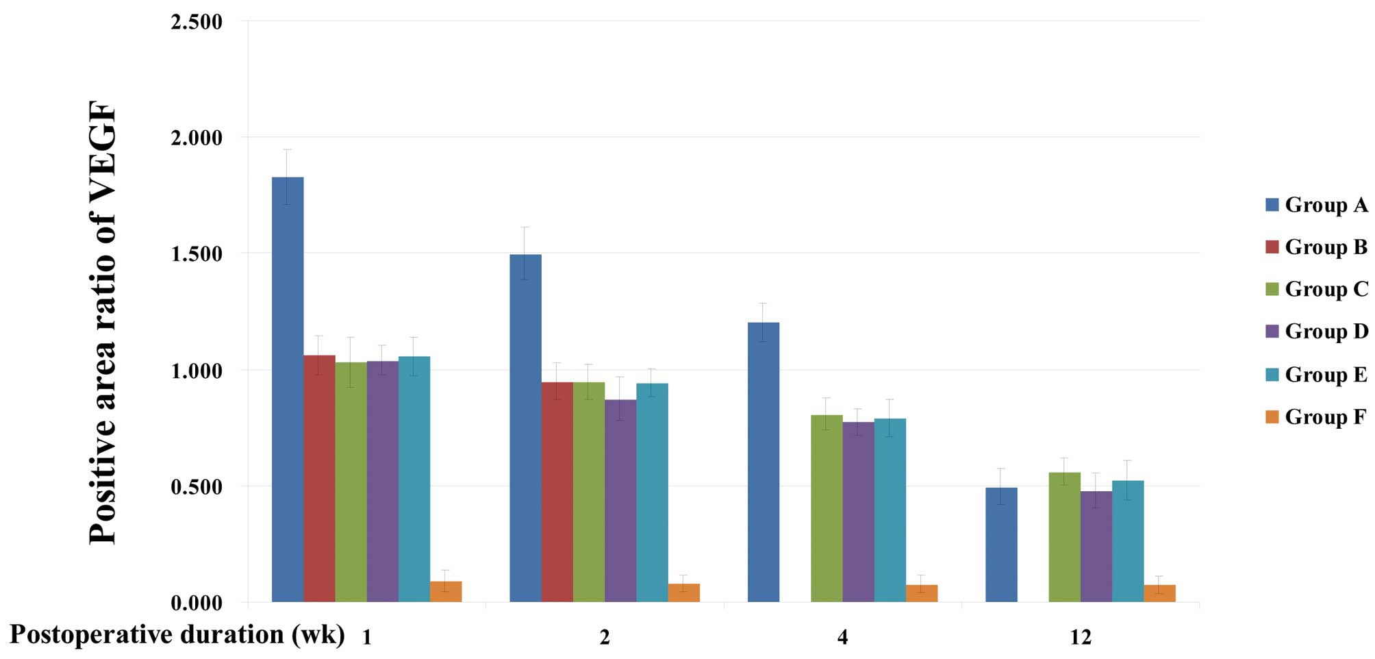
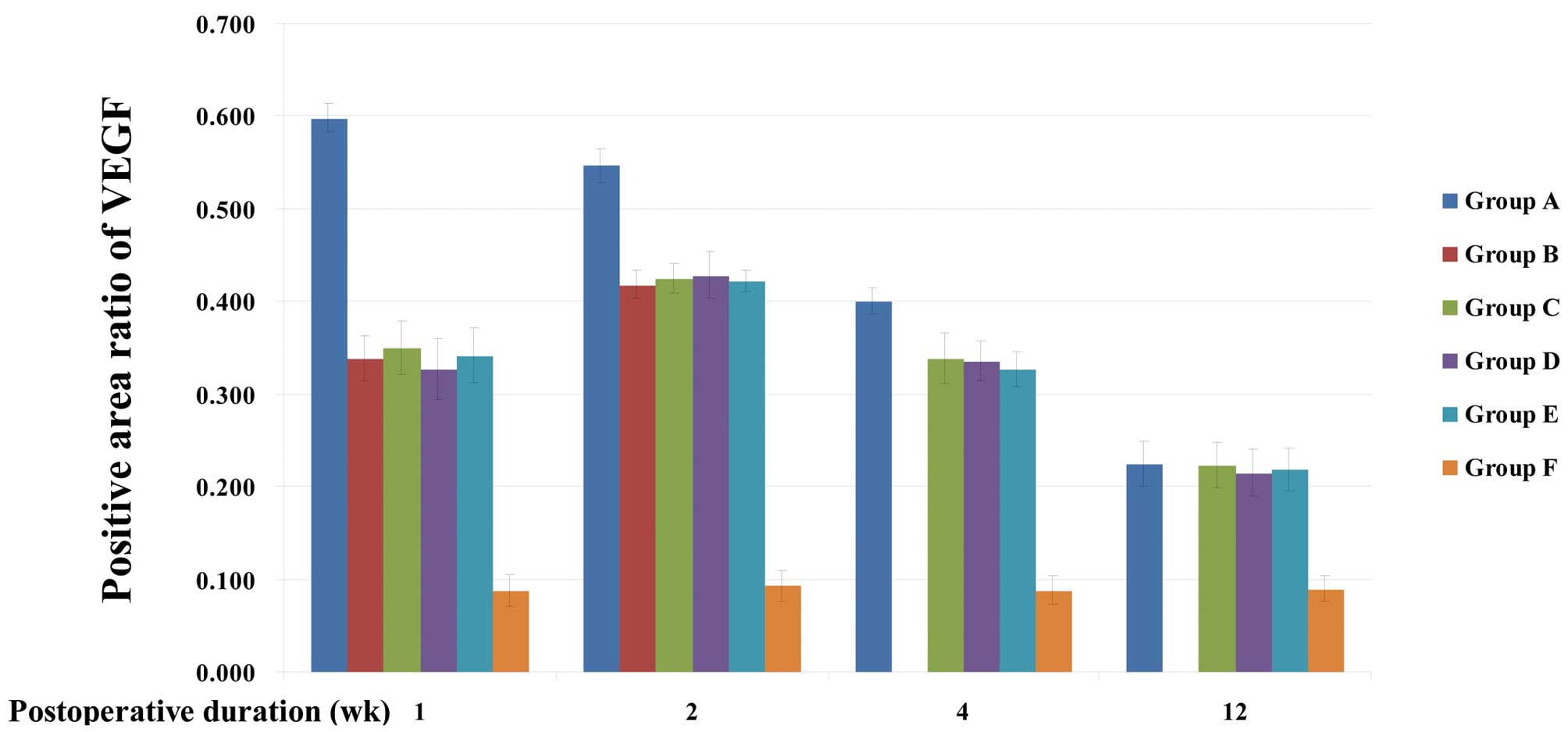
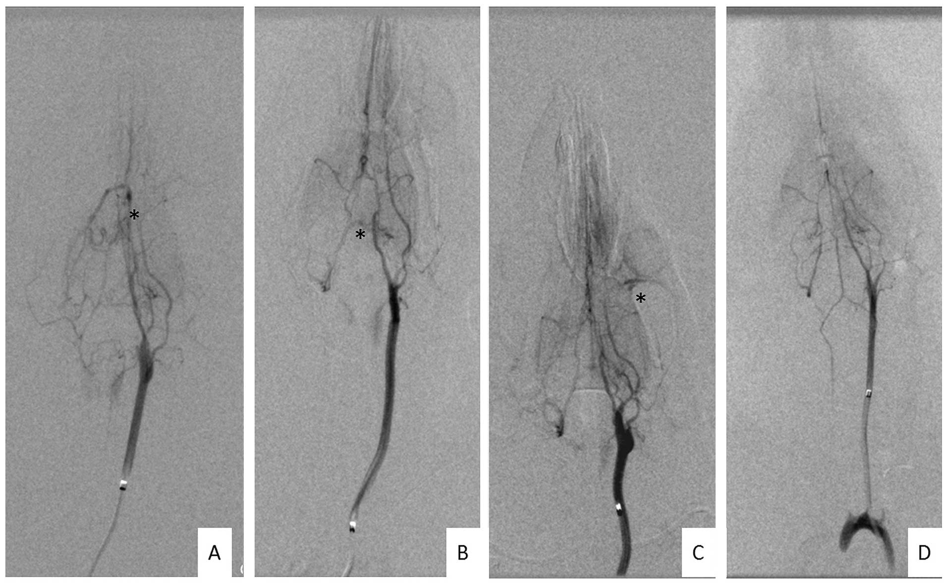
Dural arteriovenous fistulas (DAVFs) are associated with venous hypertension. Numerous studies have revealed high expression levels of vascular endothelial growth factor (VEGF) in human DAVF specimens, as well as in animal models of experimental venous hypertension. The objective of the present study was to clarify whether the VEGF signaling pathway is important in the development of DAVFs. Rats (n=216) were randomly divided into six groups. In the rats from five groups (groups A and C-E, n=45 in each group; group B, n=12), experimental venous hypertension was induced by right common carotid artery (CCA)‑external jugular vein (EJV) anastomosis, superior sinus occlusion and left transver sinus occlusion, while the remaining group (group F, n=24) underwent sham surgery. The rats in group A received a VEGF recombinant adenovirus injection into the distal section of the right EJV 30 min prior to anastomosis of the CCA and EJV. An equivalent control adenovirus was injected into the right EJV of group B rats prior to anastomosis. The rats in group C received no virus prior to anastomosis and no medicine subsequent to surgery. The group D rats were lavaged with Vatalanib, a VEGF receptor (VEGFR) inhibitor, and the group E rats were lavaged with an equal quantity of saline weekly following surgery. Six rats from groups A-E and one rat from group F were sacrificed in the first, second, fourth and twelfth weeks after surgery for immunohistochemical analysis of VEGF expression and analysis of microvessel density. Cerebral angiography was performed on the remaining rats in each group on the twelfth week after surgery. The results revealed that following transfection with VEGF recombinant adenovirus, angiogenesis in the dura mater of venous hypertensive rats was increased subsequent to the increase in the VEGF expression levels of the brain and dura mater. The rate of DAVF induction by venous hypertension was significantly reduced by the VEGFR antagonist due to reduced angiogenesis in the dura mater. In conclusion, VEGF and its receptor may be important in the formation of venous hypertension-induced DAVFs.