Effects of canine myoblasts expressing human cartilage‑derived morphogenetic protein‑2 on the repair of meniscal fibrocartilage injury
- Authors:
- Published online on: March 13, 2014 https://doi.org/10.3892/mmr.2014.2047
- Pages: 1767-1772
Metrics: Total
Views: 0 (Spandidos Publications: | PMC Statistics: )
Total PDF Downloads: 0 (Spandidos Publications: | PMC Statistics: )
Abstract
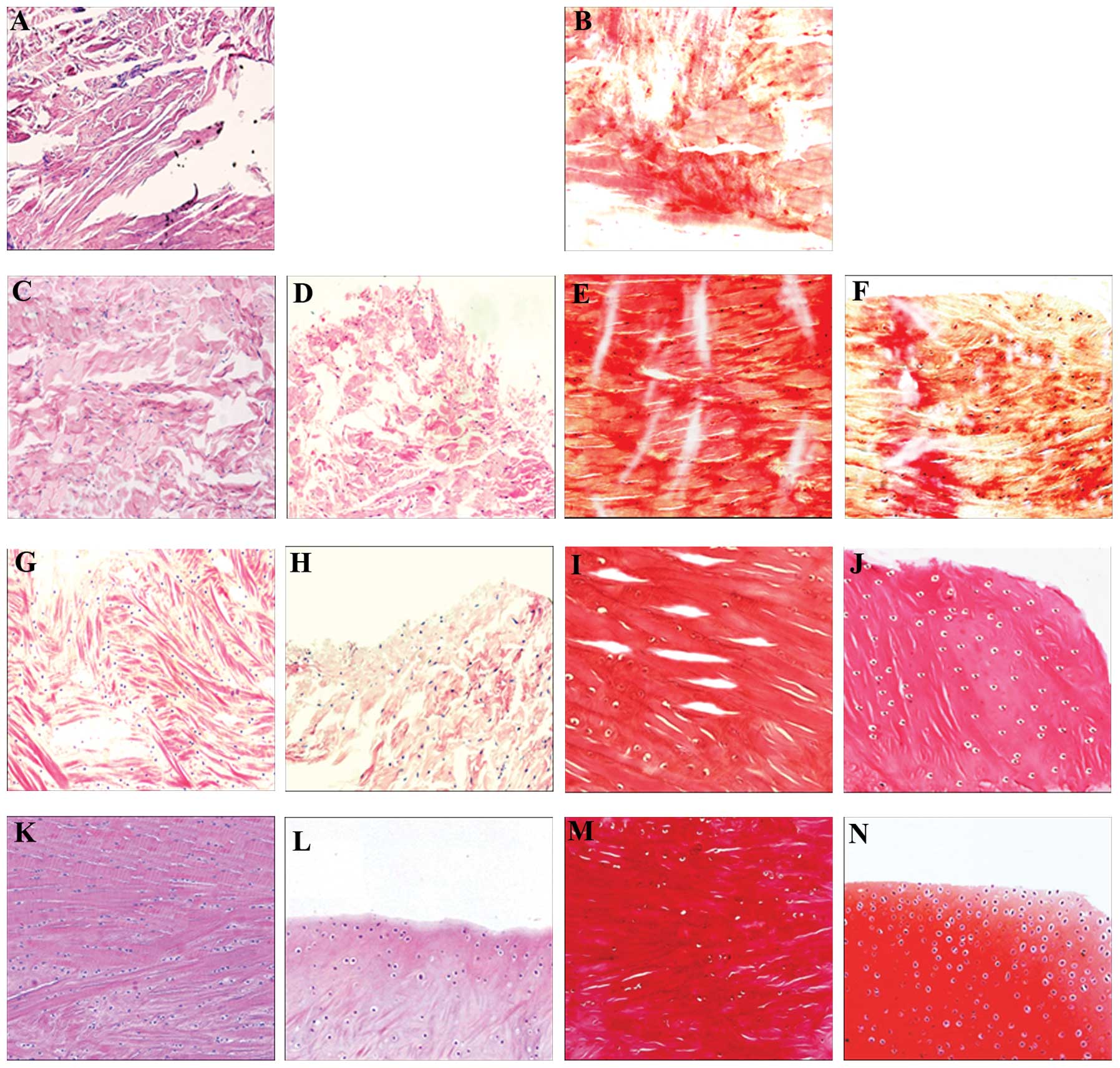
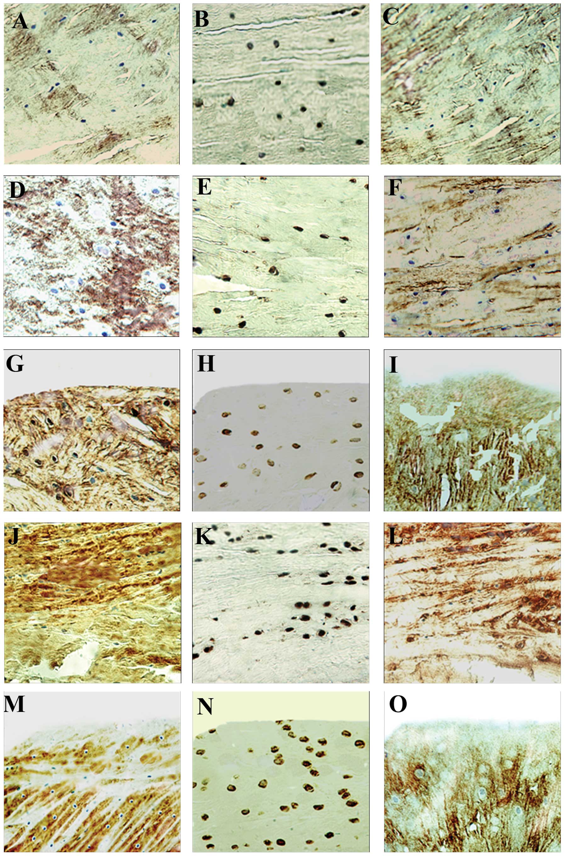
The aim of the present study was to explore the effects of human cartilage‑derived morphogenetic protein‑2 (hCDMP‑2)‑expressing canine myoblasts on the repair of meniscal fibrocartilage injury. Purified canine myoblasts were infected with lentiviruses carrying an empty vector or the hCDMP‑2 gene. The following four experimental groups were established to study the in vivo meniscal repair in a canine model of meniscal injury: Group A, suture only; group B, suture with the addition of the recombinant hCDMP‑2 on a polylactic acid/polyglycolic acid (PLA/PGA) scaffold; group C, a PLA/PGA scaffold with canine myoblasts carrying the empty vector; and group D, a PLA/PGA scaffold with canine myoblasts expressing hCDMP‑2. Samples of the regenerated tissue were extracted at weeks 3, 8 and 12 post‑repair and analyzed by morphological observation, immunohistochemistry (IHC) and quantitative analysis. At week 12 post‑repair, the scaffold material had completely dissolved in the control groups and no changes were observed at the injured area, while regenerated tissue was observed in group D only. Hematoxylin and eosin and Safranin‑O staining techniques further revealed cartilage lacunae and fibers present at the red‑red zone of the repaired tissue, while cartilage lacunae without fibers were observed at the white‑white zone in group D. In addition, IHC studies demonstrated that collagen I and II, and the S‑100 protein were expressed at the red‑red and the white‑white zones of the repaired tissue in group D. It was concluded that purified canine myoblasts expressing the hCDMP‑2 gene were able to promote meniscal fibrocartilage healing by regenerating fibrocartilage‑like tissue. The tissue in the red‑red zone was regenerated more rapidly than that in the white‑white zone. Further studies are required to identify the best way to combine hCDMP‑2 growth factor with myoblasts for use in the clinic due to the limitations regarding the clinical use of lentiviral infections.