Introduction
Hepatocellular carcinoma (HCC) is one of the most
prevalent tumor types (1). HCC
represents a major histological subtype, accounting for 70–85% of
total primary liver cancers worldwide (2), and is the third most common cause of
cancer-related mortality (3).
Hepatocarcinogenesis is a generally slow process, with initial
genomic changes progressively altering the hepatocellular phenotype
to produce cellular intermediates that evolve into HCC (4). HCC has a number of interesting
epidemiologic features, such as dynamic temporal changes and marked
variations among geographic regions and between genders. China
alone accounts for >50% of the HCC cases worldwide (5).
Orthotopic liver transplantation (OLT) is a rational
therapeutic option for patients with HCC (6), but recurrence still remains the main
cause of death in HCC patients subjected to OLT (7). To reduce recurrence, an increasing
number of physicians focus on strict selection criteria for
transplantation candidates on the basis of tumor features (8). Tumor recurrence or metastasis is
still an important and unresolved issue (9). Pharmacologic immunosuppression,
required after transplantation, can accelerate tumor growth,
although the effects of different immunosuppressive agents and
regimes on HCC recurrence following OLT remain unclear (10).
Tacrolimus (FK506), a nonsteroidal topical
immunomodulator, is an immunosuppressive drug widely used in
patients subjected to organ transplantation (11,12).
FK506 is believed to exert its immunosuppressive effect through
targeted binding and inactivation of calcineurin (13). In previous studies, FK506 was
reported to promote recurrence following lung transplantation
(14), to enhance the invasive
potential of HCC cells and to promote lymphatic metastasis
(15). However in another study,
FK506 did not promote the proliferation of rat HCC cells (16). Thus, the effects of FK506 on
proliferation of cancer cells are still unclear.
Chemokines are soluble proteins of low molecular
mass (5–15 kDa) that can mediate their corresponding effects by
binding to specific, seven-transmembrane domain, G protein-coupled
receptors (17). Chemokines and
their receptors mediate the recruitment of immune cells in a number
of diseases. In different tumor types, expression of the C-X-C
chemokine receptor type 4 (CXCR4) has been associated with tumor
dissemination and poor prognosis (18,19).
Most particularly, the protein complex CXCR4/stromal cell-derived
factor-1α (SDF-1α) is considered to play a central role in breast
carcinoma metastasis (20), and
might be involved in regulation of the metastatic behavior of tumor
cells (21), as well as in
metastasis of CXCR4+ tumor cells into the bone marrow
and lymph nodes (22). However,
whether the CXCR4/SDF-1α complex and downstream signaling has an
effect on HCC remains uncertain.
In this context, the aim of the present study was to
investigate the potential effects of FK506 and AMD3100, which is
the antagonist of CXCR4, on the Morris rat hepatoma cell line
MH3924A. Briefly, the proliferation, morphological changes and
invasiveness of MH3924A cells, as well as the expression of CXCR4
in these cells, were examined in vitro. In addition, the
growth and invasiveness of an implanted tumor, the expression of
CXCR4 in tumor tissues and the expression of SDF-1α in the adjacent
tissues to the HCC ones, were examined in vivo, in August
Copenhagen Irish rats.
Materials and methods
Cell culture
Morris rat hepatoma 3924A cells (MH3924A) were
purchased from the Institut für angewandte Zellkultur, (Munich,
Germany). Cell lines were cultured in Dulbecco’s modified Eagle’s
medium (DMEM), with 10% heat-inactivated fetal bovine serum (FBS;
Invitrogen Life Technologies, Carlsbad, CA, USA) at 37°C in a
humidified incubator with an atmosphere of 5% CO2. When
the fusion rate reached >80%, the cells were digested with 25%
trypsin and serial passages were performed at a dilution rate of
1:3.
MTT assay
Tumor cell proliferation was determined by the
3-(4,5-dimethylthiazol-2-yl)-2,5-diphenyltetrazolium bromide assay
(MTT; Sigma-Aldrich, St. Louis, MO, USA). Briefly, after MH3924A
cells had reached the logarithmic growth phase, a 0.2-ml cell
suspension at 1×10⁴ cells/ml was added into each well of
a 96-well plate and cultured in DMEM with 10% FBS, 10 μg/l vascular
endothelial growth factor and 0.1 g/l heparin for 24 h. When
adherent growth was established, different concentrations of FK506
(10, 100 and 1,000 μg/l), AMD3100 (10, 50 and 100 μg/l) and FK506
(0 and 100 μg/l) + AMD3100 (0, 10, 50 and 100 μg/l) were added into
the plates. Untreated cells cultured in medium alone were used as
controls. After culturing for 48 h, 10 μl MTT (5 g/l) were added,
and each well was incubated for 6 h; next, 150 μl/well dimethyl
sulfoxide were added, followed by measurements of the absorbance at
570 mm on a spectrophotometer reader (Dynatech MRX, Elvetec, Genas,
France). Each well was measured three times, and each sample was
assayed in triplicate.
Immunohistochemical assay and scanning
electron microscopy (SEM)
MH3924A cells were cultured on sterile cover glasses
(8×8 mm2) for 1 day, then cultured in DMEM with 100 μg/l
FK506, or 50 μg/l AMD3100, or 100 μg/l FK506 + 50 μg/l AMD3100.
When the cover glasses were covered with cells, the morphologic
changes of MH3924A cells were observed with SEM, while a number of
cover glasses were used for the immunohistochemical assay to
determine the expression of CXCR4. This assay was performed using
the anti-CXCR4 antibody (1:100 dilution; Wuhan Boshide
Biotechnology Co., Wuhan, China) as the primary antibody and a
horseradish peroxidase-conjugated secondary antibody (1:100;
DakoCytomation, Glostrup, Denmark). The formed immunocomplex was
visualized using the 3,3′-diaminobenzidine (DAB) reagent. The level
of CXCR4 was quantified by a computer-assisted image system, which
included a Leica CCD camera DFC420 connected to a Leica DM IRE2
microscope (Leica Microsystems, Wetzlar, Germany) (23,24).
Image analysis yielded integrated optical density (IOD) values.
Transwell invasion assay
Invasion assays were performed in 8-μm, 24-well,
BioCoat Matrigel invasion chambers (Corning, Cambridge, MA, USA).
Briefly, 1×10⁴ cells/ml were suspended in serum-free
medium, and 50 μl of the suspension were added to each chamber,
while 500 μl of the culture medium containing different
concentrations and combinations of FK506, AMD3100 and SDF-1α
(Table I) were added to the bottom
chamber. Cells were allowed to invade for 12 h at 37°C in a
humidified incubator with an atmosphere of 5% CO2; the
cells that migrated to the top chamber were stained with Giemsa
(Sigma-Aldrich) and were counted at a ×400 magnification under an
electron microscope (Olympus, Tokyo, Japan). Assays were performed
3 times using triplicate wells.
 | Table IThe groups in the transwell invasion
assay. |
Table I
The groups in the transwell invasion
assay.
| Group | Top chamber | Bottom chamber |
|---|
| Control | DMEM | DMEM |
| FK506 | 10 μg/l FK506 | DMEM |
| AMD3100 | 10 μg/l
AMD3100 | DMEM |
| SDF-1α | DMEM | 1 μg/l SDF-1α, 10
μg/l SDF-1α |
| FK506 +AMD3100 | 10 μg/l FK506, 10
μg/l AMD3100 | DMEM |
| FK506+SDF-1α | 10 μg/l FK506 | 10 μg/l SDF-1α |
| AMD3100
+SDF-1α | 10 μg/l
AMD3100 | 10 μg/l SDF-1α |
| FK506+AMD3100
+SDF-1α | 10 μg/l FK506, 10
μg/l AMD3100 | 10 μg/l SDF-1α |
Rat model of liver tumor
Experiments were performed in 16 healthy August
Copenhagen Irish rats (male, 16–20 weeks, weighing 240–300 g). The
animals were handled in accordance with the regulations for
laboratory animals. All animal experiments were in accordance with
national guidelines and approved by the ethical committee of
Zhongshan Hospital (Shanghai, China). The rat model of liver tumor
was established as follows: First, MH3924A cells were collected and
injected into the alar skin of rats. The tumors were removed from
alar skin when grown to 2×1×1 mm3, and intrahepatic
tumor implantation of rats was performed under aseptic conditions
as described previously (25,26).
Five days later, rats were randomly divided into two groups: one
group was subcutaneously injected with normal saline for 14 days
(NS group, n=8, 3 mg/kg/day), and the second group was
subcutaneously injected with FK506 for 14 days (FK506 group, n=8,
0.3 mg/kg/day). Forty days following implantation, rats were
sacrificed, and the weight of tumor, the volume of the fluid in the
ascites, the incidence of lymphatic metastasis in the abdominal
cavity and of abdominal wall metastasis were measured. In addition,
the lungs were irrigated with 15% Indian ink, followed by counting
of the number of metastatic nodules in the lung. The tumor and
adjacent tissues, as well as healthy liver tissues, were harvested
and preserved in 4% formalin for later use.
Immunohistochemical assay
The tumor and adjacent tissues along with healthy
liver tissues were sectioned (4 μm) according to the EliVision
method (27). First, all sections
were dried at 92°C for 30 min, treated with a 100% dimethylbenzene,
3% formalin-H2O2 solution, and washed with
phosphate-buffered saline. Second, the sections were incubated with
polyclonal antibodies of CXCR4 and SDF-1α purchased from Santa Cruz
Biotechnology, Inc. (Santa Cruz, CA, USA) and Boster Biotechnology
(Wuhan Boster Biological Technology, Ltd., Wuhan, Hubei, China),
respectively, colored with DAB reagent and counterstained with
hematoxylin and eosin. Following washing in PBS and drying and
mounting in buffered glycerol-saline (90% glycerol, 10% PBS), the
sections were observed under an inverted confocal microscope
(DMRIE2; Leica Microsystems, Wetzlar, Germany). Sections were
observed at a ×500 magnification, and 5 pictures were randomly
selected and saved. The analysis of these images yielded IOD
values.
Statistical analysis
Data with a normal distribution are expressed as
mean ± standard deviation (SD), while median values are used to
represent data that were not normally distributed. We used the
Windows version of the SPSS 16.0 software (SPSS, Inc., Chicago, IL,
USA) to perform one-way analysis of variance for the comparison of
normally distributed data, and the Mann-Whitney rank sum test
implemented in the Image-Pro Plus (IPP) v6.0 software (Media
Cybernetics, Inc., Bethesda, MD, USA) to compare the data that were
not normally distributed, as for example the IOD values from the
immunohistochemical assay. P<0.05 and P<0.01 were considered
to indicate statistically significant differences.
Results
MTT assay
As shown by the MTT assay (Table II), treatment with a low
concentration of FK506 (10 μg/l) did not significantly affect the
proliferation of MH3924A cells (P=0.135). Upon treatment with
higher concentrations of FK506 (100–1,000 μg/l), the proliferation
of MH3924A cells was significantly enhanced (P<0.01). Treatment
with AMD3100 at any concentration (10, 50 or 100 μg/l), had no
obvious effect on MH3924A cell proliferation (P>0.05). However,
when different concentrations of AMD3100 were combined with 100
μg/l FK506, the in vitro proliferation of MH3924A cells was
increased (P<0.01, Table
III). These data suggested that FK506 (≥100 μg/l) can promote
the proliferation of MH3924A cells and that AMD3100 has no effect
on the FK506-induced increase in proliferation.
 | Table IIThe absorbance values of MH3924A
cells treated with different concentrations of FK506 or
AMD3100. |
Table II
The absorbance values of MH3924A
cells treated with different concentrations of FK506 or
AMD3100.
| Group | 0 μg/l | 10 μg/l | 100 μg/l | 1,000 μg/l |
|---|
| FK506 |
| Absorbance
value | 0.80±0.17 | 1.17±0.49 | 1.53±0.39 | 1.64±0.19 |
| P | - | 0.135 | 0.006a | 0.002a |
| AMD3100 |
| Absorbance
value | 0.80±0.17 | 0.84±0.06 | 0.86±0.10 | 0.85±0.10 |
| P | - | 0.760 | 0.788 | 0.812 |
 | Table IIIThe absorbance values of MH3924A
cells treated with different concentrations of FK506 combined with
different concentrations of AMD3100. |
Table III
The absorbance values of MH3924A
cells treated with different concentrations of FK506 combined with
different concentrations of AMD3100.
| AMD3100 (μg/l) | FK506 |
|---|
|
|---|
| 0 μg/l | 100 μg/l | P |
|---|
| 0 | 0.80±0.17 | 1.53±0.39 | 0.006a |
| 10 | 0.84±0.06 | 1.79±0.10 | 0.000a |
| 50 | 0.86±0.10 | 1.46±0.27 | 0.005a |
| 100 | 0.85±0.10 | 1.55±0.31 | 0.002a |
CXCR4 expression
The effects of FK506 and AMD3100 on the expression
of CXCR4 were studied in MH3924A cells with an immunohistochemical
assay (Fig. 1). In non-treated
MH3924A cells (Fig. 1A), CXCR4
expression was detectable at medium levels. CXCR4-stained particles
were mainly distributed in the cytoplasm and the extracellular
matrix, were occasionally present on the cytomembrane, and were
absent from the nucleus. Following treatment with 100 μg/l FK506
(Fig. 1B), stronger staining of
CXCR4 was observed, although this increase was not significant
(P>0.05). By contrast, upon treatment with 50 μg/l AMD3100
(Fig. 1C), no or weaker expression
of CXCR4 was observed compared to non-treated cells (P<0.05).
The expression of CXCR4 was also negative or weaker in MH3924A
cells treated with 100 μg/l FK506 combined with 50 μg/l AMD3100,
compared to non-treated cells (P<0.05). These results suggested
that the expression of CXCR4 is marginally increased by treatment
with 100 μg/l FK506, but decreased by treatment with AMD3100 or
FK506 + AMD3100.
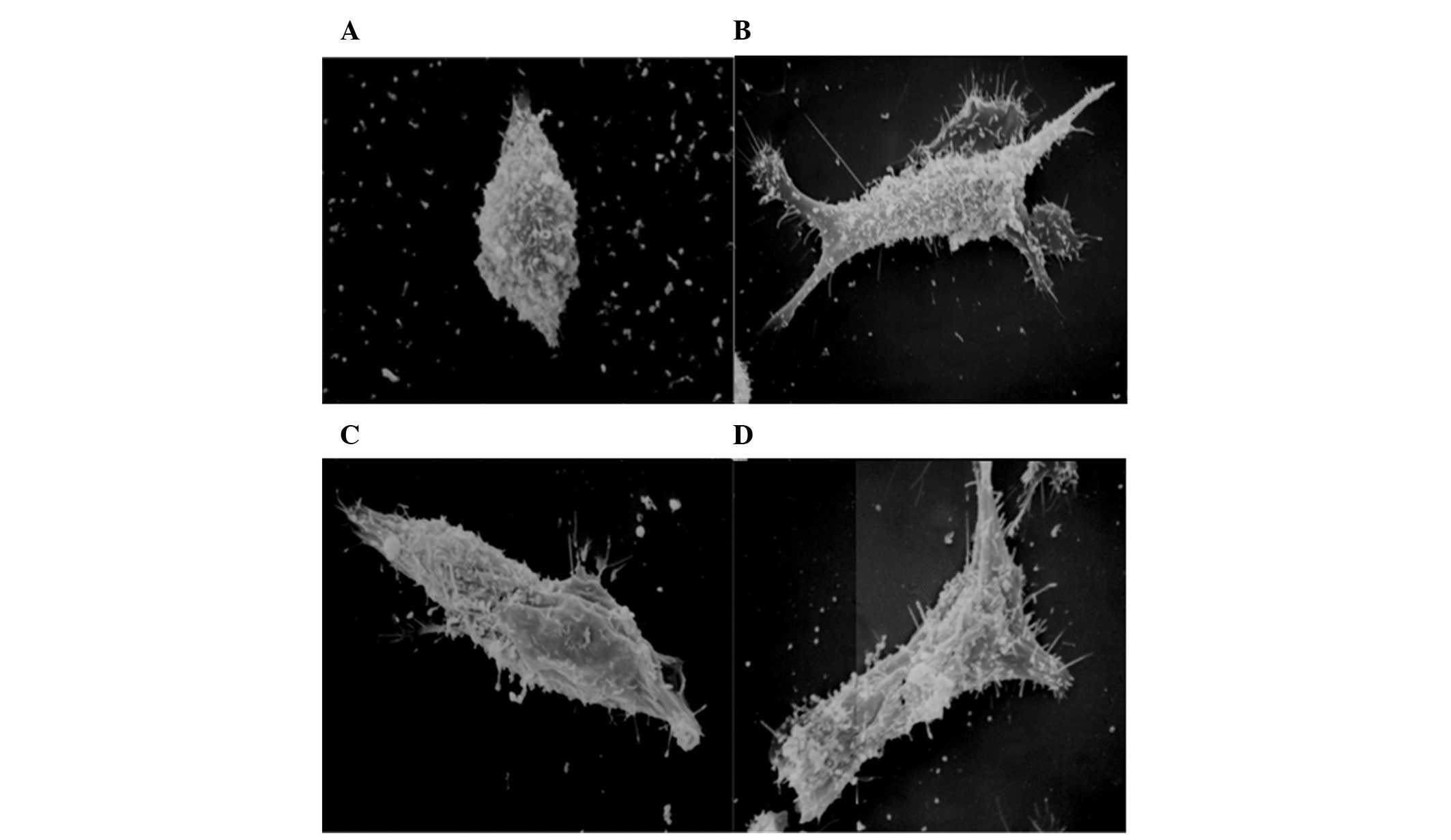
SEM
Examination of the cell morphology was performed
with SEM (Fig. 2). The shape of
non-treated MH3924A cells was regularly elliptical, and their
surface was covered with microvilli and a few immobile processes
(Fig. 2A). The MH3924A cells that
were treated with 100 μg/l FK506 displayed a higher number of
microvilli, longer immobile processes, and their shape was
irregularly expanded compared to non-treated cells (Fig. 2B), suggesting stronger invasive
ability. The cells treated with 50 μg/l AMD3100 were regularly
elliptical similar to non-treated cells, while the number of
microvilli and immobile processes did not significantly change
(Fig. 2C). In addition, the
morphology of MH3924A cells that were treated with 100 μg/l FK506
and 50 μg/l AMD3100 (Fig. 2D), was
similar to that of cells treated with 100 μg/l FK506, displaying a
higher number of microvilli and immobile processes, and irregular
expansions; this morphology indicates increased cell
invasiveness.
Invasion assay
MH3924A cells are invasive. Therefore, their
invasive ability was measured in vitro (Fig. 3). In the FK506 group, the number of
invasive cells was significantly increased compared to the NS group
(P<0.01). AMD3100 treatment had no significant effect on
invasive ability compared to the NS group (P=0.09), but it clearly
decreased the invasiveness of cells compared to treatment with
FK506 (P=0.046) or SDF-1α (P=0.032), similarly to the NS group. In
the SDF-1α group, the number of invasive cells was significantly
increased compared to the NS group (P<0.01), but no significant
difference was observed in the comparison with the FK506 group
(P=0.881).
The number of invasive cells after treatment with
the combination FK506 + AMD3100 was significantly increased
compared to the NS group (P<0.01), but was not significantly
affected compared to the FK506 (P=0.607) and SDF-1α (P=0.507)
groups. In addition, the number of invasive cells was significantly
increased in the FK506 + SDF-1α compared to the NS group
(P<0.01), and was increased compared to the FK506 and the SDF-1α
groups, but this increase was not significant (P=0.653 and P=0.548,
respectively). Moreover, the number of invasive cells in the
AMD3100 + SDF-1α group did not significantly change compared to the
NS group (P=0.864). In the FK506 + AMD3100 + SDF-1α group, the
invasive ability of MH3924A cells was significantly enhanced
compared to the NS group (P<0.01), but not significantly
enhanced in comparison to the FK506 (P=0.983) and the SDF-1α
(P=0864) groups. These results showed that both FK506 and SDF-1α
can enhance the invasive ability of MH3924A cells, and that AMD3100
can reduce the invasive ability of MH3924A cells when combined with
SDF-1α.
In vivo effects of tacrolimus
Following treatment with NS or FK506, none of the
rats died or failed to keep the tumor implant until the day of
observation. Tumor growth was examined after the implantation
(Table IV). The liver of rats of
the NS group was large, with an average weight at 15.56±11.17 g
(Fig. 4A), and the liver of rats
of the FK506 group was oversize, with an average weight at
28.19±3.89 g (Fig. 4B); no
significant difference was found between the two groups (P=0.041).
Regarding the number of metastatic nodules in the lungs, this was
significantly increased in the FK506 compared to the NS group
(6.50±4.63 vs. 1.39±1.25, P=0.012) (Fig. 4C–D). Moreover, the ascite fluid
volume was increased in rats of the FK506 group compared to the NS
group, although this change was not statistically significant
(21.25±6.94 vs. 13.13±21.87 ml, P=0.317). The rate of lymph node
metastasis, as well as the number of pulmonary nodules, were
significantly increased in the FK506 compared to the NS group
(P=0.002 and P=0.012, respectively). However, no significant
difference in the rate of abdominal wall metastasis was observed
between the two groups (P=0.442).
 | Table IVParameters of the in vivo HCC
models following treatment with FK506 and NS. |
Table IV
Parameters of the in vivo HCC
models following treatment with FK506 and NS.
| Parameters | NS | FK506 | P |
|---|
| Tumor weight
(g)a | 15.56±11.17 | 28.19±3.89 | 0.041c |
| Ascite fluid
(ml)a | 13.13±21.87 | 21.25±6.94 | 0.317 |
| Lymph node
metastasisb | 0 | 8 | 0.002c |
| Abdominal wall
metastasisb | 2 | 5 | 0.442 |
| Pulmonary nodules
(n)a | 1.39±1.25 | 6.50±4.63 | 0.012c |
| Expression of
CXCR4a | 1.48±0.29 | 2.50±0.62 | 0.048c |
| Expression of
SDF-1αa | 1.46±0.39 | 2.54±0.94 | 0.026c |
Immunohistochemical detection of CXCR4
and SDF-1α
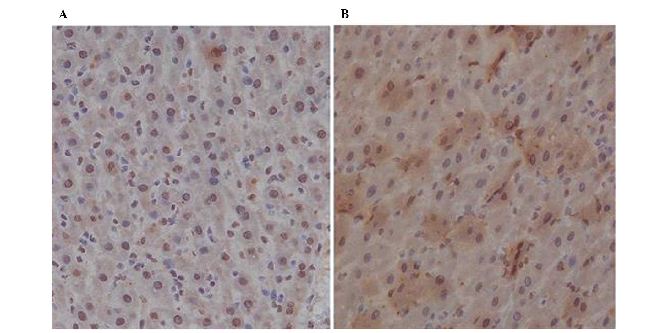
Immunohistochemical staining was performed in order
to detect the expression of CXCR4 in tumor HCC tissues and of
SDF-1α in the tissues adjacent to the HCC ones (Table IV). In the NS group, CXCR4 was
expressed at medium levels, with an average IOD at 1.48±0.29
(Fig. 5A), but in the FK506 group,
the expression of CXCR4 was higher, with an average IOD at
2.50±0.62 (Fig. 5B). The increased
expression of CXCR4 following FK506 treatment was significant
(P=0.048). In addition, the expression level of SDF-1α in tissues
adjacent to the HCC ones was medium in the NS group, with an
average IOD at 1.46±0.39 (Fig.
6A). In the FK506 group (Fig.
6B), the expression of SDF-1α in the adjacent tissues was
significantly higher compared to the NS group (P=0.026), with an
average IOD at 2.54±0.94.
Discussion
OLT is the best therapeutic option for patients with
HCC. However, the pharmacologic immunosuppression regimes required
at the post-transplantation stage can affect the recurrence of HCC
by accelerating tumor growth, and the effects of immunosuppression
on post-OLT HCC recurrence have been poorly investigated (8). Therefore, finding an effective method
to reduce the recurrence of HCC while using immunosuppressive
agents is an issue of great importance in the clinic.
In this study, we investigated the effects of the
immunosuppressive drug tacrolimus (FK506) on the proliferation of
MH3924A cells. The growth of MH3924A cells was significantly
increased following treatment with 100 μg/l FK506, while 10 μg/l
FK506 had no significant effect on cell growth, thus indicating
that high concentrations of FK506 can promote the proliferation of
MH3924A cells in vitro. Moreover, in an in vivo HCC
rat model, the metastatic rate of tumor in the abdominal wall, the
number of lymph nodes, as well as the lung size, were increased
upon FK506 treatment compared to treatment with NS. This result was
consistent with the in vitro experiments. Therefore, FK506
can promote the proliferation of MH3924A cells. Treatment with
FK506 resulted in a dose-dependent increase in the number of
pulmonary metastases in a previous study (28), and stimulated the Rho/ROCK
signaling pathway to enhance the invasiveness of HCC (16). This is evidence that FK506 may
promote the progress of HCC (29).
Moreover, the effect of the CXCR4/SDF-1α complex in
HCC was investigated in the present study. The SDF-1α chemokine and
its receptor were suggested to play an important role in metastasis
towards lymph nodes in cervical cancer (30). In our study, CXCR4 was found to be
expressed in MH3924A cells. When the cells were treated with FK506,
the expression of CXCR4 did not significantly change, while
treatment with FK506 and AMD3100 significantly decreased its
expression. These results suggest that FK506 can not alter the
expression of CXCR4 in MH3924A cells. Nevertheless, in our in
vivo experiments, the expression of CXCR4 in tumor tissues, as
well as the expression of SDF-1α in the tissues adjacent to the HCC
ones, was increased following FK506 treatment. This might be due to
the metastatic potential of MH3924A cells. In a previous study, the
HCC cell line HepG2 was found to be unresponsive to SDF stimulation
due to an unknown defect, which was identified to involve a step
after receptor binding but before the activation of the signaling
cascade (31). CXCR4 was also
reported to be ‘trapped’ in the cytoplasm and not recruited to the
cell surface in response to standard extrinsic stimuli in the
majority of HCC cell lines, resulting in a negligible response to
SDF-1 (32). Hence, these two
proteins might be involved in the HCC invasion process, although
their specific roles need to be further investigated.
The invasiveness of MH3924A cells was also studied
in vitro. FK506 treatment significantly increased the
invasiveness of MH3924A cells, while AMD3100 treatment decreased
it. Treatment with SDF-1α also clearly enhanced the invasiveness of
MH3924A cells, similar to FK506, and this increase was also
observed for the treatment combining FK506 with AMD3100 and/or
SDF-1α. By contrast, AMD3100 + SDF-1α treatment reduced cell
invasiveness to levels comparable to those of the NS group. These
results are in agreement with previous studies, reporting that the
biological effects of SDF-1 are strongly inhibited by AMD3100
(33) and that AMD3100 can block
HIV-1 entry via its antagonistic effect on CXCR4 (34). Similarly, our results indicated
that both FK506 and SDF-1α enhance the invasiveness of MH3924A
cells, and that AMD3100 attenuates the effects of FK506 by blocking
CXCR4.
In conclusion, FK506 promotes the proliferation of
MH3924A cells, increases the expression of CXCR4 in tumor tissues
and that of SDF-1α in adjacent tissues to the HCC ones, enhances
the invasiveness of MH3924A cells and significantly intensifies a
number of pathological features. Moreover, SDF-1α increases, while
AMD3100 decreases, the invasiveness of MH3924A cells in
vitro, potentially by blocking the formation of the
CXCR4/SDF-1α complex. Therefore, minimizing the use of FK506 may
reduce the recurrence of OLT, and the CXCR4/SDF-1α interaction may
play vital roles in HCC metastasis. However, whether CXCR4/SDF-1α
can be used as a new target of prevention and treatment of OLT
needs to be further investigated.
Acknowledgements
This study was supported by the National Key
Sci-Tech Special Project of China (no. 2012ZX10002-016) and grants
from the National Natural Science Foundation of China (nos.
81272574 and 81172277).
References
|
1
|
Jemal A, Siegel R, Ward E, et al: Cancer
statistics, 2008. CA Cancer J Clin. 58:71–96. 2008. View Article : Google Scholar
|
|
2
|
Jemal A, Bray F, Center MM, Ferlay J, Ward
E and Forman D: Global cancer statistics. CA Cancer J Clin.
61:69–90. 2011. View Article : Google Scholar
|
|
3
|
Cheng AL, Kang YK, Chen Z, et al: Efficacy
and safety of sorafenib in patients in the Asia-Pacific region with
advanced hepatocellular carcinoma: a phase III randomised,
double-blind, placebo-controlled trial. Lancet Oncol. 10:25–34.
2009. View Article : Google Scholar : PubMed/NCBI
|
|
4
|
Thorgeirsson SS and Grisham JW: Molecular
pathogenesis of human hepatocellular carcinoma. Nat Genet.
31:339–346. 2002. View Article : Google Scholar : PubMed/NCBI
|
|
5
|
El-Serag HB and Rudolph KL: Hepatocellular
carcinoma: epidemiology and molecular carcinogenesis.
Gastroenterology. 132:2557–2576. 2007. View Article : Google Scholar : PubMed/NCBI
|
|
6
|
Yao FY, Ferrell L, Bass NM, et al: Liver
transplantation for hepatocellular carcinoma: expansion of the
tumor size limits does not adversely impact survival. Hepatology.
33:1394–1403. 2001. View Article : Google Scholar : PubMed/NCBI
|
|
7
|
Escartin A, Sapisochin G, Bilbao I, et al:
Recurrence of hepatocellular carcinoma after liver transplantation.
Transplant Proc. 39:2308–2310. 2007. View Article : Google Scholar : PubMed/NCBI
|
|
8
|
Vivarelli M, Cucchetti A, Piscaglia F, et
al: Analysis of risk factors for tumor recurrence after liver
transplantation for hepatocellular carcinoma: key role of
immunosuppression. Liver Transpl. 11:497–503. 2005. View Article : Google Scholar : PubMed/NCBI
|
|
9
|
Leung JY, Zhu AX, Gordon FD, et al: Liver
transplantation outcomes for early-stage hepatocellular carcinoma:
results of a multicenter study. Liver Transpl. 10:1343–1354. 2004.
View Article : Google Scholar : PubMed/NCBI
|
|
10
|
Vivarelli M, Bellusci R, Cucchetti A, et
al: Low recurrence rate of hepatocellular carcinoma after liver
transplantation: better patient selection or lower
immunosuppression? Transplantation. 74:1746–1751. 2002. View Article : Google Scholar
|
|
11
|
Haufroid V, Mourad M, Van Kerckhove V, et
al: The effect of CYP3A5 and MDR1 (ABCB1) polymorphisms on
cyclosporine and tacrolimus dose requirements and trough blood
levels in stable renal transplant patients. Pharmacogenetics.
14:147–154. 2004. View Article : Google Scholar : PubMed/NCBI
|
|
12
|
McAlister VC, Gao Z, Peltekian K,
Domingues J, Mahalati K and MacDonald AS: Sirolimus-tacrolimus
combination immunosuppression. Lancet. 355:376–377. 2000.
View Article : Google Scholar : PubMed/NCBI
|
|
13
|
Dumont FJ: FK506, an immunosuppressant
targeting calcineurin function. Curr Med Chem. 7:731–748. 2000.
View Article : Google Scholar : PubMed/NCBI
|
|
14
|
Paloyan EB, Swinnen LJ, Montoya A,
Lonchyna V, Sullivan HJ and Garrity E: Lung transplantation for
advanced bronchioloalveolar carcinoma confined to the lungs.
Transplantation. 69:2446–2448. 2000. View Article : Google Scholar : PubMed/NCBI
|
|
15
|
Zhou ZJ, Dai Z, Zhou SL, et al:
Overexpression of HnRNP A1 promotes tumor invasion through
regulating CD44v6 and indicates poor prognosis for hepatocellular
carcinoma. Int J Cancer. 132:1080–1089. 2013. View Article : Google Scholar
|
|
16
|
Ogawa T, Tashiro H, Miyata Y, et al:
Rho-associated kinase inhibitor reduces tumor recurrence after
liver transplantation in a rat hepatoma model. Am J Transplant.
7:347–355. 2007. View Article : Google Scholar : PubMed/NCBI
|
|
17
|
Hatse S, Princen K, Bridger G, De Clercq E
and Schols D: Chemokine receptor inhibition by AMD3100 is strictly
confined to CXCR4. FEBS Lett. 527:255–262. 2002. View Article : Google Scholar : PubMed/NCBI
|
|
18
|
Schimanski CC, Bahre R, Gockel I, et al:
Dissemination of hepatocellular carcinoma is mediated via chemokine
receptor CXCR4. Br J Cancer. 95:210–217. 2006. View Article : Google Scholar : PubMed/NCBI
|
|
19
|
Sehgal A, Ricks S, Boynton AL, Warrick J
and Murphy GP: Molecular characterization of CXCR-4: a potential
brain tumor-associated gene. J Surg Oncol. 69:239–248. 1998.
View Article : Google Scholar : PubMed/NCBI
|
|
20
|
Muller A, Homey B, Soto H, et al:
Involvement of chemokine receptors in breast cancer metastasis.
Nature. 410:50–56. 2001. View
Article : Google Scholar : PubMed/NCBI
|
|
21
|
Kucia M, Jankowski K, Reca R, et al:
CXCR4-SDF-1 signalling, locomotion, chemotaxis and adhesion. J Mol
Histol. 35:233–245. 2004. View Article : Google Scholar : PubMed/NCBI
|
|
22
|
Libura J, Drukala J, Majka M, et al:
CXCR4-SDF-1 signaling is active in rhabdomyosarcoma cells and
regulates locomotion, chemotaxis, and adhesion. Blood.
100:2597–2606. 2002. View Article : Google Scholar : PubMed/NCBI
|
|
23
|
Gao Q, Wang XY, Qiu SJ, et al:
Overexpression of PD-L1 significantly associates with tumor
aggressiveness and postoperative recurrence in human hepatocellular
carcinoma. Clin Cancer Res. 15:971–979. 2009. View Article : Google Scholar
|
|
24
|
Gao Q, Qiu SJ, Fan J, et al: Intratumoral
balance of regulatory and cytotoxic T cells is associated with
prognosis of hepatocellular carcinoma after resection. J Clin
Oncol. 25:2586–2593. 2007. View Article : Google Scholar : PubMed/NCBI
|
|
25
|
Semela D, Piguet AC, Kolev M, et al:
Vascular remodeling and antitumoral effects of mTOR inhibition in a
rat model of hepatocellular carcinoma. J Hepatol. 46:840–848. 2007.
View Article : Google Scholar : PubMed/NCBI
|
|
26
|
Yang R, Rescorla FJ, Reilly CR, et al: A
reproducible rat liver cancer model for experimental therapy:
introducing a technique of intrahepatic tumor implantation. J Surg
Res. 52:193–198. 1992. View Article : Google Scholar : PubMed/NCBI
|
|
27
|
Zhang GQ, Han F, Fang XZ and Ma XM: CD4+,
IL17 and Foxp3 expression in different pTNM stages of operable
non-small cell lung cancer and effects on disease prognosis. Asian
Pac J Cancer Prev. 13:3955–3960. 2012.
|
|
28
|
Maluccio M, Sharma V, Lagman M, et al:
Tacrolimus enhances transforming growth factor-beta1 expression and
promotes tumor progression. Transplantation. 76:597–602. 2003.
View Article : Google Scholar : PubMed/NCBI
|
|
29
|
Gutierrez-Dalmau A and Campistol JM:
Immunosuppressive therapy and malignancy in organ transplant
recipients: a systematic review. Drugs. 67:1167–1198. 2007.
View Article : Google Scholar : PubMed/NCBI
|
|
30
|
Zhang JP, Lu WG, Ye F, Chen HZ, Zhou CY
and Xie X: Study on CXCR4/SDF-1alpha axis in lymph node metastasis
of cervical squamous cell carcinoma. Int J Gynecol Cancer.
17:478–483. 2007. View Article : Google Scholar : PubMed/NCBI
|
|
31
|
Mitra P, De A, Ethier MF, et al: Loss of
chemokine SDF-1alpha-mediated CXCR4 signalling and receptor
internalization in human hepatoma cell line HepG2. Cell Signal.
13:311–319. 2001. View Article : Google Scholar : PubMed/NCBI
|
|
32
|
Kim SW, Kim HY, Song IC, et al:
Cytoplasmic trapping of CXCR4 in hepatocellular carcinoma cell
lines. Cancer Res Treat. 40:53–61. 2008. View Article : Google Scholar : PubMed/NCBI
|
|
33
|
Sutton A, Friand V, Brule-Donneger S, et
al: Stromal cell-derived factor-1/chemokine (C-X-C motif) ligand 12
stimulates human hepatoma cell growth, migration, and invasion. Mol
Cancer Res. 5:21–33. 2007. View Article : Google Scholar : PubMed/NCBI
|
|
34
|
Arakaki R, Tamamura H, Premanathan M, et
al: T134, a small-molecule CXCR4 inhibitor, has no cross-drug
resistance with AMD3100, a CXCR4 antagonist with a different
structure. J Virol. 73:1719–1723. 1999.PubMed/NCBI
|


