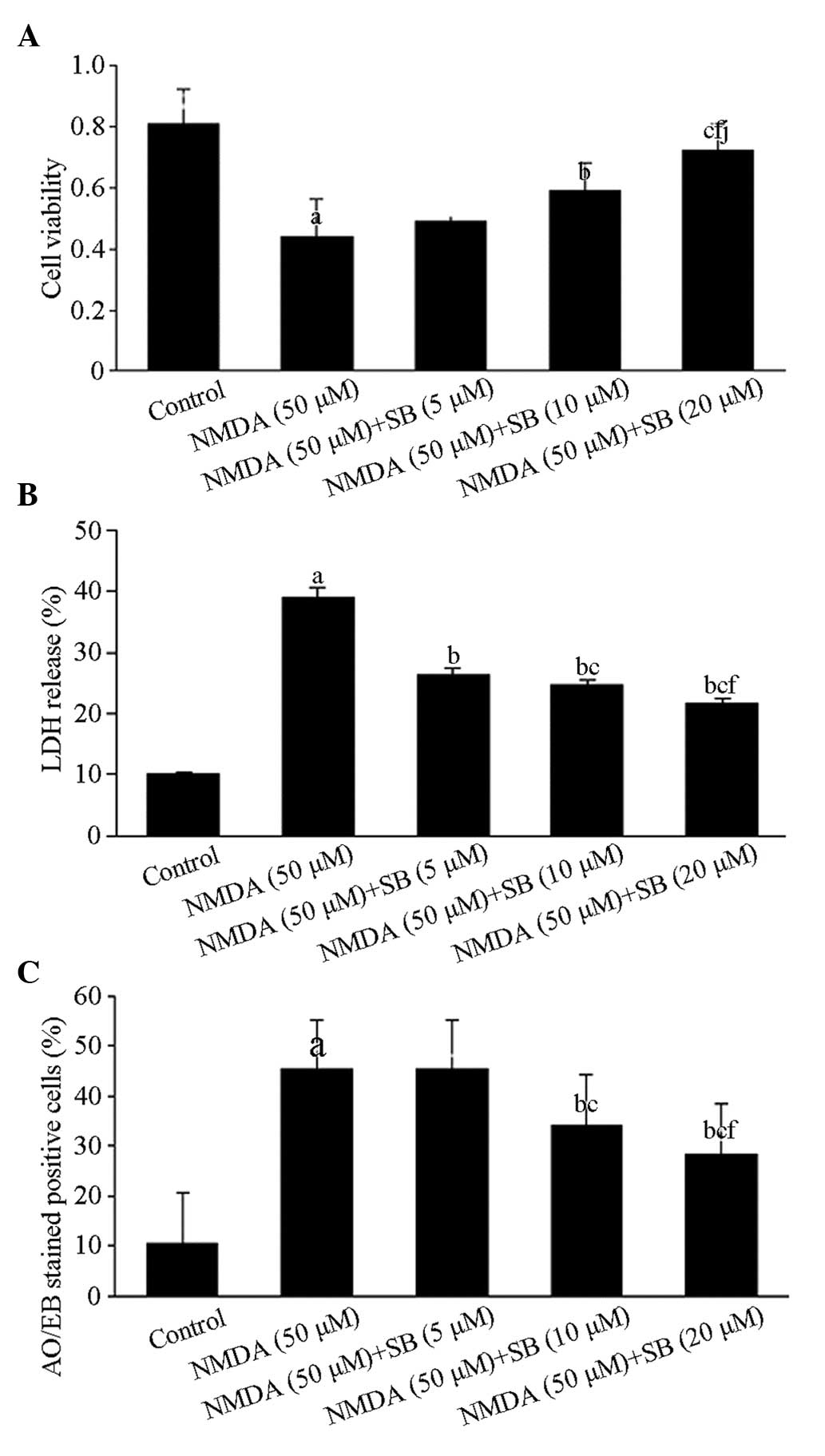
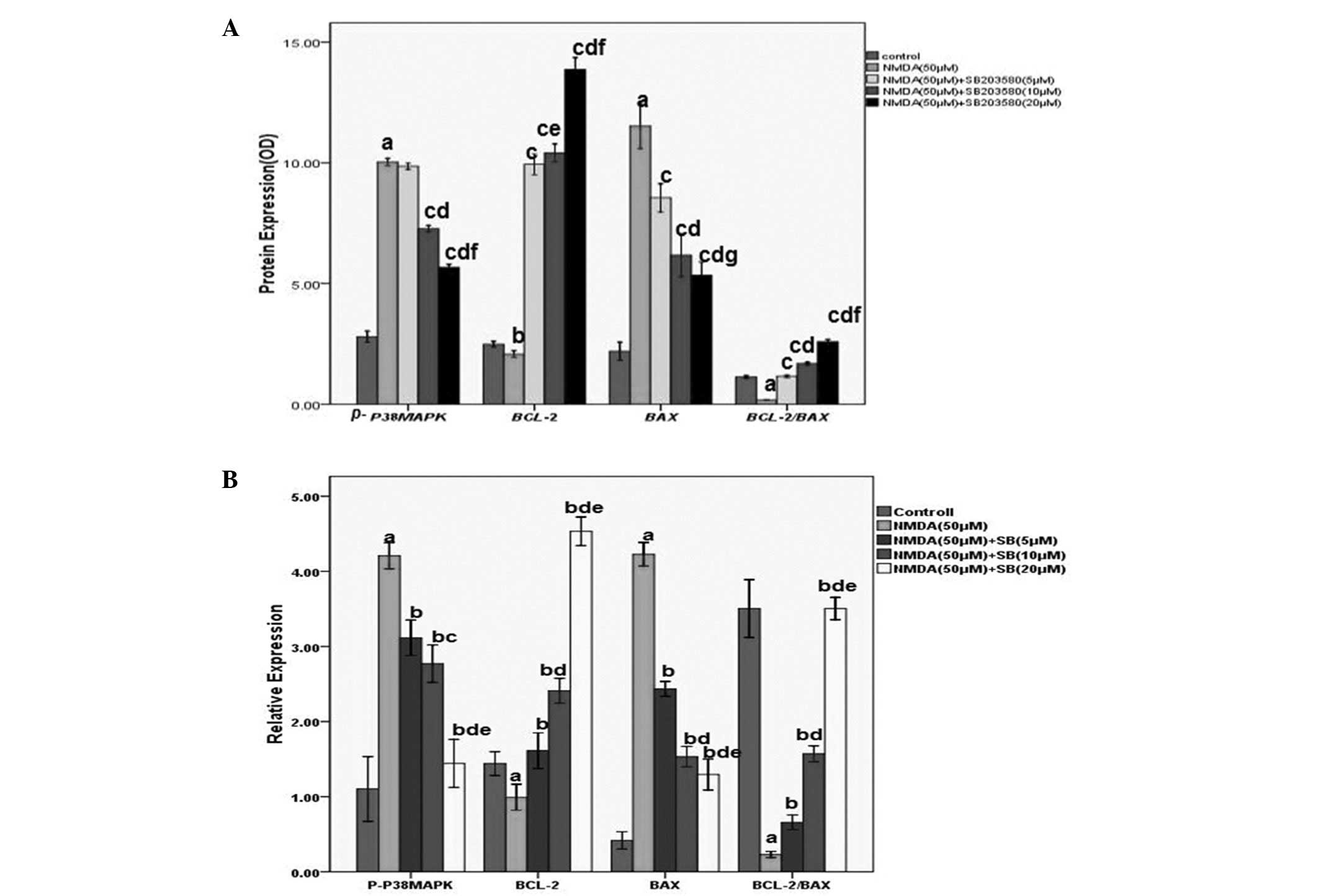
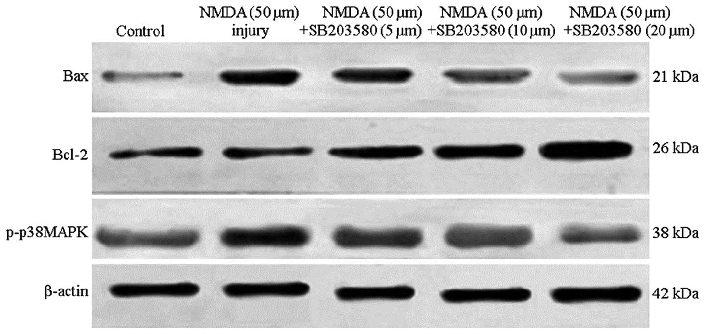
|
1
|
Molz S, Decker H, Dal-Cim T, Cremonez C,
Cordova FM, Leal RB and Tasca CI: Glutamate-induced toxicity in
hippocampal slices involves apoptotic features and p38 MAPK
signaling. Neurochem Res. 33:27–36. 2008. View Article : Google Scholar : PubMed/NCBI
|
|
2
|
Chen J, Errico SL and Freed WJ: Reactive
oxygen species and p38 phosphorylation regulate the protective
effect of Δ9-tetrahydrocannabinol in the apoptotic
response to NMDA. Neurosci Lett. 389:99–103. 2005.
|
|
3
|
Yang XR, Sun P, Qin HP, Si PP, Sun XF and
Zhang C: Involvement of MAPK pathways in NMDA-induced apoptosis of
rat cortical neurons. Sheng Li Xue Bao. 64:609–616. 2012.PubMed/NCBI
|
|
4
|
Lu TH, Hsieh SY, Yen CC, et al:
Involvement of oxidative stress-mediated ERK1/2 and p38 activation
regulated mitochondria-dependent apoptotic signals in
methylmercury-induced neuronal cell injury. Toxicol Lett.
204:71–80. 2011. View Article : Google Scholar
|
|
5
|
Zhang R, Sun L, Hayashi Y, Liu X, Koyama
S, Wu Z and Nakanishi H: Acute p38-mediated inhibition of
NMDA-induced outward currents in hippocampal CA1 neurons by
interleukin-1beta. Neurobiol Dis. 38:68–77. 2010. View Article : Google Scholar : PubMed/NCBI
|
|
6
|
Dunleavy M, Provenzano G, Henshall DC and
Bozzi Y: Kainic acid-induced seizures modulate Akt (SER473)
phosphorylation in the hippocampus of dopamine D2 receptor knockout
mice. J Mol Neurosci. 49:202–210. 2013. View Article : Google Scholar : PubMed/NCBI
|
|
7
|
Giordano G, Klintworth HM, Kavanagh TJ and
Costa LG: Apoptosis induced by domoic acid in mouse cerebellar
granule neurons involves activation of p38 and JNK MAP kinases.
Neurochem Int. 52:1100–1105. 2008. View Article : Google Scholar : PubMed/NCBI
|
|
8
|
Ola MS, Nawaz M and Ahsan H: Role of Bcl-2
family proteins and caspases in the regulation of apoptosis. Mol
Cell Biochem. 351:41–58. 2011. View Article : Google Scholar : PubMed/NCBI
|
|
9
|
Neoh CA, Wang RY, Din ZH, et al: Induction
of apoptosis by sinulariolide from soft coral through
mitochondrial-related and p38MAPK pathways on human bladder
carcinoma cells. Mar Drugs. 10:2893–2911. 2012. View Article : Google Scholar : PubMed/NCBI
|
|
10
|
Grethe S, Ares MP, Andersson T and
Pörn-Ares MI: p38 MAPK mediates TNF-induced apoptosis in
endothelial cells via phosphorylation and downregulation of
Bcl-x(L). Exp Cell Res. 298:632–642. 2004. View Article : Google Scholar : PubMed/NCBI
|
|
11
|
Liang HL, Dhar SS and Wong-Riley MT: p38
mitogen-activated protein kinase and calcium channels mediate
signaling in depolarization-induced activation of peroxisome
proliferator-activated receptor gamma coactivator-1alpha neurons. J
Neurosci Res. 88:640–649. 2010.
|
|
12
|
Yan Y, Bian JC, Zhong LX, Zhang Y, Sun Y
and Liu ZP: Oxidative stress and apoptotic changes of rat cerebral
cortical neurons exposed to cadmium in vitro. Biomed Environ Sci.
25:172–181. 2012.PubMed/NCBI
|
|
13
|
Sanchez A, Tripathy D, Yin X, Luo J,
Martinez J and Grammas P: Pigment epithelium-derived factor (PEDF)
protects cortical neurons in vitro from oxidant injury by
activation of extracellular signal-regulated kinase (ERK) 1/2 and
induction of Bcl-2. Neurosci Res. 72:1–8. 2012. View Article : Google Scholar
|
|
14
|
Guo RB, Wang GF, Zhao AP, Gu J, Sun XL and
Hu G: Paeoniflorin protects against ischemia-induced brain damages
in rats via inhibiting MAPKs/NF-kB-mediated inflammatory responses.
PLoS One. 7:e497012012. View Article : Google Scholar : PubMed/NCBI
|
|
15
|
Śmiałowska M, Gołembiowska K, Kajta M,
Zieba B, Dziubina A and Domin H: Selective mGluR1 antagonist EMQMCM
inhibits the kainate-induced excitotoxicity in primary neuronal
cultures and in the rat hippocampus. Neurotox Res. 21:379–392.
2012.PubMed/NCBI
|
|
16
|
Tiwari M, Lopez-Cruzan M, Morgan WW and
Herman B: Loss of caspase-2-dependent apoptosis induces autophagy
after mitochondrial oxidative stress in primary cultures of young
adult cortical neurons. J Biol Chem. 286:8493–8506. 2011.
View Article : Google Scholar
|
|
17
|
Liu B, Zhang H, Xu C, et al:
Neuroprotective effects of icariin on corticosterone-induced
apoptosis in primary cultured rat hippocampal neurons. Brain Res.
1375:59–67. 2011. View Article : Google Scholar : PubMed/NCBI
|
|
18
|
Xu Z, Wang BR, Wang X, Kuang F, Duan XL,
Jiao XY and Ju G: ERK 1/2 and p38 mitogen-activated protein kinase
mediate iNOS-induced spinal neuron degeneration after acute
traumatic spinal cord injury. Life Sci. 79:1895–1905. 2006.
View Article : Google Scholar
|
|
19
|
Jiang W, Van Cleemput J, Sheerin AH, et
al: Involvement of extracellular regulated kinase and p38 kinase in
hippocampal seizure tolerance. J Neurosci Res. 81:581–588. 2005.
View Article : Google Scholar : PubMed/NCBI
|
|
20
|
Segura Torres JE, Chaparro-Huerta V,
Rivera Cervantres MC, Montes-González R, Flores Soto ME and
Beas-Zárate C: Neuronal cell death due to glutamate excitotocity is
mediated by P38 activation in the rat cerebral cortex. Neurosci
Lett. 403:233–238. 2006.PubMed/NCBI
|
|
21
|
Miloso M, Scuteri A, Foudah D and Tredici
G: MAPKs as mediators of cell fate determination: an approach to
neurodegenerative diseases. Curr Med Chem. 15:538–548. 2008.
View Article : Google Scholar : PubMed/NCBI
|
|
22
|
Chen S, Xu Y, Xu B, et al: CaMKII is
involved in cadmium activation of MAPK and mTOR pathways leading to
neuronal cell death. J Neurochem. 119:1108–1118. 2011. View Article : Google Scholar : PubMed/NCBI
|
|
23
|
Zhang Y and Bhavnani BR: Glutamate-induced
apoptosis in primary cortical neurons is inhibited by equine
estrogens via down-regulation of caspase-3 and prevention of
mitochondrial cytochrome c release. BMC Neurosci. 6:132005.
View Article : Google Scholar
|
|
24
|
Liu XW, Ma C, Xing RX, et al: The
calmodulin-dependent protein kinase II inhibitor KN-93 protects rat
cerebral cortical neurons from N-methyl-D-aspartic acid-induced
injury. NRR. 8:111–120. 2013.PubMed/NCBI
|
|
25
|
Xu B, Xu Z, Deng Y, Liu W, Yang H and Wei
YG: MK-801 protects against intracellular Ca(2+)
overloading and improves N-methyl-D-aspartate receptor expression
in cerebral cortex of methylmercury-poisoned rats. J Mol Neurosci.
49:162–171. 2013.PubMed/NCBI
|