Introduction
Parkinson’s disease (PD) is a disease of the nervous
system, characterized by the loss of nigrostriatal dopaminergic
neurons (NDN) (1). PD, also termed
tremors paralysis, is a common age-associated neurodegenerative
disease, which is associated with environmental and genetic factors
(2,3), with a prevalence of 0.3% in the whole
population and an increasing incidence in the elderly (4). The main clinical manifestation of PD
includes motionless tremor, which is limited to one side of the
body. Clinically, PD features motionless tremor, muscle rigidity,
bradykinesia and postural instability (5). At present, the therapeutic methods
used for the treatment of PD involve pharmacotherapy, gene therapy,
rehabilitation and surgical treatment. In 1967, clinical studies
demonstrated that oral levodopa can improve the symptoms of PD,
establishing its current status in the treatment of PD. Levodopa
remains the most effective drug in PD therapy (6). However, treatment with levodopa is
associated with various side-effects, including a decline in
efficacy following prolonged usage, switch effect and end-of-dose
phenomenon (7). Based on current
understanding of the neural circuits of the basal ganglia (8), PD therapy predominantly requires the
use of a dopamine D1 receptor-stimulating agent (9).
microRNAs (miRNA) are small, endogenous, RNA-coding
molecules, which are important in almost all biological pathways,
particularly in the transcriptional regulation of gene expression.
As a small molecule regulator of gene transcription and expression,
miRNAs are involved in almost all important biological pathways and
disease processes in the human body (10). In recent years, there have been an
increasing number of studies regarding the association of miRNAs
with various diseases and biological functions. miRNAs are
important in cancer, inflammation and infection as well as
cardiovascular, immune and degenerative diseases acting as
molecular markers for screening, but also may be used as new
targets for drug development and the prevention and control of
severe diseases (11).
Small, gene regulating miRNAs may also be important
in PD. It has previously been reported that miRNA regulates leucine
repeat kinase-2 (LRRK2) which contributes to the etiology of
sporadic PD (12). Jewish
individuals of European and North African descent often carry a
LRRK2 mutation. These mutations are strongly associated with the
occurrence of PD, however, the specific mechanism remains to be
elucidated.
Based on data from miRNA and mRNA double expression
profiles, the present study identified a synergistic miRNA pattern
in PD and constructed an miRNA network. Through gene ontology (GO)
function and Kyoto Encyclopedia of Genes and Genomes (KEGG) pathway
annotation, the miRNA-associated pathogenesis of PD was determined.
The changes in gene expression observed under pathological
conditions assists in improving understanding of the pathogenesis
of PD and facilitates the identification of corresponding targets
for therapy. Gene expression profile analysis is a fast, high
through-put detection method for miRNA expression in tissues or
cells. Comparison of the differences in expression between patients
and healthy controls using this method, improves knowledge of the
pathogenesis and development of PD.
Materials and methods
miRNA and mRNA expression profiles of
PD
The miRNA and mRNA expression profiles of PD were
downloaded from the Gene Expression Omnibus and were termed
GSE16658 (http://www.ncbi.nlm.nih.gov/geo/query/acc.cgi?acc=GSE16658;
accessed 9th June 2013) and GSE22491 (http://www.ncbi.nlm.nih.gov/geo/query/acc.cgi?acc=GSE22491;
accessed 9th June 2013), respectively (13,14).
miRNA expression profiling was performed using the miRCURY LNA
microRNA Array platform, which included 32 samples (19 PD samples
and 13 normal control samples). The mRNA profiling was performed
using the Agilent-014850 Whole Human Genome Microarray 4×44K
(G4112F) platform (Agilent Technologies, Inc., Santa Clara, CA,
USA), which included 18 samples (10 PD samples and eight normal
control samples).
Differential expression analysis for
miRNA and mRNA expression profiling
Probes of miRNA and mRNA profiling were mapped to
the miRNA name and Entrez Gene ID in the miRBase, respectively.
When multiple probes were mapped to one miRNA or gene, the average
expression level was calculated. Log2 conversion was then performed
on the expression level.
A two-tailed student’s t-test was used to analyze
the differentially expressed miRNA and mRNA in the PD samples
compared with the normal samples when the P-value was <0.01
following false discovery rate (FDR) adjustment.
Predicted target genes of miRNA and
function and pathway enrichment analysis
Based on the seven target gene prediction algorithms
PicTar, DIANA-microT, miRanda, miRBase, RNAhybrid, RNA22 and
TargetScan, a set of predicted target genes of miRNA was
determined. In order to reduce false positive predictions, miRNA
target genes with high confidence were selected, which were
predicted by at least three prediction algorithms. The miRNA target
genes set with high confidence were then used for further
analysis.
In order to determine which biological functions the
synergistic miRNA was involved in, GO (15) was used for the miRNA regulatory
target gene enrichment analysis (P<0.001). In addition, GenMAPP
software (Gladstone Institute, San Francisco, CA, USA) (16) was used for KEGG pathway
analysis.
Identification of synergistic miRNAs
Each miRNA set with high confidence of target genes
was obtained and an accumulative hyper-geometric distribution test
was performed to locate miRNAs sharing the same target genes. These
miRNAs were defined as synergistic miRNAs. P<0.01 was considered
to indicate a statistically significant difference following
multiple FDR adjustment.
Results
Differential expression analysis
For expression analysis, 200 significantly
differentially expressed miRNAs were identified in the PD samples
compared with the normal samples. In the mRNA expression profile
analysis, 2,966 differentially expressed mRNAs were
identified.
Identification of synergistic miRNA and
functional annotation for target genes
A total of 3,860 abnormal miRNA interactions were
identified, which regulated at least one common target gene. A
total of 1,502 miRNA interactions (P≤0.01) were identified based on
super geometric distribution algorithm, including 147 miRNAs. These
significantly synergistic miRNA pairs were involved in the
regulation of 304 abnormally expressed genes.
In order to determine the biological functions that
PD-associated disordered miRNAs were involved in, 304 abnormal
target genes were used to perform GO function enrichment. A total
of 74 GO biological processes were significantly enriched,
including the biosynthetic process, the cellular biosynthetic
process, the cellular component assembly involved in morphogenesis,
mitogen-activated protein kinase (MAPK) signaling, the myometrial
relaxation and contraction pathways and calcium regulation in the
cardiac cell (Table I). In
addition, 304 target genes were significantly enriched in eight
KEGG pathways (Table II).
 | Table IGO functional annotation of
synergistic microRNA regulatory target genes. |
Table I
GO functional annotation of
synergistic microRNA regulatory target genes.
| GO ID (Biological
process) | P-value | Term |
|---|
| GO:0009058 | 1.63E-04 | Biosynthetic
process |
| GO:0044249 | 1.01E-04 | Cellular biosynthetic
process |
| GO:0010927 | 2.72E-04 | Cellular component
assembly involved in morphogenesis |
| GO:0034641 | 1.27E-04 | Cellular nitrogen
compound metabolic process |
| GO:0016265 | 6.60E-05 | Cell death |
| GO:0006310 | 8.80E-04 | DNA
recombination |
| GO:0033036 | 9.14E-06 | Macromolecule
localization |
| GO:0043412 | 3.23E-05 | Macromolecule
modification |
| GO:0008152 | 2.26E-04 | Metabolic
process |
| GO:0048519 | 5.61E-05 | Negative regulation
of biological process |
| GO:0006807 | 1.94E-04 | Nitrogen compound
metabolic process |
| GO:0008104 | 4.20E-06 | Protein
localization |
| GO:0015031 | 1.48E-05 | Protein
transport |
| GO:0050789 | 1.00E-04 | Regulation of
biological process |
| GO:0010468 | 2.26E-06 | Regulation of gene
expression |
| GO:0019219 | 1.07E-05 | Regulation of
nucleobase-containing compound metabolic process |
| GO:0080090 | 1.63E-06 | Regulation of primary
metabolic process |
| GO:0051252 | 6.76E-06 | Regulation of RNA
metabolic process |
| GO:0044281 | 3.21E-04 | Small molecule
metabolic process |
| GO:0006915 | 3.52E-04 | Apoptosis |
| GO:0065007 | 1.47E-04 | Biological
regulation |
| GO:0008219 | 6.39E-05 | Cell death |
| GO:0051641 | 7.15E-07 | Cellular
localization |
| GO:0034645 | 3.39E-06 | Cellular
macromolecule biosynthetic process |
| GO:0044265 | 2.60E-04 | Cellular
macromolecule catabolic process |
| GO:0070727 | 3.98E-07 | Cellular
macromolecule localization |
| GO:0044260 | 1.69E-08 | Cellular
macromolecule metabolic process |
| GO:0044237 | 5.70E-07 | Cellular metabolic
process |
| GO:0009987 | 2.29E-05 | Cellular process |
| GO:0044257 | 7.03E-04 | Cellular protein
catabolic process |
| GO:0034613 | 1.06E-06 | Cellular protein
localization |
| GO:0044267 | 6.43E-05 | Cellular protein
metabolic process |
| GO:0033554 | 9.44E-04 | Cellular response to
stress |
| GO:0051649 | 3.09E-05 | Establishment of
localization in cell |
| GO:0045184 | 5.25E-06 | Establishment of
protein localization |
| GO:0010467 | 3.81E-05 | Gene
expression |
| GO:0006886 | 1.58E-05 | Intracellular
protein transport |
| GO:0046907 | 3.68E-08 | Intracellular
transport |
| GO:0009059 | 8.70E-06 | Macromolecule
biosynthetic process |
| GO:0043170 | 6.81E-06 | Macromolecule
metabolic process |
| GO:0043632 | 3.94E-04 |
Modification-dependent macromolecule
catabolic process |
| GO:0019941 | 3.58E-04 |
Modification-dependent protein catabolic
process |
| GO:0016071 | 1.53E-04 | mRNA metabolic
process |
| GO:0006397 | 1.12E-05 | mRNA
processing |
| GO:0048523 | 2.05E-05 | Negative regulation
of cellular process |
| GO:0090304 | 3.62E-06 | Nucleic acid
metabolic process |
| GO:0006139 | 2.71E-05 |
Nucleobase-containing compound metabolic
process |
| GO:0006996 | 3.93E-04 | Organelle
organization |
| GO:0006596 | 6.75E-04 | Polyamine
biosynthetic process |
| GO:0051574 | 3.38E-04 | Positive regulation
of histone H3-K9 methylation |
| GO:0044238 | 2.02E-05 | Primary metabolic
process |
| GO:0012501 | 2.14E-04 | Programmed cell
death |
| GO:0071539 | 3.38E-04 | Protein
localization to centrosome |
| GO:0033365 | 6.95E-04 | Protein
localization to organelle |
| GO:0032446 | 7.82E-05 | Protein
modification by small protein conjugation |
| GO:0070647 | 1.93E-04 | Protein
modification by small protein conjugation/removal |
| GO:0006464 | 7.95E-06 | Protein
modification process |
| GO:0016567 | 3.19E-04 | Protein
ubiquitination |
| GO:0051603 | 5.37E-04 | Proteolysis
involved in cellular protein catabolic process |
| GO:0009889 | 3.13E-05 | Regulation of
biosynthetic process |
| GO:0031326 | 2.24E-05 | Regulation of
cellular biosynthetic process |
| GO:2000112 | 9.66E-07 | Regulation of
cellular macromolecule biosynthetic process |
| GO:0031323 | 1.34E-06 | Regulation of
cellular metabolic process |
| GO:0050794 | 2.66E-05 | Regulation of
cellular process |
| GO:0010556 | 2.43E-06 | Regulation of
macromolecule biosynthetic process |
| GO:0060255 | 5.68E-06 | Regulation of
macromolecule metabolic process |
| GO:0019222 | 7.90E-06 | Regulation of
metabolic process |
| GO:0051171 | 4.62E-06 | Regulation of
nitrogen compound metabolic process |
| GO:0006355 | 1.54E-05 | Regulation of
transcription, DNA-dependent |
| GO:0032774 | 7.31E-05 | RNA biosynthetic
process |
| GO:0016070 | 8.94E-06 | RNA metabolic
process |
| GO:0008380 | 2.36E-05 | RNA splicing |
| GO:0006351 | 2.61E-05 | Transcription,
DNA-dependent |
| GO:0006511 | 2.85E-04 | Ubiquitin-dependent
protein catabolic process |
| Table IIKyoto Encyclopedia of Genes and
Genomes pathway annotation of synergistic microRNA regulatory
targeted genes. |
Table II
Kyoto Encyclopedia of Genes and
Genomes pathway annotation of synergistic microRNA regulatory
targeted genes.
| Pathway | MAPP name | Adjusted
P-value |
|---|
| WP382 | Mitogen-activated
protein kinase signaling | 0 |
| WP289 | Myometrial
relaxation and contraction | 0 |
| WP536 | Calcium regulation
in the cardiac cell | 0 |
| WP481 | Insulin
signaling | 0 |
| WP23 | B cell receptor
signaling | 0 |
| WP254 | Apoptosis | 0 |
| WP707 | DNA damage
response | 0 |
| WP1591 | Heart
development | 0 |
Synergistic miRNA network
construction
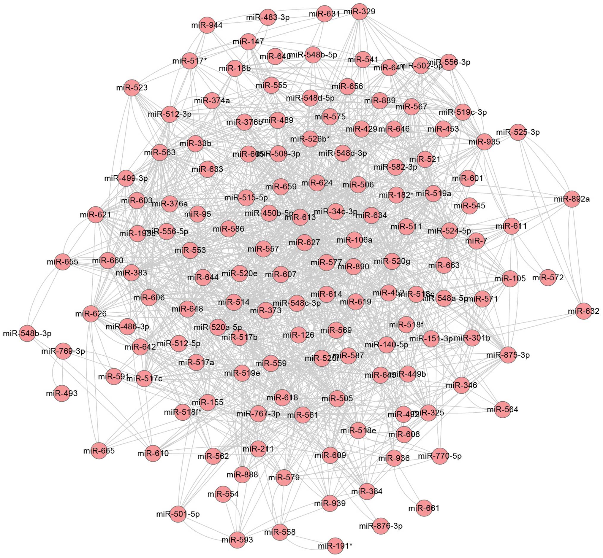
The synergistic miRNA was used to construct a
synergistic miRNA network associated with PD (Fig. 1). The miRNAs in this network
comprised all the abnormal molecules in the disease process and are
important gene regulatory factors. Therefore, the miRNAs in the
network may be beneficial to uncover the mechanism of PD. Through
binding to their target genes, miRNAs control post-transcriptional
translation or directly degrade the mRNA target genes. Degree
represents the number of interaction partners and the node with the
highest degree is essential for the stabilization of the network
(17). As shown in Table III, a high degree was observed in
miR-627, miR-634, miR-514, miR-563 and miR-613, which may be
associated with the pathogenesis of PD.
| Table IIIDegree of miRNA in the PD-associated
network. |
Table III
Degree of miRNA in the PD-associated
network.
| miRNA | Degree |
|---|
| miR-627 | 58 |
| miR-634 | 50 |
| miR-514 | 48 |
| miR-563 | 48 |
| miR-613 | 46 |
| miR-106a | 46 |
| miR-383 | 44 |
| miR-557 | 44 |
| miR-505 | 42 |
| miR-559 | 42 |
Discussion
PD is a common neurological degenerative disease in
elderly individuals. It is well established that miRNA is involved
in adjusting the target genes involved in cell proliferation,
differentiation, apoptosis and extensive biological processes
(18). In addition, miRNA is also
important in the differentiation of stem cells (19,20).
Dysfunction of miRNA may affect the development of the nervous
system, which causes diseases, including Alzheimer’s disease and PD
(21). In the present study, the
expression profiles of miRNA and mRNA were used to identify
disordered miRNA. A total of 304 abnormal miRNA regulatory target
genes were identified. GO function and KEGG pathway analysis
demonstrated that miRNA regulatory target genes were enriched in
several biosynthetic processes and pathways, among which cell
apoptosis (22,23), the MAPK signal pathway (24), calcium ion regulation (25) and insulin signals (26) have been associated with the
development or the treatment process and response to DNA damage in
PD.
In the present study, a total of 1,502 synergistic
miRNA interactions were identified and an miRNA synergistic network
was constructed. Within this network, miR-7 has been demonstrated
to regulate critical genes in the nervous system and to be involved
the processes of PD (14,27). In the synergistic network
constructed in the present study, miR-7 and another six miRNAs
regulated common target genes. In addition, through binding to the
3′UTR of amyloid precursor protein (APP), miR-147 regulated the
level of APP expression and, therefore, affected the risk of
developing Alzheimer’s disease and PD (28). The miRNA network demonstrated that
miR-627, miR-634, miR-514, miR-563 and miR-613 exhibited a high
degree. miR-627, miR-634 and miR-514 have been investigated in
human colorectal cancer cells (29), acute lymphoblastic leukemia
(30) and ovarian cancer (31). miR-563 has been reported to be
differentially expressed in Alzheimer’s disease samples (32) and targets the nuclear liver X
receptor, which is important in the metabolism and homeostasis of
cholesterol, bile acids, lipids and steroid hormones (33). However, to the best of our
knowledge, no studies have investigated the association between
these miRNAs and PD or other nerve-associated diseases. Therefore,
the present study demonstrated for the first time, to the best of
our knowledge, that miR-627, miR-634, miR-514, miR-563 and miR-613
are associated with PD. Further studies are required in order to
confirm these results.
Using the expression profile data, the entire
genomic expression situation of PD was examined. This revealed
differentially expressed miRNAs and mRNAs. In addition, miRNAs were
obtained and a synergistic miRNA network was constructed. From
this, miR-627, miR-634, miR-514, miR-563 and miR-613 were obtained,
which were newly reported in the present study. However, further
studies are required to confirm these results.
Acknowledgements
This study was supported by grants from The Major
Project of Biomedical Research of the Science and Technology
Commission of Shanghai Municipality (grant no. 10411954400) and The
National Natural Science Foundation of China General Program (grant
no. 81171296).
References
|
1
|
McCormack AL, Thiruchelvam M, Manning-Bog
AB, et al: Environmental risk factors and Parkinson’s disease:
selective degeneration of nigral dopaminergic neurons caused by the
herbicide paraquat. Neurobiol Dis. 10:119–127. 2002. View Article : Google Scholar : PubMed/NCBI
|
|
2
|
Findley L, Gresty M and Halmagyi G:
Tremor, the cogwheel phenomenon and clonus in Parkinson’s disease.
J Neurol Neurosurg Psychiatry. 44:534–546. 1981. View Article : Google Scholar : PubMed/NCBI
|
|
3
|
Parsian A, Sinha R, Racette B, Zhao JH and
Perlmutter JS: Association of a variation in the promoter region of
the brain-derived neurotrophic factor gene with familial
Parkinson’s disease. Parkinsonism Relat Disord. 10:213–219. 2004.
View Article : Google Scholar : PubMed/NCBI
|
|
4
|
de Lau LM and Breteler MM: Epidemiology of
Parkinson’s disease. Lancet Neurol. 5:525–535. 2006. View Article : Google Scholar : PubMed/NCBI
|
|
5
|
Rajput A, Rozdilsky B, Ang L and Rajput A:
Significance of parkinsonian manifestations in essential tremor.
Can J Neurol Sci. 20:114–117. 1993.PubMed/NCBI
|
|
6
|
Jankovic J: Levodopa strengths and
weaknesses. Neurology. 58:S19–S32. 2002. View Article : Google Scholar : PubMed/NCBI
|
|
7
|
Zheng H, Fridkin M and Youdim MB:
Site-activated chelators derived from anti-Parkinson drug
rasagiline as a potential safer and more effective approach to the
treatment of Alzheimer’s disease. Neurochem Res. 35:2117–2123.
2010. View Article : Google Scholar : PubMed/NCBI
|
|
8
|
Hashimoto T: Functional models of movement
disorders of basal ganglia origin and effects of functional
neurosurgery. Rinsho Shinkeigaku. 47:21–26. 2007.(In Japanese).
PubMed/NCBI
|
|
9
|
Wagg A: The review article by Wood et al
on nonmotor symptoms of Parkinson’s disease (PD) in the August 2010
edition of The American Journal of Geriatric Pharmacotherapy. Am J
Geriatr Pharmacother. 9:93–94. 2011. View Article : Google Scholar
|
|
10
|
Chen K and Rajewsky N: The evolution of
gene regulation by transcription factors and microRNAs. Nat Rev
Genet. 8:93–103. 2007. View
Article : Google Scholar : PubMed/NCBI
|
|
11
|
Zhang C: Novel functions for small RNA
molecules. Curr Opin Mol Ther. 11:641–651. 2009.
|
|
12
|
Gehrke S, Imai Y, Sokol N and Lu B:
Pathogenic LRRK2 negatively regulates microRNA-mediated
translational repression. Nature. 466:637–641. 2010. View Article : Google Scholar : PubMed/NCBI
|
|
13
|
Mutez E, Larvor L, Lepretre F, et al:
Transcriptional profile of Parkinson blood mononuclear cells with
LRRK2 mutation. Neurobiol Aging. 32:1839–1848. 2011. View Article : Google Scholar
|
|
14
|
Martins M, Rosa A, Guedes LC, et al:
Convergence of miRNA expression profiling, α-synuclein interacton
and GWAS in Parkinson’s disease. PloS One. 6:e254432011. View Article : Google Scholar
|
|
15
|
Ashburner M, Ball CA, Blake JA, et al:
Gene ontology: tool for the unification of biology. The Gene
Ontology Consortium. Nat Genet. 25:25–29. 2000. View Article : Google Scholar : PubMed/NCBI
|
|
16
|
Dahlquist KD, Salomonis N, Vranizan K,
Lawlor SC and Conklin BR: GenMAPP, a new tool for viewing and
analyzing microarray data on biological pathways. Nat Genet.
31:19–20. 2002. View Article : Google Scholar : PubMed/NCBI
|
|
17
|
Xu J, Li CX, Li YS, et al: MiRNA-miRNA
synergistic network: construction via co-regulating functional
modules and disease miRNA topological features. Nucleic Acids Res.
39:825–836. 2011. View Article : Google Scholar :
|
|
18
|
Chen Y and Stallings RL: Differential
patterns of microRNA expression in neuroblastoma are correlated
with prognosis, differentiation, and apoptosis. Cancer Res.
67:976–983. 2007. View Article : Google Scholar : PubMed/NCBI
|
|
19
|
Marson A, Levine SS, Cole MF, et al:
Connecting microRNA genes to the core transcriptional regulatory
circuitry of embryonic stem cells. Cell. 134:521–533. 2008.
View Article : Google Scholar : PubMed/NCBI
|
|
20
|
Murchison EP, Partridge JF, Tam OH,
Cheloufi S and Hannon GJ: Characterization of Dicer-deficient
murine embryonic stem cells. Proc Natl Acad Sci USA.
102:12135–12140. 2005. View Article : Google Scholar : PubMed/NCBI
|
|
21
|
Hébert SS and De Strooper B: Molecular
biology. miRNAs in neurodegeneration. Science. 317:1179–1180. 2007.
View Article : Google Scholar : PubMed/NCBI
|
|
22
|
Anglade P, Vyas S, Javoy-Agid F, et al:
Apoptosis and autophagy in nigral neurons of patients with
Parkinson’s disease. Histol Histopathol. 12:25–31. 1997.PubMed/NCBI
|
|
23
|
Tatton WG, Chalmers-Redman R, Brown D and
Tatton N: Apoptosis in Parkinson’s disease: signals for neuronal
degradation. Ann Neurol. 53:S61–S70; discussion S70–S72. 2003.
View Article : Google Scholar
|
|
24
|
Yogev-Falach M, Amit T, Bar-Am O and
Youdim MB: The importance of propargylamine moiety in the
anti-Parkinson drug rasagiline and its derivatives in
MAPK-dependent amyloid precursor protein processing. FASEB J.
17:2325–2327. 2003.PubMed/NCBI
|
|
25
|
Sheehan JP, Swerdlow RH, Parker WD, Miller
SW, Davis RE and Tuttle JB: Altered calcium homeostasis in cells
transformed by mitochondria from individuals with Parkinson’s
disease. J Neurochem. 68:1221–1233. 1997. View Article : Google Scholar : PubMed/NCBI
|
|
26
|
Bruning JC, Gautam D, Burks DJ, et al:
Role of brain insulin receptor in control of body weight and
reproduction. Science. 289:2122–2125. 2000. View Article : Google Scholar : PubMed/NCBI
|
|
27
|
Fiore R, Siegel G and Schratt G: MicroRNA
function in neuronal development, plasticity and disease. Biochim
Biophys Acta. 1779:471–478. 2008. View Article : Google Scholar : PubMed/NCBI
|
|
28
|
Delay C, Calon F, Mathews P and Hébert SS:
Alzheimer-specific variants in the 3′UTR of Amyloid precursor
protein affect microRNA function. Mol Neurodegener. 6:702011.
View Article : Google Scholar
|
|
29
|
Padi SK, Zhang Q, Rustum YM, Morrison C
and Guo B: MicroRNA-627 mediates the epigenetic mechanisms of
vitamin D to suppress proliferation of human colorectal cancer
cells and growth of xenograft tumors in mice. Gastroenterology.
145:437–446. 2013. View Article : Google Scholar : PubMed/NCBI
|
|
30
|
Rainer J, Ploner C, Jesacher S, et al:
Glucocorticoid-regulated microRNAs and mirtrons in acute
lymphoblastic leukemia. Leukemia. 23:746–752. 2009. View Article : Google Scholar : PubMed/NCBI
|
|
31
|
Boren T, Xiong Y, Hakam A, et al:
MicroRNAs and their target messenger RNAs associated with ovarian
cancer response to chemotherapy. Gynecol Oncol. 113:249–255. 2009.
View Article : Google Scholar : PubMed/NCBI
|
|
32
|
Kumar P, Dezso Z, MacKenzie C, et al:
Circulating miRNA biomarkers for Alzheimer’s disease. PloS One.
8:e698072013. View Article : Google Scholar
|
|
33
|
Ou Z, Wada T, Gramignoli R, et al:
MicroRNA hsa-miR-613 targets the human LXRα gene and mediates a
feedback loop of LXRα autoregulation. Mol Endocrinol. 25:584–596.
2011. View Article : Google Scholar : PubMed/NCBI
|