Picosecond pulsed electric fields induce apoptosis in a cervical cancer xenograft
- Authors:
- Published online on: November 14, 2014 https://doi.org/10.3892/mmr.2014.2953
- Pages: 1623-1628
-
Copyright: © Jia et al. This is an open access article distributed under the terms of Creative Commons Attribution License [CC BY_NC 3.0].
Metrics: Total
Views: 0 (Spandidos Publications: | PMC Statistics: )
Total PDF Downloads: 0 (Spandidos Publications: | PMC Statistics: )
Abstract
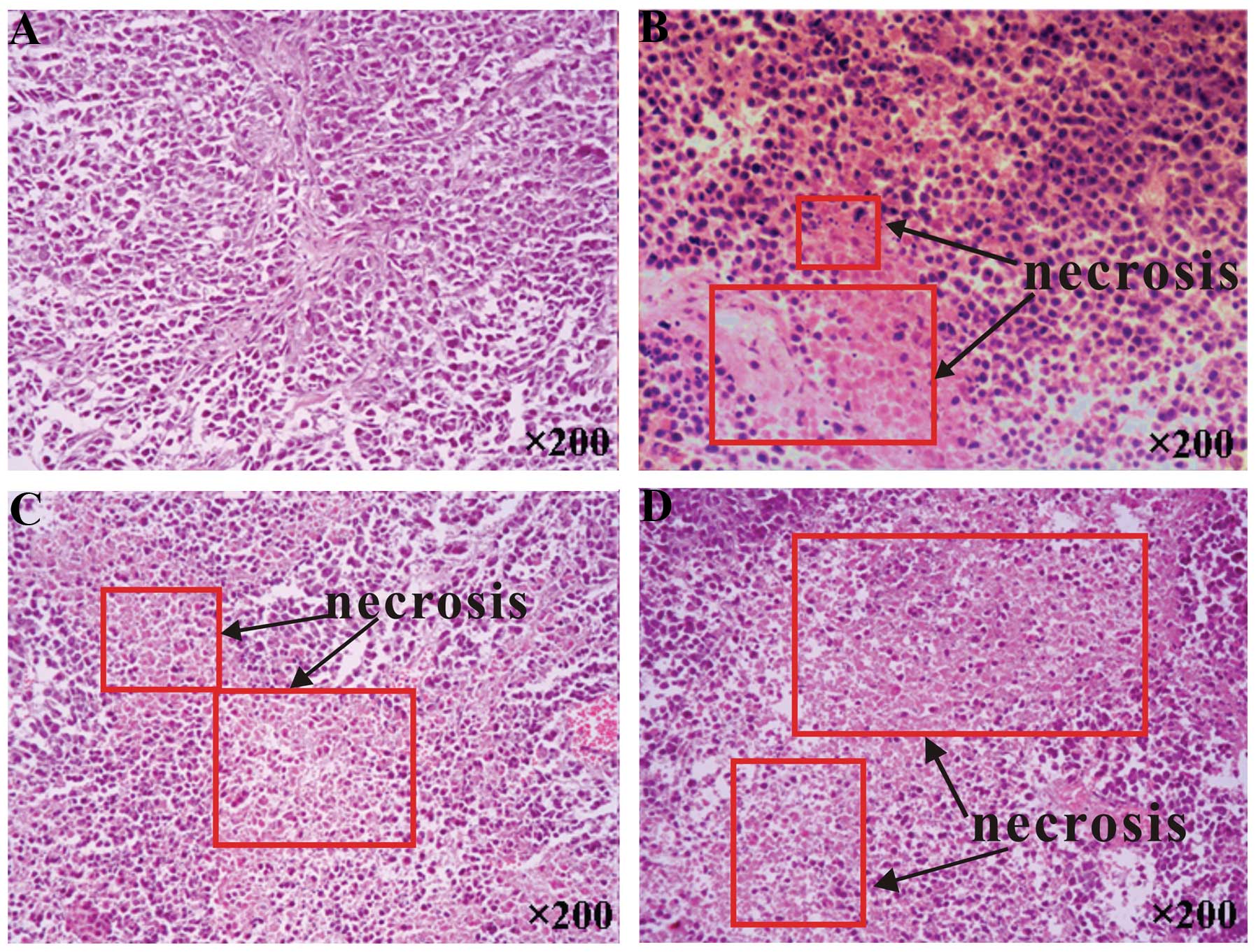
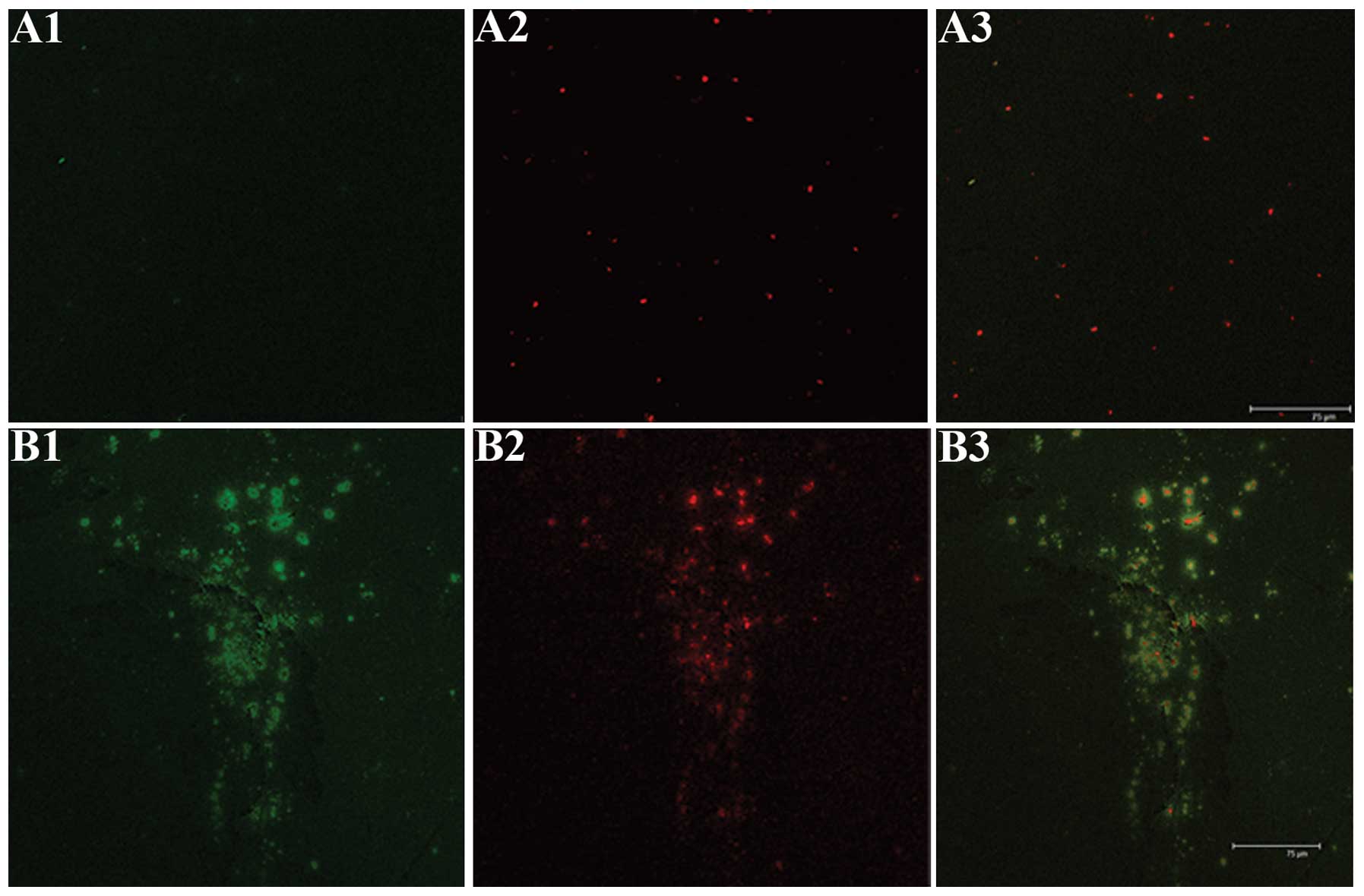
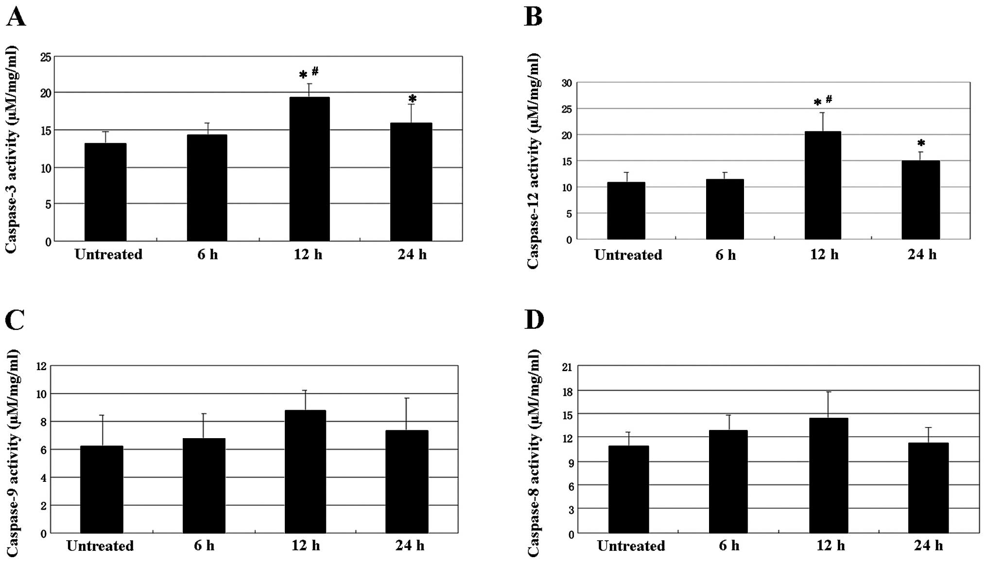
The aim of the present study was to evaluate the efficacy of picosecond pulsed electric fields (psPEF) on a cervical cancer xenograft. Human cervical cancer xenografts were established in nude mice by transplantation of HeLa cells, and the tumors were then treated with psPEF. The histological changes were observed by hematoxylin‑eosin staining and transmission electron microscopy. The rate of tumor cell apoptosis was determined using a terminal deoxynucleotidyl‑transferase‑mediated dUTP nick end labeling assay. The mitochondrial transmembrane potential of the tumor cells was detected by laser scanning confocal microscopy, and the activity of caspase‑3, ‑8, ‑9 and ‑12 was determined. The inhibitory rate seven days post‑psPEF treatment was also calculated. The results showed that exposure to psPEF led to an increased rate of apoptosis, collapse of mitochondrial transmembrane potential, and activation of caspases. The inhibitory rate was 9.11% at day 7. The results of the present study indicate that psPEF may induce apoptosis in a cervical cancer xenograft through the endoplasmic reticulum stress and caspase‑dependent signaling pathways.