Open Access

Histopathological assessment of inflammation and expression of inflammatory markers in patients with ketamine-induced cystitis

  • Authors:
    • Hsin‑Chung Lin
    • Herng‑Sheng Lee
    • Tzong‑Shi Chiueh
    • Yu‑Chieh Lin
    • Hsin‑An Lin
    • Yu‑Chun Lin
    • Tai‑Lung Cha
    • En Meng
  • View Affiliations

  • Published online on: December 18, 2014     https://doi.org/10.3892/mmr.2014.3110
  • Pages: 2421-2428
  • Copyright: © Lin et al. This is an open access article distributed under the terms of Creative Commons Attribution License [CC BY_NC 3.0].

Metrics: Total Views: 0 (Spandidos Publications: | PMC Statistics: )
Total PDF Downloads: 0 (Spandidos Publications: | PMC Statistics: )


Abstract

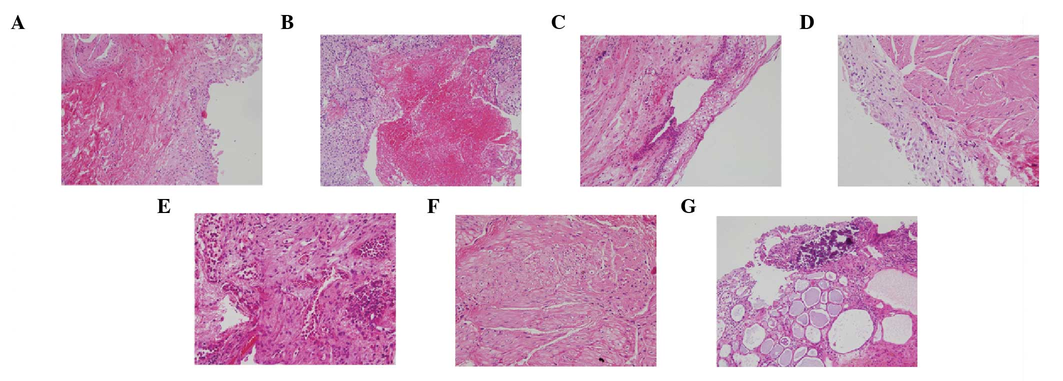
The aim of the current study was to evaluate the histopathological features of inflammation and the expression levels of inflammatory markers in tissue samples from patients with ketamine‑induced cystitis. Bladder biopsy samples for histological analysis were obtained from 23 patients (18 men and 5 women) with a self‑reported history of ketamine use and who were treated for cystitis at the Tri‑Service General Hospital of Taipei, Taiwan. Immunohistochemical staining for cyclooxygenase‑2 (COX‑2), inducible nitric oxide synthase (iNOS), matrix metallopeptidase‑9 (MMP‑9), mammalian target of rapamycin (mTOR), and phosphorylated 40S ribosomal protein S6 (Phos‑S6) was performed. The results revealed urothelial atypia in all patients, and intravascular eosinophil accumulation in 22 patients. Histopathological features included denuded urothelial mucosa, ulceration, collagen deposition, smooth muscle degeneration and vessel proliferation. Tissue samples were immunopositive for all of the inflammation markers, including the urothelium, vessel walls, and smooth muscle. COX‑2 staining revealed a significant difference between the inflammatory levels in the urothelium and smooth muscle, and iNOS staining differed significantly between inflammatory levels in smooth muscle (p=0.029). A positive correlation was observed between the percentage of Phos‑S6‑positive cells and the levels of inflammation in the urothelium. These results add to the descriptive literature on the histopathological aspects of ketamine‑induced cystitis, emphasizing the inflammatory nature and a possible role for proteins such as COX‑2, iNOS and Phos‑S6 in the degree of inflammation.
View Figures
View References

Related Articles

Journal Cover

April-2015
Volume 11 Issue 4

Print ISSN: 1791-2997
Online ISSN:1791-3004

Sign up for eToc alerts

Recommend to Library

Copy and paste a formatted citation
x
Spandidos Publications style
Lin HC, Lee HS, Chiueh TS, Lin YC, Lin HA, Lin YC, Cha TL and Meng E: Histopathological assessment of inflammation and expression of inflammatory markers in patients with ketamine-induced cystitis. Mol Med Rep 11: 2421-2428, 2015.
APA
Lin, H., Lee, H., Chiueh, T., Lin, Y., Lin, H., Lin, Y. ... Meng, E. (2015). Histopathological assessment of inflammation and expression of inflammatory markers in patients with ketamine-induced cystitis. Molecular Medicine Reports, 11, 2421-2428. https://doi.org/10.3892/mmr.2014.3110
MLA
Lin, H., Lee, H., Chiueh, T., Lin, Y., Lin, H., Lin, Y., Cha, T., Meng, E."Histopathological assessment of inflammation and expression of inflammatory markers in patients with ketamine-induced cystitis". Molecular Medicine Reports 11.4 (2015): 2421-2428.
Chicago
Lin, H., Lee, H., Chiueh, T., Lin, Y., Lin, H., Lin, Y., Cha, T., Meng, E."Histopathological assessment of inflammation and expression of inflammatory markers in patients with ketamine-induced cystitis". Molecular Medicine Reports 11, no. 4 (2015): 2421-2428. https://doi.org/10.3892/mmr.2014.3110