Introduction
In the United States, an improved regulation of the
vascular risk factors associated with strokes, together with
advances made in acute stroke care, have contributed towards a
decline in the stroke mortality rate, such that strokes have fallen
from third to fourth position in the leading causes of mortality
(1). The age-adjusted mortality
rate from strokes declined from 60.9 per 100,000 to 36.9 per
100,000 (2,3). However, in spite of this decline in
the stroke mortality rate, strokes remain a leading cause of
serious long-term disability in the United States (4). The total direct annual
stroke-associated medical costs are expected to increase from
$71.55 billion to $183.13 billion between 2012 and 2030 (5). By contrast, strokes are the leading
cause of mortality in China (6),
which, in comparison with the world average, has a higher stroke
mortality rate (126.1/100,000 higher), a greater number of years of
life lost (614/100,000 higher), and a greater number of
disability-adjusted life years (618/100,000 higher) (7), thereby imposing a heavy burden on
patients and their families. The Chinese government has been taking
several measures to prevent strokes and provide improved stroke
care (8). Although a larger number
of patients are surviving following a stroke, the burden of
post-stroke disability is becoming increasingly important as a
priority for public health (9).
Opposing needling (ON) is an important ancient
acupuncture technique, comprising the selection of acupuncture
points contralateral to the diseased side. ON has been used to
treat patients with stroke in China for a long time (10). A body of accumulating evidence from
clinical trials and experimental research revealed that ON exerts
protective effects against ischemic injury and is able to improve
motor function (11,12). However, the targets of ON and the
underlying mechanism remain to be fully elucidated.
Following a stroke, the release of excitatory amino
acids, including glutamate, exerts a key role in mediating
excitotoxic neuronal damage (13),
which consequently affects post-stroke rehabilitation.
γ-Aminobutyric acid (GABA) is the most important inhibitory
neurotransmitter in the mammalian central nervous system, which may
reverse the excitotoxic neuronal damage. A total of ~30% neurons in
the brain produce GABA, and almost every neuron is able to respond
to GABA (14). GABA class A, B and
C (GABAA, GABAB and GABAC)
receptors are the three types of receptor which have been
identified, and GABAB receptors are G-protein-coupled
receptors composed of the receptor subclasses GABAB1 and
GABAB2 (15), which act
presynaptically and postsynaptically. GABAB receptors
have been widely used in the treatment of neurological and
psychiatric disorders (16,17).
A number of previous studies demonstrated that enhancing the
activity of GABAB receptors exerted neuroprotection
against cerebral ischemia injury (18,19).
In addition, the underlying mechanism may be associated with the
adenylyl cylcase (AC)/cyclic AMP (cAMP)/protein kinase A (PKA)/cAMP
responsive element-binding protein (CREB) signal transduction
pathway, and a our previous study determined that ON is able to
increase the levels of cAMP and PKA (20). cAMP acts a secondary messenger,
which is generated by the action of transmembrane-bound enzyme AC
and is able to activate PKA. CREB in the nucleus is phosphorylated
and activated by PKA, and consequently binds to the cAMP response
element of target genes, which are considered to be involved in the
recovery of motor function and in neuroprotection against
ischemia.
The aims of the present study were: (i) To observe
the effects of ON on gait impairment in transient middle cerebral
artery occlusion (MCAO) rats; (ii) to identify whether the
cAMP/PKA/CREB signal transduction pathway and the GABAB
receptors were involved in neuroprotection against ischemia and
motor function recovery induced by ON.
Materials and methods
Animals
Sprague-Dawley rats were reported to develop
ischemic damage faster compared with Wistar-Kyoto rats, and they
also develop larger ischemic lesions compared with Wistar rats in
the MCAO model (21). Adult male
Sprague-Dawley rats (n=80) weighing 250–280 g were obtained from
the Shanghai SLAC Laboratory Animal Co., Ltd. (Shanghai, China).
The rats were caged with a standard day and night cycle (12 h/12 h)
in a temperature-controlled environment (21±2°C). The relative
humidity level was 55±5%, and the animals were provided with
adequate food and water. After 7 days of adaptation to the
experimental conditions, the rats were randomly divided into the
sham-operation (SC) group, the ischemia (IS) group, the ON group
and the ON + CGP35384 group (n=20/group). This study was approved
by the ethics committee of the Fujian University of Traditional
Chinese Medicine (Fuzhou, China). All animal procedures were
performed in accordance with the Institutional Animal Care and Use
Committee of the Fujian University of Traditional Chinese Medicine.
All efforts were made to minimize the number of animals used in the
present study and the suffering of animals.
Transient MCAO
Rats in the IS, ON and ON + CGP35384 groups were
subjected to MCAO surgery, as previously described (22), with minor modifications. Briefly,
the rats were anesthetized with 10% chloral hydrate (250 mg/kg;
Wuhan Hechang Chemical Co., Ltd., Wuhan, China) by intraperitoneal
injection. A 2–2.5 cm incision was made in the centre of the neck.
The left common carotid artery, the external carotid artery and the
internal carotid artery (ICA) were exposed and isolated. A
heparinized nylon filament (diameter 0.26 mm) was gently introduced
into the ICA in order to block the origin of the left middle
cerebral artery. The incision was subsequently ligatured and
sutured. Gentamicin sulphate was administered to prevent
postoperative infections or dehydration following surgery.
Reperfusion was induced 2 h following the MCAO surgery using the
filament withdrawal technique (23). The SC group received the identical
surgical procedure without placing the filament into the ICA. The
rectum temperature of the rats was maintained at 37±1°C during the
surgery using a heating pad. The extent of neurological impairment
was assessed 1 day following the MCAO surgery by determining the
neurological deficit score (22)
in order to confirm a successful occlusion. Animals exhibiting no
behavioral impairment were excluded from the study and no
postoperative mortality occurred.
Behavioral measurements
A five-point scale (0–4) was employed for the
neurological deficit score, and each point was characterized by the
following attributes: 0, no neurological deficit observed; 1, mild
focal neurological deficit, presented by a failure to extend the
left forepaw fully; 2, a moderate focal neurological deficit,
presented by circling to the left; 3, a severe focal deficit,
presented by an inability to bear weight on the left; 4, an
inability to walk spontaneously, with a depressed level of
consciousness. Neurological evaluation was performed by assessing
the neurological deficit score (22) on days 1, 2 and 7 following the MCAO
surgery.
For the Catwalk test, a CatWalk XT 10.0 system
(Noldus Information Technology, Wageningen, The Netherlands) was
used. The CatWalk XT 10.0 system is a video-based, advanced gait
analysis system designed to dynamically measure the footprints of
voluntarily moving animals and to assess any locomotor deficits and
the pain syndrome in animal models (24,25),
which is widely used in experimental stroke studies (26,27).
It is considered to be a useful tool for studying motor impairment
in stroke animals (28). The
'Catwalk' apparatus consisted of a plexglass walkway, fluorescent
lights, a video camera and CatWalk software. The operational
principle of CatWalk was a walkway made of plexiglass, on which the
rats traversed from one side of the glass plate to the other, and
the entire walkway was illuminated and completely internally
reflected by fluorescent lights. Since the light was restricted to
the plexiglass walkway, light was able to escape only at those
areas where the animal made a contact with the plexiglass walkway
(typically, the animal's paws). Therefore, the manner in which the
animal ran across the walkway was detected by a video camera
mounted underneath the walkway, and by means of capturing the
illuminated areas, the video images were transmitted to the
connected computer. Subsequently, all the data were acquired,
compressed, stored and eventually analyzed using the CatWalk
software program.
Prior to the first test, the rats were trained for 2
days to acclimatize to the walkway environment. The home cages of
the rats were placed at one side of the walkway, and the rats were
placed at the opposite side. The rats were trained to make
consecutive runs over the walkway towards their home cages, without
any interruption. Food was used as a reward during the rats'
training. A trial was considered to be successful if rats were able
to transverse the walkway without interruption within a duration of
10 sec. Any unsuccessful trial (marked by the animal stopping on
the walkway, walking backwards or rearing during the run) was
repeated, until the required number of trials was achieved. The
camera recorded three consecutive complete runs across the walkway.
The average of three trials was taken for subsequent statistical
analysis, as performed using the Catwalk XT 10.0 Software. An
experienced researcher labeled each paw on the recorded video, in a
blinded manner, and the paw-associated parameters were reported.
Descriptions of the gait-associated parameters (25), as measured using the CatWalk
system, were defined, as shown in Table I. The gait of the rats was assessed
on days 1 and 7 following the surgery. All rats in the present
study were able to accomplish the Catwalk task.
 | Table IExplanation of the Catwalk
parameter. |
Table I
Explanation of the Catwalk
parameter.
| Paw parameter | Explanation |
|---|
| Stance duration
(sec) | The speed between
two consecutive initial contacts of the same paw: stand +
swing |
| Stride length
(cm) | The amount of light
reflected by a paw as an indirect measure of weight bearing |
| Maximum contact
area (cm2) | The total floor
area contacted by the paw |
| Swing speed
(cm/sec) | The speed of the
paw during the swing phase |
| Swing duration
(sec) | The distance
between either the front or the hind paws |
Animal grouping and intervention
A total of 80 rats were randomized into four groups
(the SC, IS, ON and ON + CGP35384 groups; n=20/group). The rats in
the IS, ON and ON + CGP35384 groups were subjected to transient
MCAO surgery. The IS and SC groups served as no-intervention
controls for the ON group. The rats in the ON and ON + CGP35384
groups received electroacupuncture treatment for 30 min daily over
a continuous period of 7 days. For each session, acupuncture was
applied bilaterally. The Hegu (LI4), Waiguan (TE5), Yanglingquan
(GB34) and Zusanli (ST36) acupuncture points were punctured
perpendicularly at intervals of 2–3 mm. Subsequently, four
auxiliary needles were punctured at 2 mm intervals lateral to each
acupuncture point. Two types of Hwato disposable steel needle
(Suzhou Hua Tuo Medical Instruments, Suzhou, China) were used
(length, 25, diameter, 0.25 mm and length, 13, diameter, 0.18 mm),
and transcutaneous electric acupoint stimulation (HANS; Han's
acupoints nerve stimulator, HANS-200, Nanjing, China) was applied
to stimulate the acupoints for 30 min at a frequency of 1–20 Hz.
For the ON + CGP35384 group, the GABAB receptor
antagonist, CGP35384 (1 mg/ml solvent/kg body weight; Wuhan Hechang
Chemical Co., Ltd.), was injected intraperitoneally 30 min prior to
the surgery to inhibit GABAB receptor activation
(29). All rats were sacrificed by
decapitation 7 days following the MCAO surgery for subsequent
cerebral infarct volume, western blot analysis and reverse
transcription-quantitative polymerase chain reaction (RT-qPCR). The
experimental protocols are illustrated in Fig. 1.
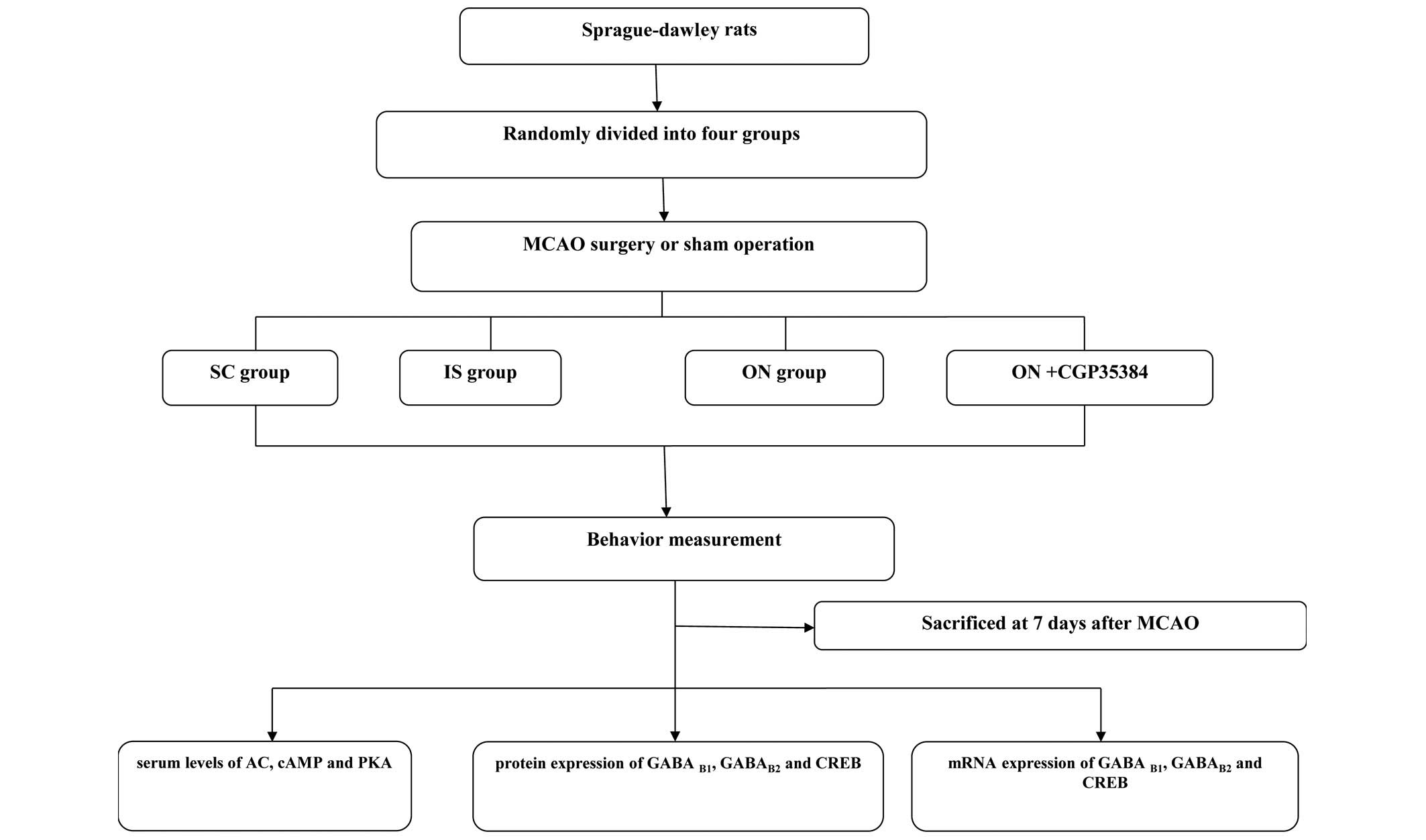 | Figure 1A flow chart illustrating the
experimental procedures employed in the present study. A total of
80 adult Sprague-dawley rats were randomly divided into the SC, IS,
ON and ON + CGP35384 groups. The behavior of the rats was assessed,
and the animals were subsequently sacrificed at 7 days following
MCAO surgery or the sham operation prior to enzyme-linked
immunosorbent assay, western blotting and reverse
transcription-quantitative polymerase chain reaction analysis. SC,
sham-operation; IS, ischemia; ON, opposing needling; MCAO, middle
cerebral artery occlusion; AC, adenylyl cyclase; cAMP, cyclic
adenosine 3′,5′-monophosphate; GABA, γ-aminobutyric acid; PKA,
protein kinase A; CREB, cAMP-responsive element binding
protein. |
Infarct volume measurement
The infarct volumes were measured using
2,3,5-triphenyltetrazolium chloride (TTC) staining (30). On day 7 following MCAO surgery, all
the rats were deeply anesthetized and decapitated. The brains were
swiftly removed, snap-frozen and stored at −20°C for 30 min. The
brains were sectioned into six 2 mm-thick slices. All slices were
stained with 1% TTC phosphate buffer solution (pH 7.4) and
incubated at 37°C for 15 min in order to optimize the TTC staining.
Subsequently, the slices were transferred to 12% formalin solution
for fixation for 2 min. Finally, images of the TTC-stained slices
were captured using an Olympus FE-240 digital camera (Shanghai
Pooher Photoelectric Technology Co., Ltd., Shanghai, China) and
analyzed using ImagePro® Plus 6.0 analysis software
(Media Cybernetics, Inc., Rockville, MD, USA). The infarction
volume was expressed as the infarction ratio (a percentage of the
total ipsilateral hemispheric volume), which was calculated
according to the following equation (31,32):
[(contralateral hemispheric volume − ipsilateral hemispheric
volume)/contra-lateral hemispheric volume] × 100%.
Western blot analysis
The left cortex surrounding the infarct was isolated
prior to homogenization in radioimmunoprecipitation buffer
(Beyotime Institute of Biotechnology, Shanghai, China), containing
a protease inhibitor cocktail (Beyotime Institute of
Biotechnology), and centrifugation at 15,984 × g for 20 min at 4°C.
The supernatants were collected and the protein concentration was
determined using a bicinchoninic acid kit (Beyotime Institute of
Biotechnology); the absorbance was measured using an automatic
enzyme instrument (ELx800; Biotek Instruments, Inc., Winooski, VT,
USA). Subsequently, the proteins were separated using 12% sodium
dodecyl sulfate-polyacrylamide gel electrophoresis (Beyotime
Institute of Biotechnology) and transferred onto polyvinylidene
difluoride membranes (EMD Millipore, Billerica, MA, USA). Following
1 h of blocking with 5% non-fat dry milk in Tris-buffered saline,
the membranes were incubated with the following primary antibodies:
Anti-CREB (1:1,000; cat. no. 9197S; Cell Signaling Technology,
Inc., Danvers, MA, USA) anti-GABAB1 (1:1,000; cat. no.
ab55051) and GABAB2 (1:1,000; cat. no. ab75838) (Abcam,
Cambridge, UK) overnight at 4°C. Following incubation with the
primary antibody, the membranes were rinsed and incubated with the
horseradish peroxidase-conjugated secondary antibody (1:2,000; cat.
no. A0545-1ML; Sigma-Aldrich, St. Louis, MO, USA). The protein
bands were visualized using an enhanced chemiluminescence kit
(Santa Cruz Biotechnology, Inc., Santa Cruz, CA, USA), and the
levels of protein present were analyzed using the integral optical
density detection technique (QuantityOne® software;
Bio-Rad, Hercules, CA, USA) and normalized against β-actin
(1:1,000; cat. no. 4970S; Cell Signaling Technology, Inc.) as the
control.
RT-qPCR
The left cortex surrounding the infarct was
dissected and frozen at −80°C. RNA isolation was performed using
standard procedures (33) and
Invitrogen TRIzol® reagent (Thermo Fisher Scientific,
Inc., Waltham, MA, USA). The total RNA was run on a 2% agarose gel
and quantified by densitometric analysis using the Fluor-S
MultiImager® imaging system (Bio-Rad Laboratories,
Milan, Italy). The total RNA (1 µg) was reverse-transcribed
using the first-strand synthesis system for RT-qPCR (Invitrogen
SuperScript®; Thermo Fisher Scientific, Inc.) in a final
volume of 20 µl for 1 h at 42°C, according to the
manufacturer's protocol. RT-qPCR was performed in a Real-Time PCR
detector using the SYBR Green real-time PCR Master mix (Toyobo Co.,
Ltd., Osaka, Japan), according to the manufacturer's protocol.
Specific primers were designed for the rat GABAB1,
GABAB2, CREB and glyceraldehyde-3-phosphate
dehydrogenase (GAPDH) genes, as shown in Table II. The basic protocol used for the
qPCR analysis was an initial denaturation at 95°C for 10 min,
followed by 45 cycles of amplification. For the cDNA amplification,
the cycles consisted of denaturation at 95°C for 10 min, annealing
at 95°C for 15 sec and elongation at 60°C for 60 sec. The SYBR
green signal was detected using the iQ5 real-time PCR detection
system (Bio-Rad). The PCR products were analyzed using gel
electrophoresis, and melting curve analysis was used to confirm the
specificity of the amplifications. The mRNA expression levels were
normalized against that of the housekeeping gene, GAPDH, and the
levels of the transcripts were quantified using the
2−ΔΔCq method (34).
 | Table IINucleotide sequences of the primers
used for real-time PCR. |
Table II
Nucleotide sequences of the primers
used for real-time PCR.
| Gene | Primer |
|---|
|
GABAB1 | Forward:
5′-CAGACACGTAGATCCTCCGC-3′ |
| Reverse:
5′-CGTACGCGTATACCAAGGCT-3′ |
|
GABAB2 | Forward:
5′-GGAAACCCGCAATGTGAGC-3′ |
| Reverse:
5′-GGCACAAACACCAGGCAFAG-3′ |
| CREB | Forward:
5′-AAGAATATGGCTCCGATTGC-3′ |
|
Reverse:5′-TGAGGATCTCATGGTAAACAGC-3′ |
| GAPDH | Forward:
5′-GGCAAGTTCAACGGCACAG-3′ |
| Reverse:
5′-CGCCAGTAGACTCCACGAC-3′ |
Enzyme-linked immunosorbent assay (ELISA)
for AC, cAMP and PKA
Blood was obtained from the abdominal aorta of the
rats using a hemospast, prior to centrifugation at 1,000 × g for 10
min at 4°C. The serum was subsequently collected and stored at
−80°C prior to assaying. The titers of AC, cAMP and PKA were
measured using ELISA, and the results were expressed as optical
density (OD) units, according to previously published methods
(35). Briefly, the plates were
coated with the capture antibody overnight at 4°C, following
washing with 0.05% Tween 20 in phosphate buffered saline. The
standards and samples were added prior to incubation in the coated
plates at room temperature for 2 h. The plates were subsequently
washed, and the detection antibody was added and incubated for 1 h,
prior to a subsequently washing step. Streptavidin-horseradish
peroxidase-conjugated antibody was added at room temperature for an
incubation of 1 h. Subsequently, tetramethylbenzidine substrate
solution (Clinical Science Products, Inc., Mansfield, MA, USA) was
added to produce a blue-colored solution. The enzyme substrate
reaction was stopped by the addition of sulfuric acid
(H2SO4), with the solution thereby turning
yellow. The OD of each well was immediately determined using a
microplate reader (Bio-Rad) set at 450 nm.
Statistical analysis
All data are continuous and are expressed as the
mean ± standard deviation. Within-group and between-group
differences were detected using analysis of co-variance (ANCOVA),
which was adjusted for baseline parameters. A post-hoc analysis of
ANCOVA was performed using Tukey's test and P<0.05 was
considered to indicate a statistically significant difference. R
software (version 3.1.1; http://www.r-project.org/) was used to conduct
statistical analyses.
Results
ON acupuncture reduced the score of the
neurological deficit
The neurological score is a 5 point scale, in which
a higher score indicated a higher loss of neurological function.
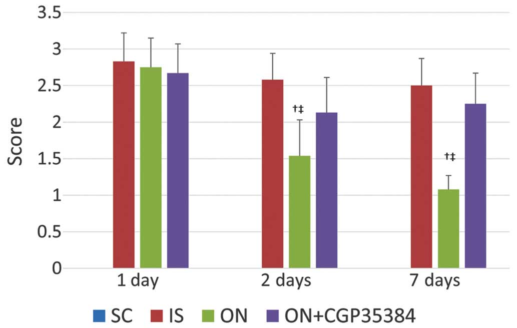
The rats in the IS group exhibited an increased neurological score
1 day following ischemia-reperfusion (IR; 2.83±0.39), which
indicated a successful implementation of the MCAO model (Fig. 2). The score was not significantly
reduced on days 2 and 7 following IR (2.58±0.36 and 2.50±0.37,
respectively), indicating no marked improvement in the animals'
condition. However, the ON acupuncture treatment significantly
reduced the neurological deficit score on day 2 following IR
(1.54±0.49, compared with 1 day following IR; P<0.05), and also
on day 7 following IR (1.08±0.19, compared with 1 day following IR;
P<0.05). However, this effect was reversed by administering the
antagonist, CGP35384, prior to the acupuncture. A small, although
insignificant reduction of the neurological deficit score was
observed (1 day following IR, 2.67±0.40; 2 days following IR,
2.13±0.48; 7 days following IR, 2.25±0.42). A significant
difference among the groups was identified: The ON group exhibited
a lower score compared with the IS and the ON + CGP35384 groups
(P<0.05). Further details are shown in Fig. 2.
ON acupuncture improves the total score
of the Catwalk system
The Catwalk system was used to assess the gait
performance. At day 1, the maximum contact area and stance duration
were significantly decreased in the right forepaw (RF; IS, vs. SC
group, 0.64, vs. 0.74 and 0.38, vs. 0.60, respectively; Table III) Following ON therapy, at day
7, the stride length and swing speed were significantly improved in
the (RF; ON, vs. IS group, 7.23, vs. 6.09, 39.82, vs. 32.1,
respectively). This effect was partly reversed by the antagonist
for the stride length in the RF (ON, vs. ON + CGP35384 group, 7.23,
vs. 6.12). Similar results were identified with respect to the
right hindpaw, as shown in Table
III.
 | Table IIIImprovement of Catwalk measurements
following ON. |
Table III
Improvement of Catwalk measurements
following ON.
A, RF
|
|---|
| Catwalk
parameter | Day 1 (mean ±
standard deviation)
| Day 7 (mean ±
standard deviation)
|
|---|
| SC | IS | ON | ON + CGP35384 | SC | IS | ON + CG ON | P35384 |
|---|
| Maximum contact
area | 0.74±0.06 |
0.64±0.1a | 0.56±0.14 | 0.58±0.1 | 0.67±0.16 |
0.41±0.12a | 0.46±0.10 | 0.38±0.09 |
| Stance
duration | 0.6±0.06 |
0.38±0.18a | 0.43±0.15 | 0.47±0.18 | 0.66±0.16 | 0.48±0.21 | 0.51±0.22 | 0.41±0.19 |
| Stride length | 7.33±1.1 | 5.87±0.87 | 5.76±1.05 | 5.98±0.99 | 7.24±1.38 | 6.09±0.91 |
7.23±1.05b |
6.12±1.1c |
| Swing duration | 0.16±0.04 | 0.21±0.08 | 0.2±0.09 | 0.18±0.12 | 0.23±0.15 | 0.26±0.17 | 0.22±0.10 | 0.25±0.95 |
| Swing speed | 49.07±12.55 |
32.19±8.87a | 34.6±12.69 | 35.22±15.23 | 49.59±15.94 |
32.1±9.17a |
39.82±6.61b | 40.82±7.61 |
B, RH
|
|---|
| Catwalk
parameter | Day 1 (mean ±
standard deviation)
| Day 7 (mean ±
standard deviation)
|
|---|
| SC | IS | ON | ON + CGP35384 | SC | IS | ON | ON + CG P35384 |
|---|
| Maximum contact
area | 0.86±0.1 | 0.93±0.21 | 0.81±0.18 | 0.89±0.15 | 0.78±0.13 | 0.54±0.13a | 0.57±0.14 | 0.51±0.1 |
| Stance
duration | 0.4±0.22 | 0.6±0.2 | 0.51±0.17 | 0.52±0.18 | 0.37±0.2 | 0.66±0.34a | 0.53±0.33 | 0.55±0.31 |
| Stride length | 7.51±0.63 | 6.05±0.74a | 6.12±0.84 | 5.89±0.88 | 7.61±0.87 | 6.52±0.67a | 7.92±0.89b | 6.06±0.73c |
| Swing duration | 0.12±0.01 | 0.14±0.04 | 0.16±0.09 | 0.18±0.07 | 0.12±0.02 | 0.14±0.01 | 0.14±0.02 | 0.12±0.02 |
| Swing speed | 62.03±8.08 | 49.5±15.56a | 48.92±8.06 | 47.6±18.22 | 67.71±12.42 | 46.55±7.31a | 53.16±8.52b | 42.33±6.66c |
ON acupuncture decreased the infarct
sizes of the rats' brains
The infarct sizes of the rat brains were measured at
7 days following the MCAO surgery; a small size indicated an
improved outcome. The rats in the IS group showed a significantly
higher infarct size compared with those in the SC group (mean
infarct size, 34.23, vs. 0; P<0.001; Fig. 3). The ON acupuncture reduced the
infarct size following MCAO surgery (mean infarct size for the ON,
vs. the IS group, 18.35, vs. 34.23; P<0.001); however, this
effect was reversed when the GABAB antagonist, CGP35384,
was administered prior to the ON acupuncture (mean infarct size for
the ON + CGP35384, vs. the ON group, 37.82, vs. 18.35; P<0.001;
Fig. 3).
ON acupuncture increased the protein and
mRNA expression levels of CREB, GABAB1 and
GABAB2 in the cerebral tissue
It was hypothesized that an increased mRNA
expression of the GABAB receptors would lead to a
suppression of the abnormal excitation of the nervous and muscular
systems, and therefore a higher level of expression of the
GABAB receptors would indicate an improvement in the
neurological and the motor functions. In the present study, the
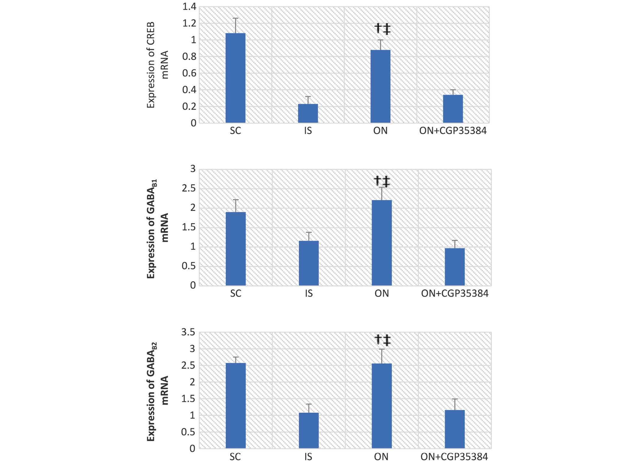
CREB mRNA decreased in the cerebral tissue of rats 7 days post-MCAO
surgery (IS, vs. SC group, 0.23±0.09, vs 1.08±0.18; P<0.05;
Fig. 4). A similar decrease in
mRNA expression was observed with GABAB1 (IS, vs. SC
group, 1.16±0.22, vs. 1.90±0.32; P<0.05), and a similar result
was also obtained for GABAB2 (IS, vs. SC group,
1.08±0.26, vs. 2.57±0.18; P<0.05). ON acupuncture significantly
increased the mRNA expression levels of CREB, GABAB1 and
GABAB2 (CREB, 0.88±0.12; GABAB1, 2.21±0.33;
P<0.001; GABAB2, 2.56±0.43, P<0.001) following
MCAO surgery. This effect was reversed by the antagonist, CGP35384
(CREB, 0.34±0.06; GABAB1, 0.97±0.2; P<0.05;
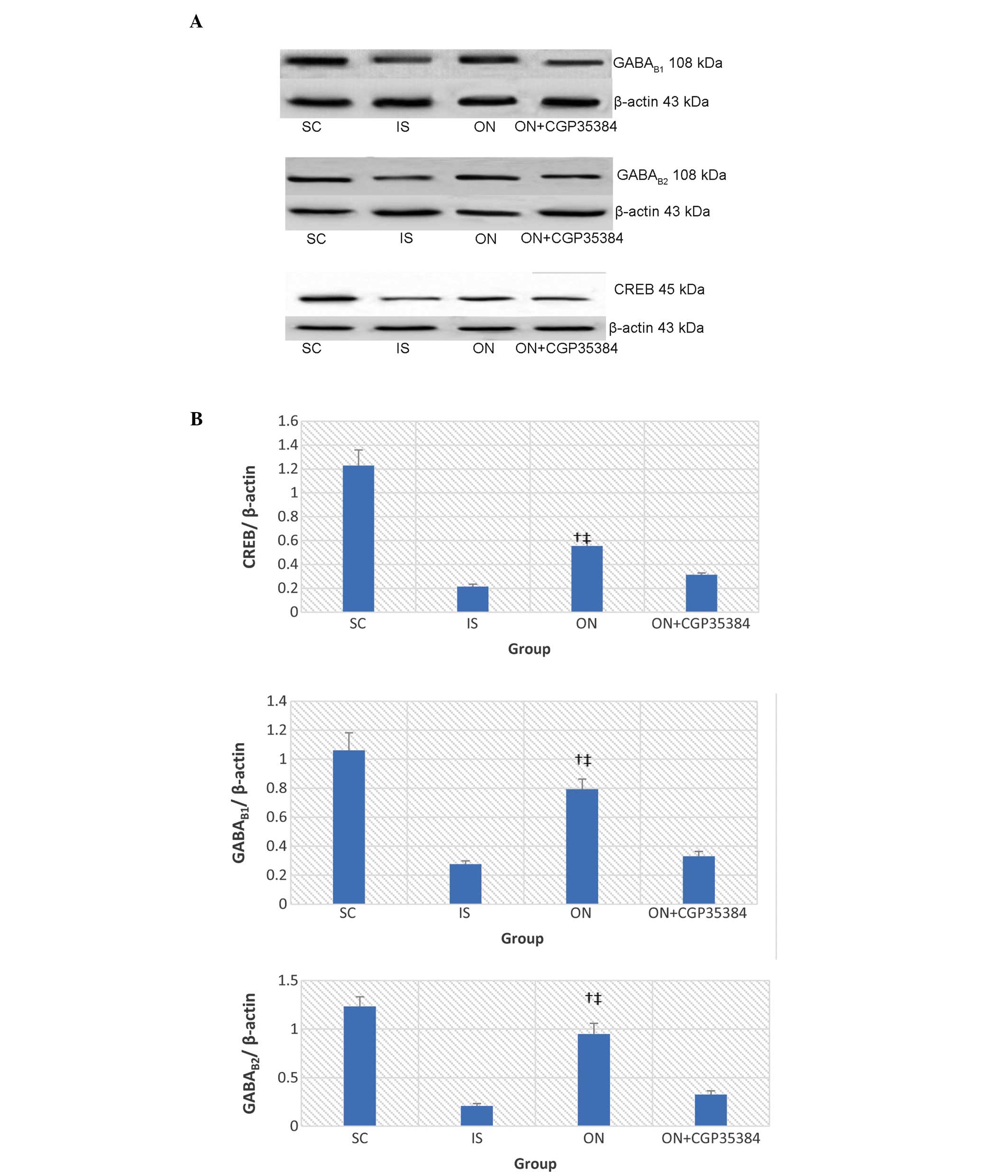
GABAB2, 1.16±0.33; P<0.05). Similar results were
obtained regarding the protein expression levels of CREB,
GABAB1 and GABAB2, as detected by western
blot analysis (Fig. 5)
ON acupuncture positively regulates the
AC/cAMP/PKA signaling pathway
The cAMP/PKA/CREB pathway operates downstream of the
activation of the GABA receptors, which may provide the putative
mechanism to account for the prevention of IR damage by the ON
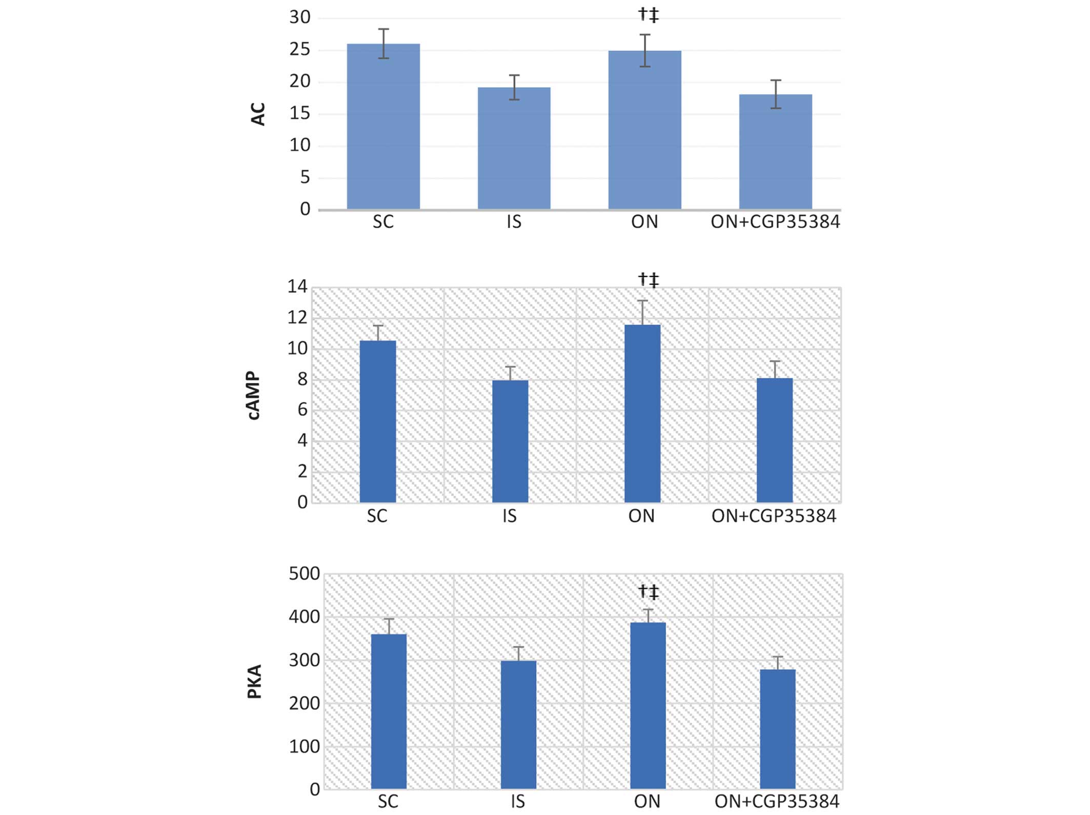
acupuncture. Following the MCAO surgery and reperfusion for 7 days,
the levels of AC, cAMP and PKA in the serum were significantly
reduced (IS, vs. SC group: AC, 19.21±1.9, vs. 26.05±2.3; cAMP,
7.98±0.89, vs. 10.55±0.98; PKA, 298.85±31.88, vs. 360.48±35.43;
P<0.05; Fig. 6). The ON
acupuncture significantly increased the levels of cAMP, PKA and
CREB in the cerebral tissue (ON, vs. IS group: AC, 24.95±2.5, vs.
19.21±1.9; cAMP, 11.58±1.56, vs. 7.98±0.89; PKA, 387.47±29.78, vs.
298.85±31.88; P<0.01). However, administering the antagonist,
CGP35384, prior to the ON acupuncture reversed this effect (ON +
CGP35384, vs. ON group: AC, 18.13±2.2, vs. 24.95±2.5; cAMP,
8.12±1.1, vs. 11.58±1.56; PKA, 278.66±29.85, vs. 387.47±29.78;
P<0.01; Fig. 6).
Discussion
The results of the present study revealed that ON
acupuncture significantly reduced the extent of cerebral infarction
and improved the motor function of the MCAO rats. These findings
also demonstrated that, compared with the IS group, ON acupuncture
resulted in an increase in the protein and mRNA expression levels
of CREB, GABAB1 and GABAB2. Using ELISA, it
was further determined that the activity of AC, cAMP and PKA
increased in the ON group, whereas the activity of AC, cAMP and PKA
decreased in the IS group. However, these effects were reversed by
administering the GABAB antagonist, CGP35384.
IR proceeds via the mechanisms associated with
excitotoxic neurotoxicity, which, following induction by excitatory
amino acids, includes the processes of intracellular
Ca2+ overload, an increase in Na+ influx
increase and the co-transport of Cl− and water,
resulting in further cytotoxic brain edema and neuronal damage
(36,37). However, these processes are
counteracted by certain inhibitory amino acids, particularly GABA,
one of the major inhibitory neurotransmitters, which is able to
increase chloride conductance, diminish the effects of
depolarization, open the calcium channel, and decrease the ATP
consumption and cell apoptosis that is induced by cerebral ischemia
(38). Therefore, increasing the
inhibitory neurotransmitter activity or increasing the activity of
the GABA receptor activity provides an effective means to exert
neuroprotection against ischemia (39). Dave et al (40) demonstrated that increased levels of
GABA release in preconditioned animals following ischemia may
provide one of the factors responsible for neuroprotection. The
specific activation of the GABAB receptors greatly
contributes towards neuroprotection against ischemia. The present
study also demonstrated that an increase in the mRNA expression
levels of GABAB1 and GABAB2 was associated
with improvements in the motor function and neuroprotection against
IR in the MCAO rats.
It is known that the cAMP/PKA/CREB signaling pathway
exerts an important role in synaptic plasticity and long-term
memory formation (41,42). However, a subsequent study
(43) and a previous report from
our laboratory (20) suggested a
possible connection between the cAMP/PKA/CREB signal transduction
system and neuroprotection. The previous report from our laboratory
revealed that ON acupuncture exerted neuroprotective effects,
promoted the activity of AC, cAMP and PKA, and increased the mRNA
expression of CREB in the cerebral tissue (20), whereas such observations were not
made in ischemic rats. Therefore, it was hypothesized that the
neuroprotection afforded by ON may be mediated through the
cAMP/PKA/CREB signaling pathway, and the present study was
performed in an attempt to confirm this hypothesis.
In the present study, experimental evidence was
provided to demonstrate that ON exerts a neuroprotective effect,
alleviates neural functional damage and improves the gait
impairment of MCAO rats. A previous study reported that the
GABAB receptor increases AC activity mediated by
Gi/Go protein (44), thereby leading to an increase in
the levels of cAMP. cAMP is a representative secondary messenger,
which is generated by the activity of the transmembrane-bound
enzyme, AC, and which is able to activate PKA. Subsequently, CREB
in the nucleus is phosphorylated and activated by PKA. CREB was
shown to regulate a number of aspects of neuronal functioning,
including neuronal development and synaptic plasticity. A
burgeoning body of evidence suggests that CREB may also be involved
in the active process of neuroprotection, and its disruption in the
brain may lead to neurodegeneration (45,46),
suggesting that CREB exerts a pivotal role in neuroprotection.
Therefore, the ON acupuncture treatment may have activated
GABAB and increased the formation of cAMP in the brain,
which in turn activated PKA and led to the phosphorylation of
CREB.
If the neuroprotection induced by ON is mediated
through the cAMP/PKA/CREB signaling pathway, identifying the
upstream target is of importance. The subsequent assays performed
in the present study revealed that CREB phosphorylation increased
following ON. Pretreatment with the GABAB antagonist,
CGP35384, effectively inhibited the ON-induced activation of CREB.
The neuroprotective effects induced by ON were also effectively
inhibited by the GABAB antagonist, thereby indicating
that the GABAB receptor is involved in neuroprotection
induced by ON acupuncture. Therefore, the protective effect of ON
against ischemia, mediated by the cAMP/PKA/CREB signaling pathway,
may be due to its action on the GABAB1 and
GABAB2 receptors. The activation of the
GABAB1 and GABAB2 receptors may lead to an
increase in the levels of cAMP, which subsequently promoted the
activity of PKA and the phosphorylation of CREB, thereby leading to
neuroprotection and the promotion of motor function recovery in
MACO rats.
GABAB may provide specific protective
effects against cerebral ischemic damage, which may be associated
with the inhibition of an excessive efflux of excitatory amino
acids, an increase in the levels of inhibitory amino acids and a
decrease in the Ca2+ level in the cortex. However, the
present study has certain limitations. The present study was not
concerned with monitoring the levels of excitatory amino acids,
including glutamate. Furthermore, it is unknown whether the motor
function recovery and the neuroprotective effects are associated
with the concentration of GABA, and further studies are required to
address these questions.
In conclusion, the results of the present study
demonstrated that ON improved the motor function in transient MCAO
rats and exerted neuroprotection against ischemia, and such effects
may be mediated through the GABAB receptor-cAMP/PKA/CREB
signal transduction pathway. These results may assist in furthering
our understanding of the mechanisms underlying ON, and in providing
a theoretical basis for clinical rehabilitation.
Acknowledgments
The present study was supported by a grant from the
National Natural Science Foundation of China (no. 81102628).
References
|
1
|
Minino AM, Xu J and Kochanek KD: Deaths:
preliminary data for 2008. Natl Vital Stat Rep. 59:1–52. 2008.
|
|
2
|
Xu J, Kochanek KD, Murphy SL and Arias E:
Mortality in the United States, 2012. NCHS Data Brief. 168:1–8.
2012.
|
|
3
|
Go AS, Mozaffarian D, Roger VL, Benjamin
EJ, Berry JD, Borden WB, Bravata DM, Dai S, Ford ES, Fox CS, et al:
Heart disease and stroke statistics - 2013 update: A report from
the American Heart Association. Circulation. 127:e6–e245. 2013.
View Article : Google Scholar
|
|
4
|
Prevalence and most common causes of
disability among adults - United States, 2005. MMWR Morb Mortal
Wkly Rep. 58:421–426. 2009.
|
|
5
|
Ovbiagele B, Goldstein LB, Higashida RT,
Howard VJ, Johnston SC, Khavjou OA, Lackland DT, Lichtman JH, Mohl
S, Sacco RL, et al: Forecasting the future of stroke in the United
States: A policy statement from the American Heart Association and
American Stroke Association. Stroke. 44:2361–2375. 2013. View Article : Google Scholar : PubMed/NCBI
|
|
6
|
Sun H and Zou X: A survey of the current
status in china. J Stroke. 15:109–114. 2013. View Article : Google Scholar : PubMed/NCBI
|
|
7
|
Xu G, Zhang Z, Lv Q, Li Y, Ye R, Xiong Y,
Jiang Y and Liu X: NSFC health research funding and burden of
disease in China. PLoS One. 9:e1114582014. View Article : Google Scholar : PubMed/NCBI
|
|
8
|
Liu L, Wang D, Wong KS and Wang Y: Stroke
and stroke care in China: Huge burden, significant workload, and a
national priority. Stroke. 42:3651–3654. 2011. View Article : Google Scholar : PubMed/NCBI
|
|
9
|
Towfighi A and Saver JL: Stroke declines
from third to fourth leading cause of death in the United States:
Historical perspective and challenges ahead. Stroke. 42:2351–2355.
2011. View Article : Google Scholar : PubMed/NCBI
|
|
10
|
Sun Liuhe WY: Study on applied law of
opposing needling. Zhongguo Zhenjiu. 23:540–542. 2003.In
Chinese.
|
|
11
|
Gangting L: Effects of opposing needling
with large needle on rheoencephalogram, hemorheology and blood
lipids in the patient of cerebral infarction. Zhongguo Zhenjiu.
24:701–703. 2004.In Chinese.
|
|
12
|
Xiaohui Y: Clinical observation on
post-stroke hemiplegic patients with the treatment of Ju-ci
method's massage. Chin Arch Tradit Chin Med. 25:1741–1742. 2007.In
Chinese.
|
|
13
|
Rothman SM and Olney JW: Glutamate and the
pathophysiology of hypoxic - ischemic brain damage. Ann Neurol.
19:105–111. 1986. View Article : Google Scholar : PubMed/NCBI
|
|
14
|
Gillani Q, Iqbal S, Arfa F, Khakwani S,
Akbar A, Ullah A, Ali M and Iqbal F: Effect of GABAB receptor
antagonist (CGP35348) on learning and memory in albino mice.
Scientific World Journal. 2014(983651)2014. View Article : Google Scholar : PubMed/NCBI
|
|
15
|
Bettler B and Tiao JY: Molecular
diversity, trafficking and subcellular localization of GABAB
receptors. Pharmacol Ther. 110:533–543. 2006. View Article : Google Scholar : PubMed/NCBI
|
|
16
|
Li CJ, Lu Y, Zhou M, Zong XG, Li C, Xu XL,
Guo LJ and Lu Q: Activation of GABAB receptors ameliorates
cognitive impairment via restoring the balance of HCN1/HCN2 surface
expression in the hippocampal CA1 area in rats with chronic
cerebral hypoperfusion. Mol Neurobiol. 50:704–720. 2014. View Article : Google Scholar : PubMed/NCBI
|
|
17
|
Cryan JF and Slattery DA: GABAB receptors
and depression. Current status. Adv Pharmacol. 58:427–451. 2010.
View Article : Google Scholar : PubMed/NCBI
|
|
18
|
Cimarosti H, Kantamneni S and Henley JM:
Ischaemia differentially regulates GABA(B) receptor subunits in
organotypic hippocampal slice cultures. Neuropharmacology.
56:1088–1096. 2009. View Article : Google Scholar : PubMed/NCBI
|
|
19
|
Cheng CY, Su SY, Tang NY, Ho TY, Lo WY and
Hsieh CL: Ferulic acid inhibits nitric oxide-induced apoptosis by
enhancing GABA(B1) receptor expression in transient focal cerebral
ischemia in rats. Acta Pharmacol Sin. 31:889–899. 2010. View Article : Google Scholar : PubMed/NCBI
|
|
20
|
Jiang Y, Yang S and Tao J: Exploration of
neuroprotective mechanism of opposing needling on the
ischemia-reperfusion injured rats. Chin J Rehab Med. 29:605–609.
2014.In Chinese.
|
|
21
|
Bardutzky J, Shen Q, Henninger N, Bouley
J, Duong TQ and Fisher M: Differences in ischemic lesion evolution
in different rat strains using diffusion and perfusion imaging.
Stroke. 36:2000–2005. 2005. View Article : Google Scholar : PubMed/NCBI
|
|
22
|
Longa EZ, Weinstein PR, Carlson S and
Cummins R: Reversible middle cerebral artery occlusion without
craniectomy in rats. Stroke. 20:84–91. 1989. View Article : Google Scholar : PubMed/NCBI
|
|
23
|
Lipsanen A and Jolkkonen J: Experimental
approaches to study functional recovery following cerebral
ischemia. Cell Mol Life Sci. 68:3007–3017. 2011. View Article : Google Scholar : PubMed/NCBI
|
|
24
|
Bozkurt A, Deumens R, Scheffel J, O'Dey
DM, Weis J, Joosten EA, Führmann T, Brook GA and Pallua N: CatWalk
gait analysis in assessment of functional recovery after sciatic
nerve injury. J Neurosci Methods. 173:91–98. 2008. View Article : Google Scholar : PubMed/NCBI
|
|
25
|
Deumens R, Jaken RJ, Marcus MA and Joosten
EA: The CatWalk gait analysis in assessment of both dynamic and
static gait changes after adult rat sciatic nerve resection. J
Neurosci Methods. 164:120–130. 2007. View Article : Google Scholar : PubMed/NCBI
|
|
26
|
Encarnacion A, Horie N, Keren-Gill H,
O'Dey DM, Weis J, Joosten EA, Führmann T, Brook GA and Pallua N:
Long-term behavioral assessment of function in an experimental
model for ischemic stroke. J Neurosci Methods. 196:247–257. 2011.
View Article : Google Scholar : PubMed/NCBI
|
|
27
|
Balkaya M, Kröber J, Gertz K, Peruzzaro S
and Endres M: Characterization of long-term functional outcome in a
murine model of mild brain ischemia. J Neurosci Methods.
213:179–187. 2013. View Article : Google Scholar : PubMed/NCBI
|
|
28
|
Parkkinen S, Ortega FJ, Kuptsova K,
Huttunen J, Tarkka I and Jolkkonen J: Gait impairment in a rat
model of focal cerebral ischemia. Stroke Res Treat.
2013(410972)2013.PubMed/NCBI
|
|
29
|
Rasoulpanah M, Kharazmi F and Hatam M:
Evaluation of GABA Receptors of Ventral Tegmental Area in
Cardiovascular Responses in Rat. Iran J Med Sci. 39:374–381.
2014.PubMed/NCBI
|
|
30
|
Li XL, Fan NX, Meng HZ, Shi MX, Luo D and
Zhang NY: Specific neuroprotective effects of manual stimulation of
real acupoints versus non-acupoints in rats after middle cerebral
artery occlusion. Afr J Tradit Complement Altern Med. 10:186–195.
2013.PubMed/NCBI
|
|
31
|
O'Donnell ME, Chen YJ, Lam TI, Taylor KC,
Walton JH and Anderson SE: Intravenous HOE-642 reduces brain edema
and Na uptake in the rat permanent middle cerebral artery occlusion
model of stroke: Evidence for participation of the blood-brain
barrier Na/H exchanger. J Cereb Blood Flow Metab. 33:225–234. 2013.
View Article : Google Scholar :
|
|
32
|
Luo D, Fan X, Ma C, Fan T, Wang X, Chang
N, Li L, Zhang Y, Meng Z, Wang S and Shi X: A Study on the Effect
of Neurogenesis and Regulation of GSK3beta/PP2A Expression in
Acupuncture Treatment of Neural Functional Damage Caused by Focal
Ischemia in MCAO Rats. Evid Based Complement Alternat Med.
2014:9623432014. View Article : Google Scholar
|
|
33
|
Chen A: The mechanism of
electroacupuncture for cerebral ischemia reperfusion injury by
means of PI3K/AKT signal transduction pathway Master's thesis.
Fujian TCM University; 2013
|
|
34
|
Livak KJ and Schmittgen TD: Analysis of
relative gene expression data using real-time quantitative PCR and
the 2(−Delta Delta C(T)) Method. Methods. 25:402–408. 2001.
View Article : Google Scholar
|
|
35
|
Du Y, Yan L, Wang J, Zhan W, Song K, Han
X, Li X, Cao J and Liu H: β1-Adrenoceptor autoantibodies from DCM
patients enhance the proliferation of T lymphocytes through the
β1-AR-cAMP/PKA and p38 MAPK pathways. PLoS One. 7:e529112012.
View Article : Google Scholar
|
|
36
|
Choi DW: Glutamate neurotoxicity in
cortical cell culture is calcium dependent. Neurosci Lett.
58:293–297. 1985. View Article : Google Scholar : PubMed/NCBI
|
|
37
|
Smith WS: Pathophysiology of focal
cerebral ischemia: a therapeutic perspective. J Vasc Interv Radiol.
15:S3–S12. 2004. View Article : Google Scholar : PubMed/NCBI
|
|
38
|
Ouyang C, Guo L, Lu Q and Qu L: Effects of
γ-aminobutyric acid on amino acids and calcium levels in rat brain
of acute incomplete global cerebral ischemia. Chin J Pharmacol
Toxicol. 18:248–252. 2004.
|
|
39
|
Nelson RM, Hainsworth AH, Lambert DG,
Jones JA, Murray TK, Richards DA, Gabrielsson J, Cross AJ and Green
AR: Neuroprotective efficacy of AR-A008055, a clomethiazole
analogue, in a global model of acute ischaemic stroke and its
effect on ischaemia-induced glutamate and GABA efflux in vitro.
Neuropharmacology. 41:159–166. 2001. View Article : Google Scholar : PubMed/NCBI
|
|
40
|
Dave KR, Lange-Asschenfeldt C, Raval AP,
Prado R, Busto R, Saul I and Pérez-Pinzón MA: Ischemic
preconditioning ameliorates excitotoxicity by shifting
glutamate/gamma-aminobutyric acid release and biosynthesis. J
Neurosci Res. 82:665–673. 2005. View Article : Google Scholar : PubMed/NCBI
|
|
41
|
Takeo S, Niimura M, Miyake-Takagi K,
Nagakura A, Fukatsu T, Ando T, Takagi N, Tanonaka K and Hara J: A
possible mechanism for improvement by a cognition-enhancer
nefiracetam of spatial memory function and cAMP-mediated signal
transduction system in sustained cerebral ischaemia in rats. Br J
Pharmacol. 138:642–654. 2003. View Article : Google Scholar : PubMed/NCBI
|
|
42
|
Wu W, Yu X, Luo XP, Yang SH and Zheng D:
Tetramethylpyrazine protects against scopolamine-induced memory
impairments in rats by reversing the cAMP/PKA/CREB pathway. Behav
Brain Res. 253:212–216. 2013. View Article : Google Scholar : PubMed/NCBI
|
|
43
|
Nu LC, Zhang YH, Li CQ, Liu B, Jiang Y and
Li LL: Effects of rehabilitation training on motor function
recovery and cAMP-PKA signal transduction pathway after ischemic
stroke in rats. Acta Laboratorium Animalis Scientia Sinica. 21:
View Article : Google Scholar : 2014.
|
|
44
|
Bowery NG, Bettler B, Froestl W, Gallagher
JP, Marshall F, Raiteri M, Bonner TI and Enna SJ: International
Union of Pharmacology. XXXIII. Mammalian gamma-aminobutyric acid(B)
receptors: Structure and Function. Pharmacol Rev. 54:247–264. 2002.
View Article : Google Scholar : PubMed/NCBI
|
|
45
|
Dawson TM and Ginty DD: CREB family
transcription factors inhibit neuronal suicide. Nat Med. 8:450–451.
2002. View Article : Google Scholar : PubMed/NCBI
|
|
46
|
Ao H, Ko SW and Zhuo M: CREB activity
maintains the survival of cingulate cortical pyramidal neurons in
the adult mouse brain. Mol Pain. 2(15)2006. View Article : Google Scholar : PubMed/NCBI
|


