Introduction
The endoplasmic reticulum (ER) is the main organelle
involved in protein modification and folding, lipid biogenesis, and
is an intracellular Ca2+ store, and is important for the
maintenance of homeostasis. Perturbations in ER homeostasis, for
example hypoxia, nutrient deprivation, acidosis and a reduction in
luminal Ca2+ concentration, can lead to an accumulation
of unfolded proteins, which results in the ER stress response, also
known as the unfolded protein response (UPR) (1,2). The
UPR aims to re-establish homeostasis by the inhibition of new
protein synthesis, the induction of ER chaperones and the
activation of the ER-associated degradation (ERAD) system, which
translocates and removes misfolded proteins by proteasomal
degradation (3–5). In doing so, UPR signaling acts to
promote survival and adaptation to ER stress. However, persistent
and severe ER stress activates apoptotic pathways and therefore
contributes to organ damage. ER stress has been implicated in the
pathogenesis of diseases including cardiovascular diseases,
neurodegenerative diseases, diabetes and liver disease (6–9).
This suggests that screening drugs that can reverse ER stress may
be an effective strategy in the treatment of diseases such as
Parkinson's disease, Alzheimer's disease and diabetes.
Natural products are recognized as rich resources
for drug discovery in disease treatment. Natural compounds can
offer powerful leads with favorable absorption, distribution,
metabolism, excretion and toxicity characteristics (10). Additionally, they are frequently
more cost-effective, easily preserved and standardized.
Furthermore, their effects on inhibiting and activating the
function of specific proteins are often reversible and may be
finely tuned by varying the concentration (11,12).
Selenoprotein S (SelS; also known as SEPS1, VIMP,
Tanis and SELENOS) was identified by Walder et al (13) in 2002. Previous studies have
reported the role of SelS in ER stress. In the ER membrane, SelS
forms a complex with Derlin-1 and the p97 ATPase. The complex
mediates the retrotranslocation of misfolded proteins out of the ER
towards cytosolic degradation, a process also known as ERAD, and
thereby reduces ER stress (14).
SelS expression was immediately and markedly increased by ER stress
agents such as tunicamycin (TM), thapsigargin, dithiothreitol
(DTT), cycloheximide, staurosporine, β-mercaptoethanol and sodium
selenite during ER stress (15,16).
Conversely, SelS expression was observed to be markedly
downregulated following a reduction in ER stress (15). Taken together, this indicates SelS
to be a sensitive and ideal marker of ER stress for the screening
of natural compounds that are able to attenuate ER stress.
In the current study, a firefly luciferase reporter
screening system driven by SelS promoter was established, and
greater than 300 purified natural compounds were screened, from
which paclitaxel (PTX) was identified to effectively inhibit
TM-induced upregulation of SelS at the mRNA and protein levels in
HepG2 and HEK293T cells. Furthermore, PTX was able to efficiently
inhibit the expression levels of a marker of ER stress,
glucose-regulated protein 78 (GRP78), in ER stress. These results
suggest that PTX is a novel small molecule able to reduce ER
stress, and is a potential drug for the treatment of diseases
associated with ER stress.
Materials and methods
Cell lines and cell culture
HepG2 human hepatocellular carcinoma cells and
HEK293T human embryonic kidney cells were obtained from the Chinese
Academy of Sciences Shanghai Institute for Biological Sciences Cell
Resource Center (Shanghai, China). Cells were cultured in
Dulbecco's modified Eagle's medium (DMEM; Invitrogen; Thermo Fisher
Scientific, Inc., Waltham, MA, USA), which was supplemented with
10% fetal bovine serum (FBS; Invitrogen; Thermo Fisher Scientific,
Inc.), 100 U penicillin and 100 µg/ml streptomycin
(Ameresco, LLC Solon, OH, USA) at 37°C with 5% CO2.
Reagents and natural compounds
DTT,
3-(4,5-dimethyl-thiazol-2-yl)-2,5-diphenyl-2H-tetrazolium bromide
(MTT) and TM were obtained from Sigma-Aldrich (St. Louis, MO, USA).
PTX was purchased from the National Institutes for Food and Drug
Control (lot number 100382-201102; Beijing, China), and the purity
of PTX was 99.6%. The other natural compounds used in the study
were extracted from plants and animals in our laboratory and the
purity was greater than 95%. All compounds were dissolved in 100%
dimethyl sulfoxide (DMSO; Sigma-Aldrich) as a 10 mg/ml stock.
Screening of potential inhibitors of SelS
expression
A pSelS-luc reporter plasmid was constructed as
described previously (17). In the
primary screening assay, HEK293T cells were plated at
6×10⁵ cells/well in a 6-well plate. After 24 h, cells
were transfected with 3–4 µg of the pSelS-luc reporter
plasmid using the Calcium Phosphate Cell Transfection kit (Beyotime
Institute of Biotechnology, Shanghai, China), and were maintained
in DMEM. After 4 h, the transfected cells were replated in 96-well
plates. At 24 h later, cells were treated with the compounds at
final concentration of 5 µg/ml in DMEM containing 3% FBS
(v/v) (to reduce the complex interference caused by the composition
of serum) for 24 h. Luciferase activity was measured as described
previously (18).
In the secondary screening assay, HEK293T cells were
plated at a concentration of 1×10⁵ cells/well in a
24-well plate. After 24 h, cells were transfected with 1 µg
of pSelS-luc plasmids or 1–1.5 µg of pGL3-basic vector
plasmids per well plus 0.1 µg of pCMV-β-galactosidase
plasmids using Calcium Phosphate Cell Transfection kit according to
the manufacturer's instructions. The cells were incubated for 24 h
and then treated with the compounds at a final concentration of 5
µg/ml or 0.05% DMSO for 24 h. Subsequently, luciferase
activity was measured and normalized to the β-galactosidase
activity using a FLUOstar OPTIMA system (BMG Labtech, Offenburg,
Germany).
RNA extraction and reverse
transcription-polymerase chain reaction (RT-PCR)
HepG2 cells were plated at a concentration of
5×10⁵ cells/well in a 6-well plate. Following culture
for 24 h, cells were treated with TM (5 µg/ml) and PTX (the
identified concentrations) in 2 ml DMEM containing 3% FBS (v/v) for
12 h. Total RNA was prepared from the cells using Trizol reagent
(Invitrogen; Thermo Fisher Scientific, Inc.) following the
manufacturer's instructions. RNA was quantified by measuring the
absorbance at 260/280 nm (E-SPECT, Malcom Co., Ltd., Tokyo, Japan).
A total of 1 µg of total RNA was reverse transcribed by
oligo (dT) primers using the Reverse Transcription System (Takara
Biotechnology, Co., Ltd., Dalian, China). The RT-PCR kit was
obtained from Beijing TransGen Biotech Co., Ltd. (Beijing, China).
SelS mRNA was amplified from cDNA templates using the following
primer sequences: Sense, 5′-GTTGCGTTGAATGATGTCTTCCT-3′ and
antisense, 5′-AGAAACAAACCCCATCAACTGT-3′. The sequences of the
primers of the β-actin internal control were: Sense,
5′-TCGTGCGTGACATTAAGGAG-3′ and antisense,
5′-ATGCCAGGGTACATGGTGGT-3′. PCR was performed for 25 cycles, with
each cycle consisting of 94°C for 30 sec, 55°C for 40 sec, and 72°C
for 30 sec. The PCR products were analyzed by electrophoresis on a
1% agarose gel (Sigma-Aldrich) stained with cyanine (Nanjing KeyGEN
Biotech Co., Ltd., Nanjing, China) and visualized using UV light
(DNR Bio-imaging Systems Ltd., Jerusalem, Israel).
Protein preparation and western blot
analysis
To determine the expression of associated proteins,
the whole-cell lysates were prepared and western blotting was
performed. Briefly, cells were harvested, resuspended in cell lysis
buffer (1% Triton X-100, 0.015 M NaCl, 10 mM Tris-HCl, 1 mM
ethylenediaminetetraacetic acid, 1 mM phenylmethylsulfonyl
fluoride, 10 µg/ml leupeptin and 10 µg/ml pepstain A)
and then incubated on ice for 30 min. The cell lysates were
centrifuged at 12,000 x g for 10 min at 4°C, and the supernatants
were mixed with one quarter volume of 4x sodium dodecyl sulfate
(SDS) sample buffer, boiled for 10 min, and then separated by 12%
SDS-polyacrylamide gel electrophoresis. Following electrophoresis,
proteins were transferred to polyvinylidene fluoride membranes
(Beyotime Institute of Biotechnology) and blocked with 5% non-fat
dry milk in Tris-buffered saline-Tween (TBST) buffer (20 mM
Tris-HCl pH 7.6, 150 mM NaCl and 0.05% Tween-20) for 2 h at room
temperature. Subsequently, the membranes were probed with the
following primary antibodies in TBST buffer overnight at 4°C: Mouse
monoclonal anti-SelS (1:1,000; sc-365498; Santa Cruz Biotechnology,
Inc., Dallas, TX, USA); rabbit polyclonal anti-GRP78 (1:1,000;
sc-13968; Santa Cruz Biotechnology, Inc.); mouse monoclonal
anti-glyceraldehyde-3-phosphate dehydrogenase (GAPDH; 1:5,000;
KC-5G4; Kangcheng Biotech, Shanghai, China). The membranes were
then washed three times with TBST, and incubated with horseradish
peroxidase-conjugated goat anti-mouse (1:2,000; A0216; Beyotime
Institute of Biotechnology) and goat anti-rabbit (1:2,000; A0208;
Beyotime Institute of Biotechnology) secondary antibodies for 30
min at room temperature, and washed extensively with TBST. Finally,
protein bands were identified using enhanced chemiluminescence
detection (ECL-Plus kit, Beyotime Institute of Biotechnology) and
were digitally captured (MicroChemi, DNR Bio-imaging Systems,
Ltd.). Differences in protein loading were controlled by blotting
for GAPDH.
Immunoreactive bands were quantified by densitometry
of unsaturated images with background density subtracted (ImageJ;
National Institutes of Health, Bethesda, MD, USA). GRP78 and SelS
immunoblotting intensities were normalized by dividing by the
corresponding GAPDH immunoblotting intensities from the same sample
labeled on the same gel. Each experiment was repeated a minmum of
three times to obtain the mean values and standard deviations. The
normalized intensity values were statistically analyzed with SPSS
software, version 17.0 (SPSS, Inc., Chicago, IL, USA) using one-way
analysis of variance followed by Dunnett's post-hoc tests.
MTT assay
HepG2 or HEK293T cells were seeded into 96-well
plates at a concentration of 1×10⁴ cells/well for 24 h
at 37°C and then treated with PTX (0.0625, 0.125, 0.25, 0.5, 1, 2
and 4 µg/ml) in the presence of 3% FBS (v/v) medium for 44 h
at 37°C. Subsequently, 20 µl MTT solution (5 µg/ml in
phosphate-buffered saline) was added to each well and incubated for
4 h at 37°C. The culture medium was removed and 100 µl DMSO
was added and agitated for 10 min at room temperature. Cell
viability was determined by measuring the absorbance at 570 nm by
plate reader (Bio-Rad Laboratories, Inc., Hercules, CA, USA).
Statistical analysis
All experiments were repeated a minimum of three
times. SPSS software, version 17.0 (SPSS, Inc.) was used for
statistical analysis. Student's t-test and one-way analysis of
variance followed by Dunnett's multiple comparisons tests were used
to compare the results from each group. P<0.05 was considered to
indicate a statistically significant difference. All data are
presented as the mean ± standard deviation.
Results
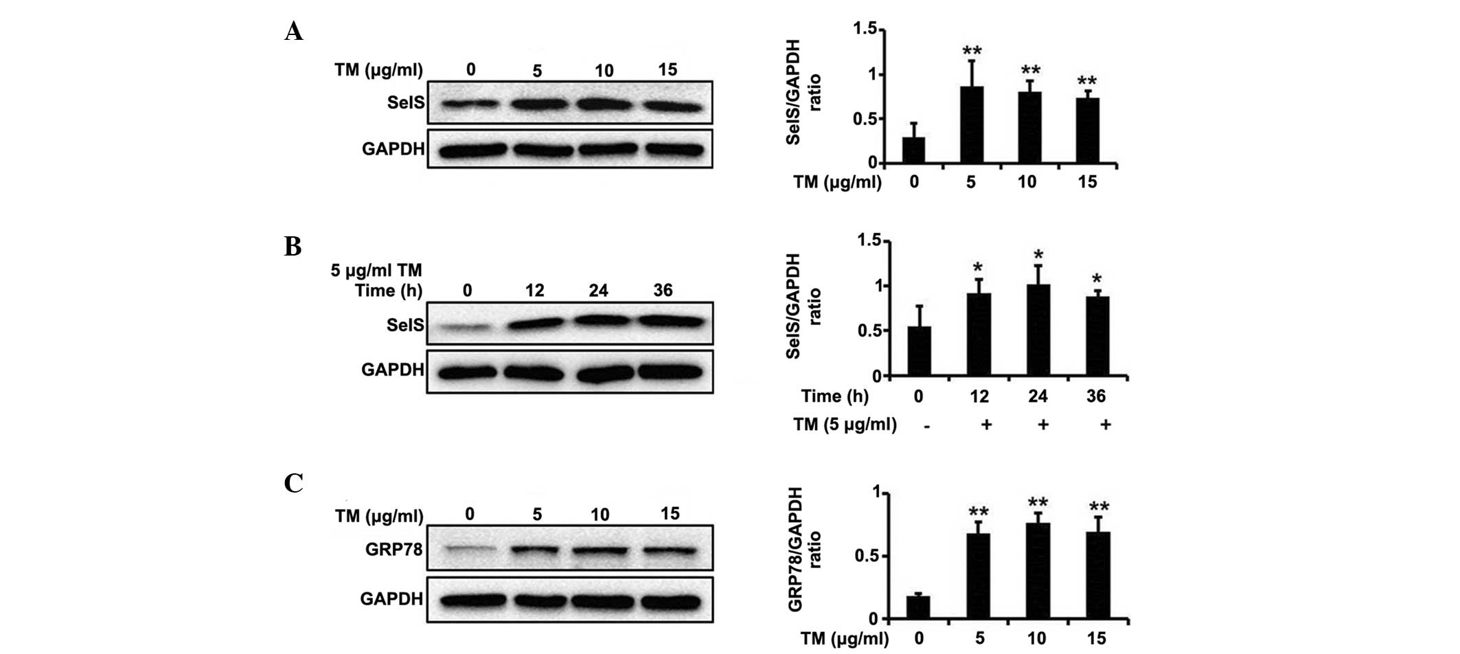
TM upregulates SelS expression and
induces ER stress
Prior to the screening of ER stress inhibitors, an
ER stress model was generated using TM. TM, which is an inhibitor
of protein glycosylation, has been widely used as an inducer of ER
stress (19). As presented in
Fig. 1A and B, TM was able to
significantly promote SelS expression at different concentrations
(5, 10 and 15 µg/ml) and at varying durations of treatment
(12, 24 and 36 h).
GRP78 (also known as BiP or heat shock 70kDa protein
5) is a major ER chaperone protein, which is able to increase the
protein folding capacity of the ER and regulate the activation of
ER transmembrane signaling molecules (20). GRP78 has an anti-apoptotic function
to prevent ER stress-induced cell death and is widely used as a
marker of ER stress (21). As
presented in Fig. 1C, TM markedly
increased GRP78 expression at different concentrations (5, 10 and
15 µg/ml), suggesting that TM results in significant ER
stress.
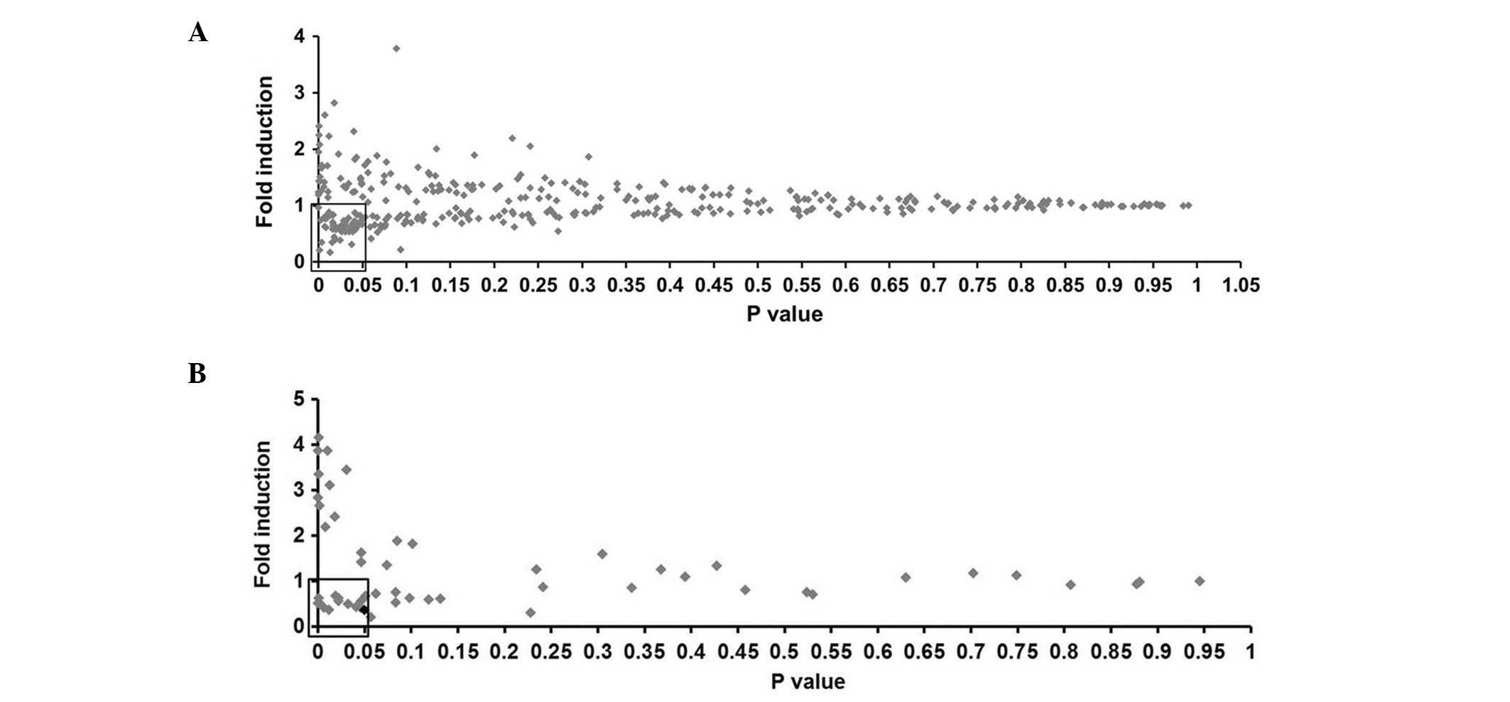
Screening of SelS expression
inhibitors
In order to directly test SelS expression, a firefly
luciferase reporter screening system driven by SelS promoter
(pSelS-luc) was generated, and greater than 300 natural compounds
were screened in HEK239T cells. Fig.
2A presents the fold induction and P-value of each compound.
With the thresholds of fold induction <1.00 and P<0.05, 54 of
the tested compounds were selected for further analysis.
In the secondary screening assay, HEK239T cells were
plated in 24-well plates. After 24 h, cells were transfected with
pSelS-luc plasmids or pGL3-basic vector plasmids plus
pCMV-β-galactosidase. The cells were incubated for 24 h prior to
treatment with the identified compounds at final concentration of 5
µg/ml or 0.05% DMSO for 24 h. The fold induction and P-value
of each compound are presented in Fig.
2B. PTX (Fig. 3A) exhibited a
fold induction of 0.368 (P<0.05) and the most marked inhibitory
effect on SelS promoter activity. Therefore, the subsequent
investigations focused on PTX.
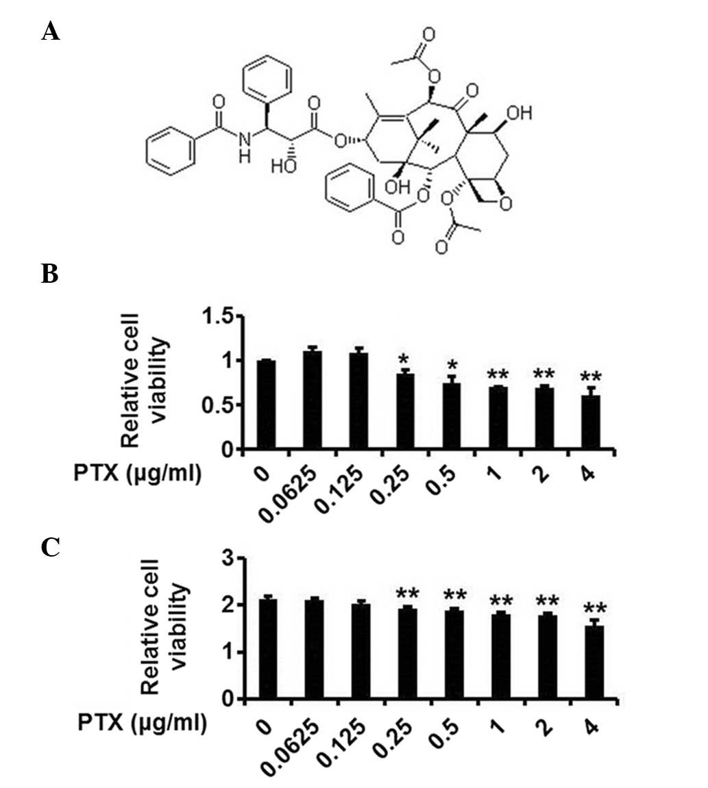 | Figure 3The structure and cytotoxicity of PTX.
(A) The structure of PTX. (B) The cytotoxicity of PTX to HepG2
cells. HepG2 cells were treated with 0.0625, 0.125, 0.25, 0.5, 1, 2
and 4 µg/ml PTX and cell viability was assessed. (C) The
cytotoxicity of PTX to HEK293T cells. HEK293T cells were treated
with 0.0625, 0.125, 0.25, 0.5, 1, 2 and 4 µg/ml PTX and cell
viability was assessed. The data are presented as the mean ±
standard deviation (n=3; *P<0.05,
**P<0.01 vs. the control group). PTX, paclitaxel. |
The cytotoxicity of PTX was subsequently
investigated using an MTT assay in HepG2 and HEK293T cells. As
presented in Fig. 3B and C, PTX at
the concentrations of 0.0625 and 0.125 µg/ml was not toxic
to HepG2 and HEK293T cells.
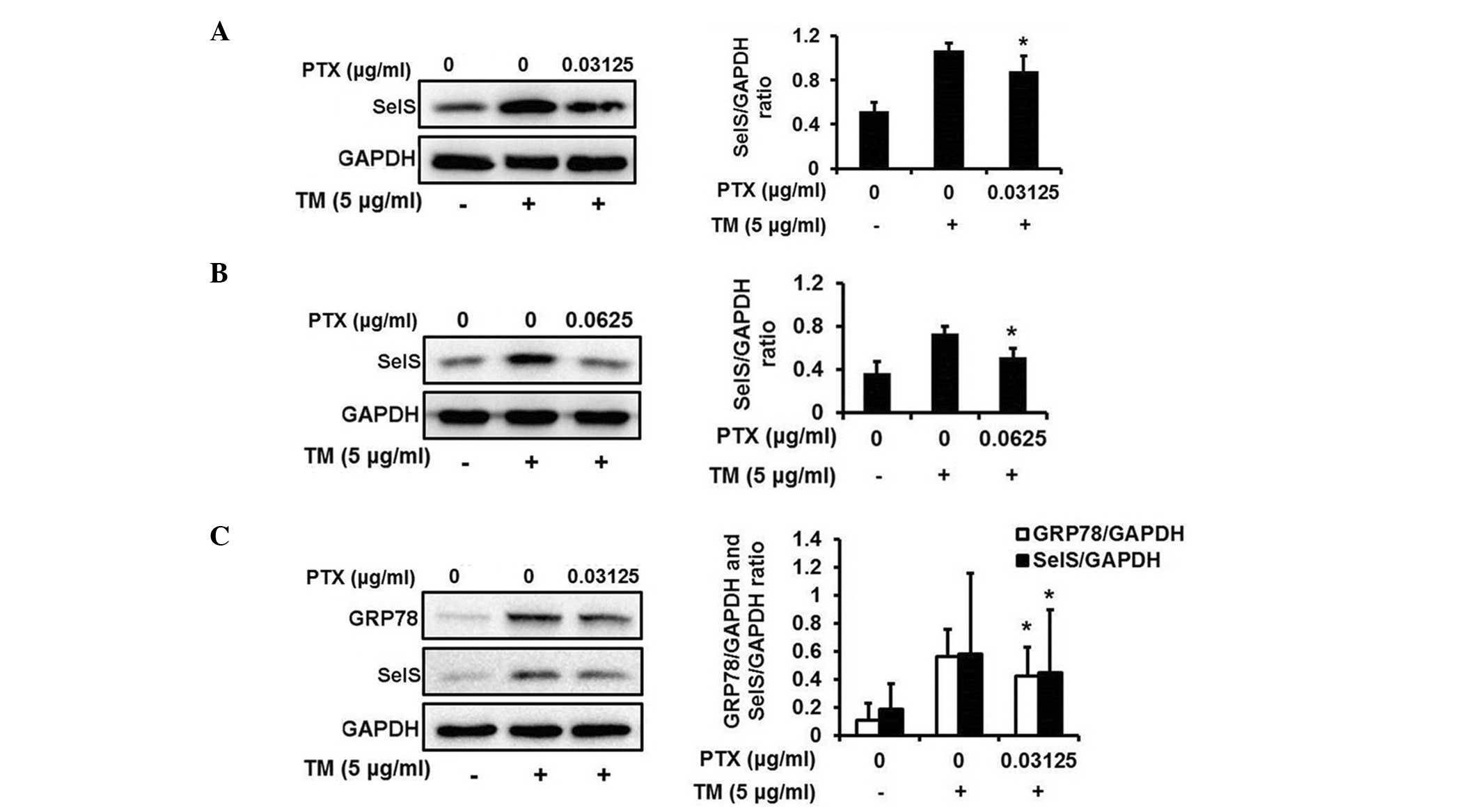
PTX inhibits TM-induced upregulation of
SelS in HepG2 cells
To confirm whether PTX is able to reverse the
TM-induced upregulation of SelS expression, the expression levels
of SelS were measured by RT-PCR in HepG2 cells. As presented in
Fig. 4A–C, TM increased SelS
expression, and treatment with PTX reversed the upregulation of
SelS induced by TM at different concentrations (0.03125, 0.0625 and
0.125 µg/ml). Western blotting also indicated that PTX
markedly reversed the upregulation of SelS at different
concentrations (0.0625 and 0.125 µg/ml) in HepG2 cells
(Fig. 4D and E). Taken together,
these results indicate that PTX may reverse the TM-induced
upregulation of SelS, and that PTX may be an ER stress
inhibitor.
PTX attenuates ER stress in HepG2
cells
Considering that PTX is able to inhibit SelS
expression, it was subsequently investigated whether PTX was able
to reverse ER stress. As presented in Fig. 4D and E, TM induced ER stress as
indicated by the upregulation of GRP78 levels. Following treatment
with PTX (0.0625 and 0.125 µg/ml), the expression levels of
GRP78 were significantly reduced, demonstrating that PTX is able to
reverse TM-induced ER stress. As presented in Fig. 4F, PTX was additionally able to
inhibit DTT-induced upregulation of GRP78, suggesting that PTX may
attenuate DTT-induced ER stress.
PTX inhibits TM-induced upregulation of
SelS and attenuates ER stress in HEK293T cells
In agreement with the observation in HepG2 cells,
PTX markedly attenuated TM-induced upregulation of SelS expression
at different concentrations (0.03125 and 0.0625 µg/ml) in
HEK293T cells (Fig. 5A and B).
Additionally, PTX was able to inhibit the TM-induced upregulation
of GRP78 (Fig. 5C), suggesting
that PTX may reverse ER stress in HEK293T cells.
Discussion
ER stress is involved in the pathogenesis of a
number of diseases including neurodegenerative diseases, liver
diseases, cardiovascular diseases and diabetes (22–25).
The identification of compounds that can attenuate ER stress may be
beneficial to patients. Natural products, characterized by high
chemical diversity and biochemical specificity, have been widely
explored as potential therapeutics for a variety of diseases
(10). As SelS is a sensitive
marker of ER stress (26,27) it was selected as a marker for the
screening of compounds that are able to reverse ER stress.
The liver and the kidneys are important organs in
the human body, and perform numerous essential functions. ER stress
in hepatocytes and nephrocytes is induced in a number of serious
diseases, including viral hepatitis, hepatocellular carcinoma,
glomerular injury and renal ischemia reperfusion injury (28,29).
The identification of drugs that can attenuate ER stress in liver
and kidney cells will be advantageous, therefore, liver and kidney
cell lines (HepG2 and HEK293T cells) were selected for the
screening compounds able to reduce ER stress.
The pSelS-luci reporter plasmid was constructed by
subcloning the SelS promoter region (−1073 to +39 bp) into the
pGL3-basic plasmid. This region contains an ER stress response
element that is activated by ER stress (17). Previous studies have indicated that
a number of transcription factor binding sites are located in the
SelS promoter region and that the promoter is important for
regulating the basal transcription of SelS (26). Therefore, a firefly luciferase
reporter driven by the SelS gene promoter was used in the present
study to screen the compounds that were able to inhibit SelS gene
promoter activity. Following screening of >300 compounds, PTX
was observed to markedly inhibit the activity of the SelS promoter
(Fig. 2A and B). Further results
revealed that PTX was able to inhibit the TM-induced upregulation
of SelS mRNA and protein at different concentrations (Fig. 4). However, the possibility that PTX
may influence the activity of other genes in the screening assay
cannot be excluded.
The current study demonstrated that PTX at
concentrations <0.125 µg/ml were able to attenuate ER
stress in HepG2 and HEK293T cells (Figs. 4 and 5), and that these concentrations of PTX
did not exhibit cytotoxicity, as presented in Fig. 3B and C. These results suggest that
PTX may attenuate ER stress at sub-toxic concentrations in HepG2
and HEK293T cells. However, whether PTX can attenuate ER stress at
higher (toxic) concentrations in these cells requires further
investigation.
The ER stress response, also known as the UPR, is an
adaptive response used to protect ER function. The mechanisms of
the UPR reduce the load of unfolded protein and restore ER
homeostasis by reducing protein translation and inducing the
transcription of components of the ER machinery involved in
folding, N-glycosylation, ERAD, quality control, redox and lipid
biogenesis (30). The mechanisms
by which PTX inhibits ER stress is unknown and requires further
study to be fully elucidated.
In conclusion, the current study demonstrates the
role of PTX in inhibiting SelS expression during ER stress and
attenuating ER stress. The present study identifies a novel natural
small molecule able to attenuate ER stress, and provides new
insight into the function and therapeutic application of PTX.
Acknowledgments
The present study was supported by the Fundamental
Research Funds for the Central Universities (grant no. 11QNJJ021),
and the Research Foundation of Jilin Provincial Science &
Technology Development (grant no. 20110938).
References
|
1
|
Gentile CL, Frye M and Pagliassotti MJ:
Endoplasmic reticulum stress and the unfolded protein response in
nonalcoholic fatty liver disease. Antioxid Redox Signal.
15:505–521. 2011. View Article : Google Scholar :
|
|
2
|
Marciniak SJ and Ron D: Endoplasmic
reticulum stress signaling in disease. Physiol Rev. 86:1133–1149.
2006. View Article : Google Scholar : PubMed/NCBI
|
|
3
|
Meusser B, Hirsch C, Jarosch E and Sommer
T: ERAD: The long road to destruction. Nat Cell Biol. 7:766–772.
2005. View Article : Google Scholar : PubMed/NCBI
|
|
4
|
Sozen E, Karademir B and Ozer NK: Basic
mechanisms in endoplasmic reticulum stress and relation to
cardiovascular diseases. Free Radic Biol Med. 78:30–41. 2015.
View Article : Google Scholar
|
|
5
|
Zhao L and Ackerman SL: Endoplasmic
reticulum stress in health and disease. Curr Opin Cell Biol.
18:444–452. 2006. View Article : Google Scholar : PubMed/NCBI
|
|
6
|
Halliday M and Mallucci GR: Targeting the
unfolded protein response in neurodegeneration: A new approach to
therapy. Neuropharmacology. 76:169–174. 2014. View Article : Google Scholar
|
|
7
|
Imai Y, Soda M, Inoue H, Hattori N, Mizuno
Y and Takahashi R: An unfolded putative transmembrane polypeptide,
which can lead to endoplasmic reticulum stress, is a substrate of
Parkin. Cell. 105:891–902. 2001. View Article : Google Scholar : PubMed/NCBI
|
|
8
|
Kaufman RJ: Orchestrating the unfolded
protein response in health and disease. J Clin Invest.
110:1389–1398. 2002. View Article : Google Scholar : PubMed/NCBI
|
|
9
|
Minamino T and Kitakaze M: ER stress in
cardiovascular disease. J Mol Cell Cardiol. 48:1105–1110. 2010.
View Article : Google Scholar
|
|
10
|
Sulaiman RS, Basavarajappa HD and Corson
TW: Natural product inhibitors of ocular angiogenesis. Exp Eye Res.
129:161–171. 2014. View Article : Google Scholar : PubMed/NCBI
|
|
11
|
Corson TW and Crews CM: Molecular
understanding and modern application of traditional medicines:
Triumphs and trials. Cell. 130:769–774. 2007. View Article : Google Scholar : PubMed/NCBI
|
|
12
|
Hou P, Li Y, Zhang X, Liu C, Guan J, Li H,
Zhao T, Ye J, Yang W, Liu K, et al: Pluripotent stem cells induced
from mouse somatic cells by small-molecule compounds. Science.
341:651–654. 2013. View Article : Google Scholar : PubMed/NCBI
|
|
13
|
Walder K, Kantham L, McMillan JS,
Trevaskis J, Kerr L, De Silva A, Sunderland T, Godde N, Gao Y,
Bishara N, et al: Tanis: A link between type 2 diabetes and
inflammation? Diabetes. 51:1859–1866. 2002. View Article : Google Scholar : PubMed/NCBI
|
|
14
|
Ye Y, Shibata Y, Yun C, Ron D and Rapoport
TA: A membrane protein complex mediates retro-translocation from
the ER lumen into the cytosol. Nature. 429:841–847. 2004.
View Article : Google Scholar : PubMed/NCBI
|
|
15
|
Du S, Liu H and Huang K: Influence of SelS
gene silence on beta-Mercaptoethanol-mediated endoplasmic reticulum
stress and cell apoptosis in HepG2 cells. Biochim Biophys Acta.
1800:511–517. 2010. View Article : Google Scholar : PubMed/NCBI
|
|
16
|
Kim KH, Gao Y, Walder K, Collier GR,
Skelton J and Kissebah AH: SEPS1 protects RAW264.7 cells from
pharmacological ER stress agent-induced apoptosis. Biochem Biophys
Res Commun. 354:127–132. 2007. View Article : Google Scholar : PubMed/NCBI
|
|
17
|
Gao Y, Feng HC, Walder K, Bolton K,
Sunderland T, Bishara N, Quick M, Kantham L and Collier GR:
Regulation of the seleno-protein SelS by glucose deprivation and
endoplasmic reticulum stress-SelS is a novel glucose-regulated
protein. FEBS Lett. 563:185–190. 2004. View Article : Google Scholar : PubMed/NCBI
|
|
18
|
Zhang Y, Bao YL, Wu Y, Yu CL, Sun Y and Li
YX: Identification and characterization of the human SLC5A8 gene
promoter. Cancer Genet Cytogenet. 196:124–132. 2010. View Article : Google Scholar : PubMed/NCBI
|
|
19
|
Lee AS: The glucose-regulated proteins:
Stress induction and clinical applications. Trends Biochem Sci.
26:504–510. 2001. View Article : Google Scholar : PubMed/NCBI
|
|
20
|
Iwasa K, Nambu Y, Motozaki Y, Furukawa Y,
Yoshikawa H and Yamada M: Increased skeletal muscle expression of
the endoplasmic reticulum chaperone GRP78 in patients with
myasthenia gravis. J Neuroimmunol. 273:72–76. 2014. View Article : Google Scholar : PubMed/NCBI
|
|
21
|
Ma KX, Chen GW, Shi CY, Cheng FF, Dou H,
Feng CC and Liu DZ: Molecular characterization of the
glucose-regulated protein 78 (GRP78) gene in planarian Dugesia
japonica. Comp Biochem Physiol B Biochem Mol Biol. 171:12–17. 2014.
View Article : Google Scholar : PubMed/NCBI
|
|
22
|
Dara L, Ji C and Kaplowitz N: The
contribution of endoplasmic reticulum stress to liver diseases.
Hepatology. 53:1752–1763. 2011. View Article : Google Scholar : PubMed/NCBI
|
|
23
|
Liu H, Cao MM, Wang Y, Li LC, Zhu LB, Xie
GY and Li YB: Endoplasmic reticulum stress is involved in the
connection between inflammation and autophagy in type 2 diabetes.
Gen Comp Endocrinol. 210:124–129. 2015. View Article : Google Scholar
|
|
24
|
Luo T, Kim JK, Chen B, Abdel-Latif A,
Kitakaze M and Yan L: Attenuation of ER stress prevents
post-infarction-induced cardiac rupture and remodeling by
modulating both cardiac apoptosis and fibrosis. Chem Biol Interact.
225:90–98. 2015. View Article : Google Scholar :
|
|
25
|
Torres M, Matamala JM, Duran-Aniotz C,
Cornejo VH, Foley A and Hetz C: ER stress signaling and
neurodegeneration: At the intersection between Alzheimer's disease
and Prion-related disorders. Virus Res. 207:69–75. 2015. View Article : Google Scholar : PubMed/NCBI
|
|
26
|
Gao Y, Hannan NR, Wanyonyi S,
Konstantopolous N, Pagnon J, Feng HC, Jowett JB, Kim KH, Walder K
and Collier GR: Activation of the selenoprotein SEPS1 gene
expression by pro-inflammatory cytokines in HepG2 cells. Cytokine.
33:246–251. 2006. View Article : Google Scholar : PubMed/NCBI
|
|
27
|
Speckmann B, Gerloff K, Simms L, Oancea I,
Shi W, McGuckin MA, Radford-Smith G and Khanna KK: Selenoprotein S
is a marker but not a regulator of endoplasmic reticulum stress in
intestinal epithelial cells. Free Radic Biol Med. 67:265–277. 2014.
View Article : Google Scholar
|
|
28
|
Malhi H and Kaufman RJ: Endoplasmic
reticulum stress in liver disease. J Hepatol. 54:795–809. 2011.
View Article : Google Scholar
|
|
29
|
Taniguchi M and Yoshida H: Endoplasmic
reticulum stress in kidney function and disease. Curr Opin Nephrol
Hypertens. 24:345–350. 2015. View Article : Google Scholar : PubMed/NCBI
|
|
30
|
Cnop M, Foufelle F and Velloso LA:
Endoplasmic reticulum stress, obesity and diabetes. Trends Mol Med.
18:59–68. 2012. View Article : Google Scholar
|