Staging recurrent ovarian cancer with 18FDG PET/CT

  • Authors:
    • Sanja Dragosavac
    • Sophie Derchain
    • Nelson M.G. Caserta
    • Gustavo De Souza
  • View Affiliations

  • Published online on: December 13, 2012     https://doi.org/10.3892/ol.2012.1075
  • Pages: 593-597
Metrics: Total Views: 0 (Spandidos Publications: | PMC Statistics: )
Total PDF Downloads: 0 (Spandidos Publications: | PMC Statistics: )


Abstract

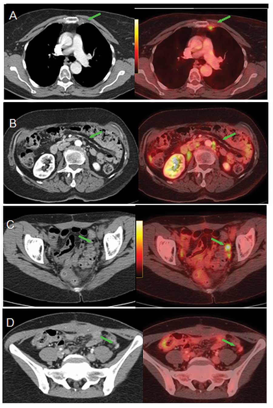
The aim of the present study was to evaluate the use of 2-deoxy-2-(18F)-fluoro-D-glucose (18FDG) positron emission tomography (PET)/computed tomography (CT) in patients with suspected ovarian cancer recurrence and describe the distribution of metastasis. A total of 45 female patients who underwent PET/CT scan due to raised CA-125 levels, clinical suspicion of ovarian cancer recurrence or alterations detected on ultrasound (US), CT or magnetic resonance imaging (MRI) were included in this retrospective study. PET/CT results were compared with histological findings (n=15) or clinical, laboratory and repeated imaging techniques during subsequent follow-up for at least six months (n=30). CA-125 was elevated in 34 patients, 14 patients had clinical symptoms of disease and 23 presented with alterations on US, CT and MRI. A total of 42 patients were confirmed to have ovarian cancer recurrence, all with abnormal findings on PET/CT. Three patients remained free of disease during clinical follow‑up, all with normal PET/CT findings. There were 11 patients with raised CA-125 levels and normal conventional imaging, all with positive PET/CT. Among the 11 patients with normal CA-125 levels, eight presented with positive PET/CT scan. Lymph nodes were the most frequent site of relapse of disease, followed by peritoneal implants. Distant sites of metastasis included the liver, spleen, pleura, lung and bone. PET/CT detected unsuspected lesions in 20/45 patients (44.4%). 18FDG PET/CT was a useful tool for evaluating the extent of ovarian cancer recurrence. In the current series, lymph nodes were the most frequent site of relapse of disease, with supradiaphragmatic lymph node metastasis in a large number of cases.
View Figures
View References

Related Articles

Journal Cover

February 2013
Volume 5 Issue 2

Print ISSN: 1792-1074
Online ISSN:1792-1082

Sign up for eToc alerts

Recommend to Library

Copy and paste a formatted citation
x
Spandidos Publications style
Dragosavac S, Derchain S, Caserta NM and De Souza G: Staging recurrent ovarian cancer with 18FDG PET/CT. Oncol Lett 5: 593-597, 2013.
APA
Dragosavac, S., Derchain, S., Caserta, N.M., & De Souza, G. (2013). Staging recurrent ovarian cancer with 18FDG PET/CT. Oncology Letters, 5, 593-597. https://doi.org/10.3892/ol.2012.1075
MLA
Dragosavac, S., Derchain, S., Caserta, N. M., De Souza, G."Staging recurrent ovarian cancer with 18FDG PET/CT". Oncology Letters 5.2 (2013): 593-597.
Chicago
Dragosavac, S., Derchain, S., Caserta, N. M., De Souza, G."Staging recurrent ovarian cancer with 18FDG PET/CT". Oncology Letters 5, no. 2 (2013): 593-597. https://doi.org/10.3892/ol.2012.1075