Contrast-enhanced ultrasonography of the rabbit VX2 tumor model: Analysis of vascular pathology
- Authors:
- Yanjun Liu
- Weidong Ren
- Caigang Liu
- Kun Huang
- Yueqin Feng
- Xuemei Wang
- Yuzhang Tong
View Affiliations
Affiliations: Department of Ultrasound, First Affiliated Hospital, China Medical University, Shenyang 110001, P.R. China, Department of Breast Surgery, General Surgery, The First Hospital of China Medical University, Shenyang 110001, P.R. China, Department of Regional Anatomy, China Medical University, Shenyang 110001, P.R. China
- Published online on: July 24, 2012 https://doi.org/10.3892/ol.2012.819
-
Pages:
685-690
Metrics: Total
Views: 0 (Spandidos Publications: | PMC Statistics: )
Metrics: Total PDF Downloads: 0 (Spandidos Publications: | PMC Statistics: )
This article is mentioned in:
Abstract
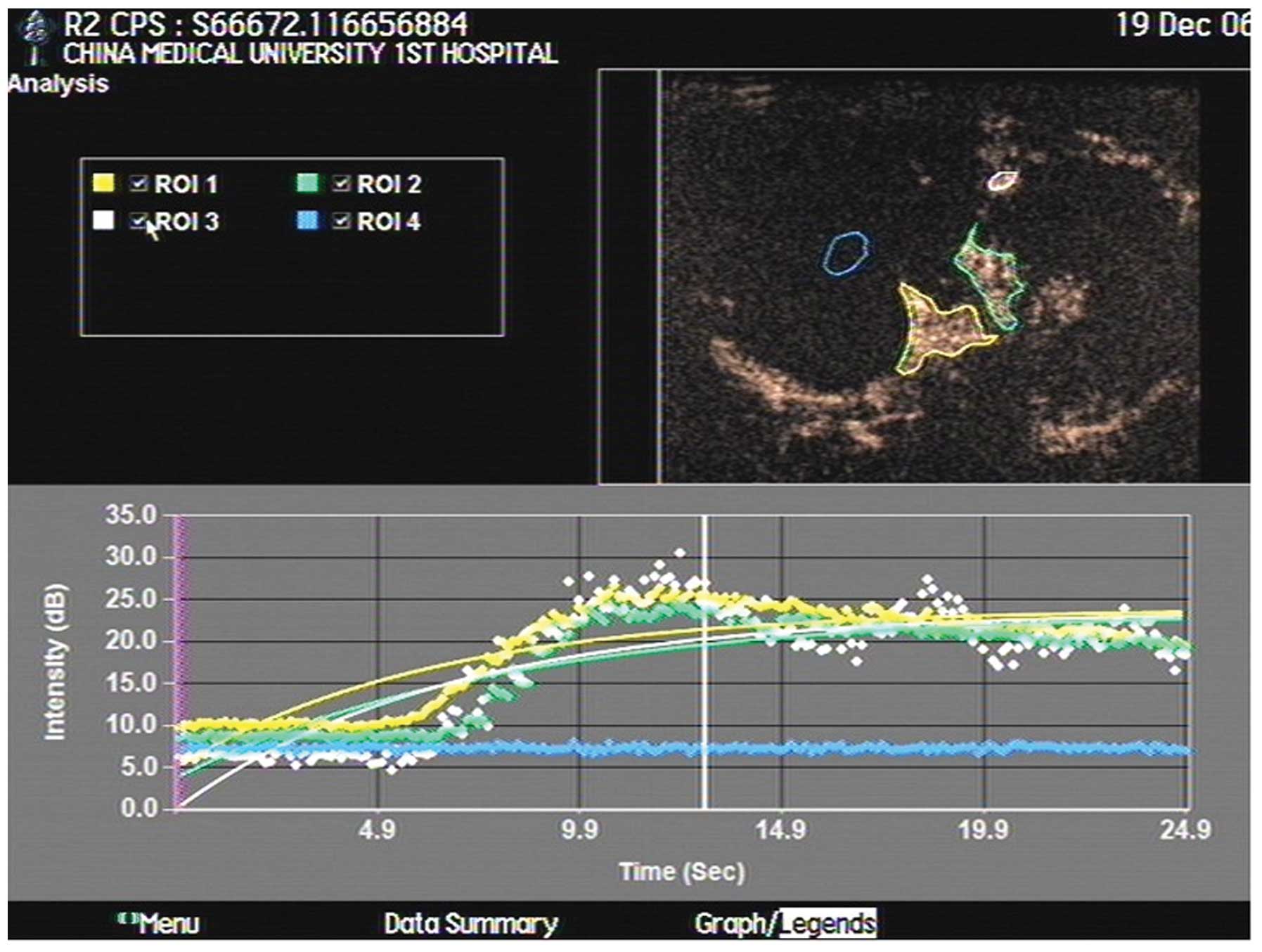
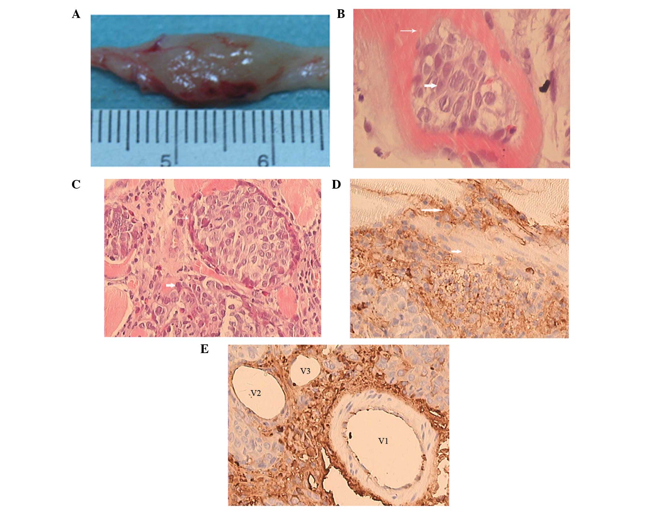
The accuracy of diagnosing tumors may be improved significantly by detecting the microvascular distribution. Indeed, contrast-enhanced ultrasonography (CEUS) has shown a distinct advantage in detecting microvasculature. This study aimed to determine the angiogenic characteristics of VX2 tumors in rabbits using CEUS. A total of 17 rabbits were injected with 0.5 ml VX2 cell suspension into the muscles of both hind legs to prepare the VX2 tumor models. At 14, 21, 28 and 35 days after tumor inoculation, CEUS was performed on the rabbits with 0.3 ml SonoVue following a local anesthesia. The pathological findings of the tumors were compared. A total of 12 rabbits survived after being inoculated with the tumor cells and developed a total of 38 tumors. The size of the tumors ranged from 1.12 to 10.85 cm. Using CEUS, all tumors demonstrated rim enhancement with some unenhanced regions. Enhancement began from the peripheral region and quickly showed internal reticular vessels. Regardless of the tumor size or the presence of necrosis, no complete enhancement of the tumors was observed. On microscopic examination, VX2 tumor cells were detected in striated muscles, immature blood capillaries and fibrosis tissues scattered in tumor nests. Immunohistochemical examination revealed that CD34+ cells appeared mainly in the muscles adjacent to vessels. In conclusion, CEUS may be an efficient method to evaluate angiogenesis and blood perfusion in VX2 tumors.
View References
1.
|
E GeorgesF BreitburdN JibardG OrthTwo
Shope papillomavirus-associated VX2 carcinoma cell lines with
different levels of keratinocyte differentiation and
transplantabilityJ Virol552462501985
|
2.
|
C LiW WangH DingB HuangJ CaoF MaoZ JiValue
of contrast-enhanced sonography in the diagnosis of peripheral
intrahepatic cholangiocarcinomaJ Clin
Ultrasound39447453201110.1002/jcu.2079721626512
|
3.
|
E DomenechD Berná-Serna JdeL PoloM ReusD
Berná-Mestre JdeM CanterasEffect of SonoVue on the synovial
membrane in rabbit kneesJ Ultrasound Med3012411246201121876095
|
4.
|
S TaoZ QinW HaoL YongquanY LanhuiY
LeiUsefulness of gray-scale contrast-enhanced ultrasonography
(SonoVue®) in diagnosing hepatic alveolar
echinococcosisUltrasound Med
Biol3710241028201110.1016/j.ultrasmedbio.2011.04.01421640477
|
5.
|
KC LeeWK MoonJW ChungAssessment of lymph
node metastases by contrast-enhanced MR imaging in a head and neck
cancer modelKorean J
Radiol8914200710.3348/kjr.2007.8.1.917277558
|
6.
|
A SonodaN NittaA Nitta-SekoTime-course
studies of implanted rabbit VX2 liver tumors to identify the
appropriate time for starting hepatic arterial embolization in
animal modelsOncology809296201110.1159/00032876321677452
|
7.
|
AA DünneR MandicA RamaswamyLymphogenic
metastatic spread of auricular VX2 carcinoma in New Zealand white
rabbitsAnticancer Res22327332792002
|
8.
|
AA DünneA SchmidtC KuropkatA RamaswamyS
SchulzJA WernerThe auricular VX2 carcinoma--an animal model for
sentinel node conceptIn Vivo17457461200314598609
|
9.
|
XF LiuLR RenGY SuEstablishment and
characterization of a rabbit tumor cell line VX2Zhonghua Bing Li
Xue Za Zhi346616632005(In Chinese)
|
10.
|
R MandicAA DünneN EikelkampExpression of
MMP-3, MMP-13, TMP-2 and TMP-3 in the VX2 carcinoma of the New
Zealand white rabbitAnticancer Res2232813284200212530076
|
11.
|
J LiB DongX YuC LiUltrasonographic
portography with low mechanical index gray-scale imaging in hepatic
VX2 tumorUltrasound Med
Biol32641647200610.1016/j.ultrasmedbio.2006.01.00816677923
|
12.
|
H MaruyamaS MatsutaniH SaishoN KamiyamaY
MineT HirataM SasamataSonographic shift of hypervascular liver
tumor on blood pool harmonic images with definity: time-related
changes of contrast-enhanced appearance in rabbit VX2 tumor under
extra-low acoustic powerEur J
Radiol566065200510.1016/j.ejrad.2005.04.004
|
13.
|
DL SimmonsAB SatterthwaiteDG TenenB
SeedMolecular cloning of a cDNA encoding CD34, a sialomucin of
human hematopoietic stem cellsJ Immunol14826727119921370171
|
14.
|
N XingZL CaiSH ZhaoL YangBX XuFL WangThe
use of CT perfusion to determine microvessel density in lung
cancer: Comparison with FDG-PET and pathologyChinese J Cancer
Res23118122201110.1007/s11670-011-0118-z23483098
|
15.
|
IJ FidlerLM EllisNeoplastic angiogenesis -
not all blood vessels are created equalN Engl J
Med351215216200410.1056/NEJMp04808015254281
|
16.
|
S ElagozR EgilmezA KoyuncuA
MuslehiddinogluS AriciThe intratumoral microvessel density and
expression of bFGF and nm23-H1 in colorectal cancerPathol Oncol
Res122127200610.1007/BF0289342716554912
|