Evaluation of photodynamic therapy in adhesion protein expression
- Authors:
- Published online on: May 16, 2014 https://doi.org/10.3892/ol.2014.2149
- Pages: 714-718
Metrics: Total
Views: 0 (Spandidos Publications: | PMC Statistics: )
Total PDF Downloads: 0 (Spandidos Publications: | PMC Statistics: )
Abstract
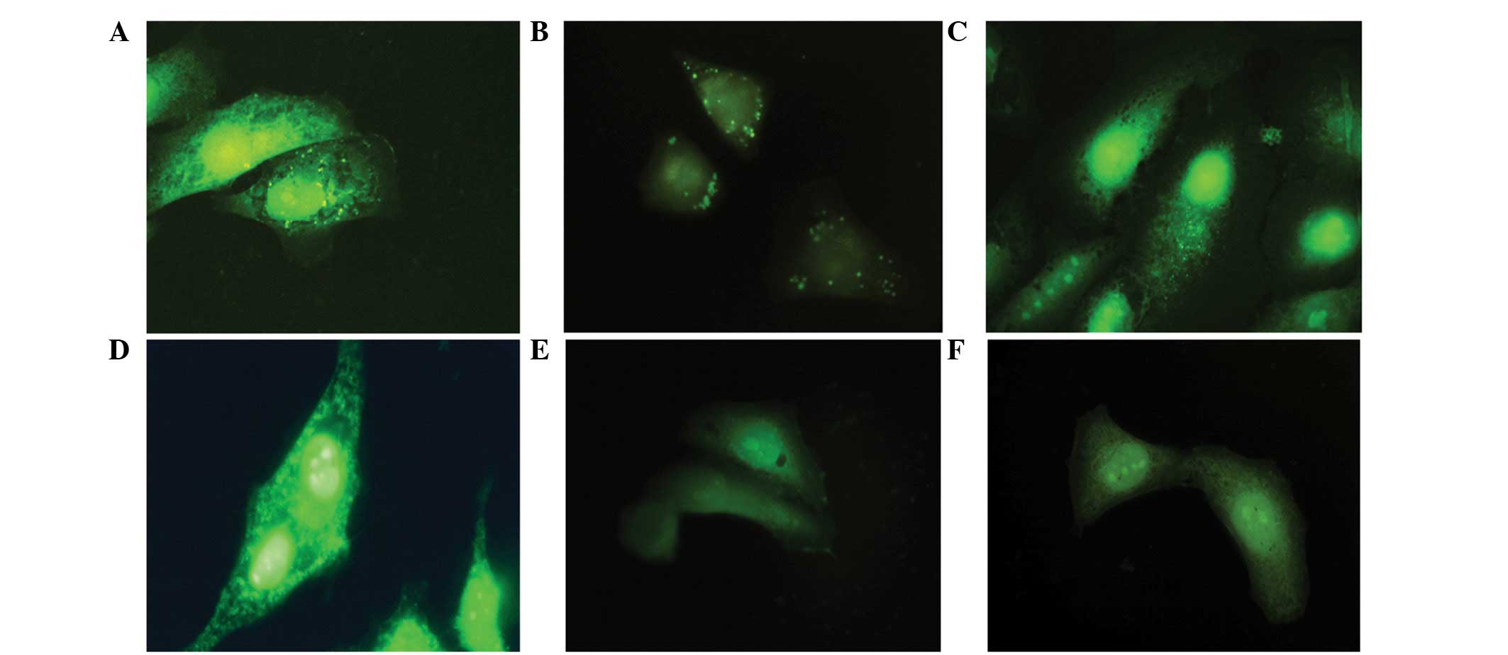
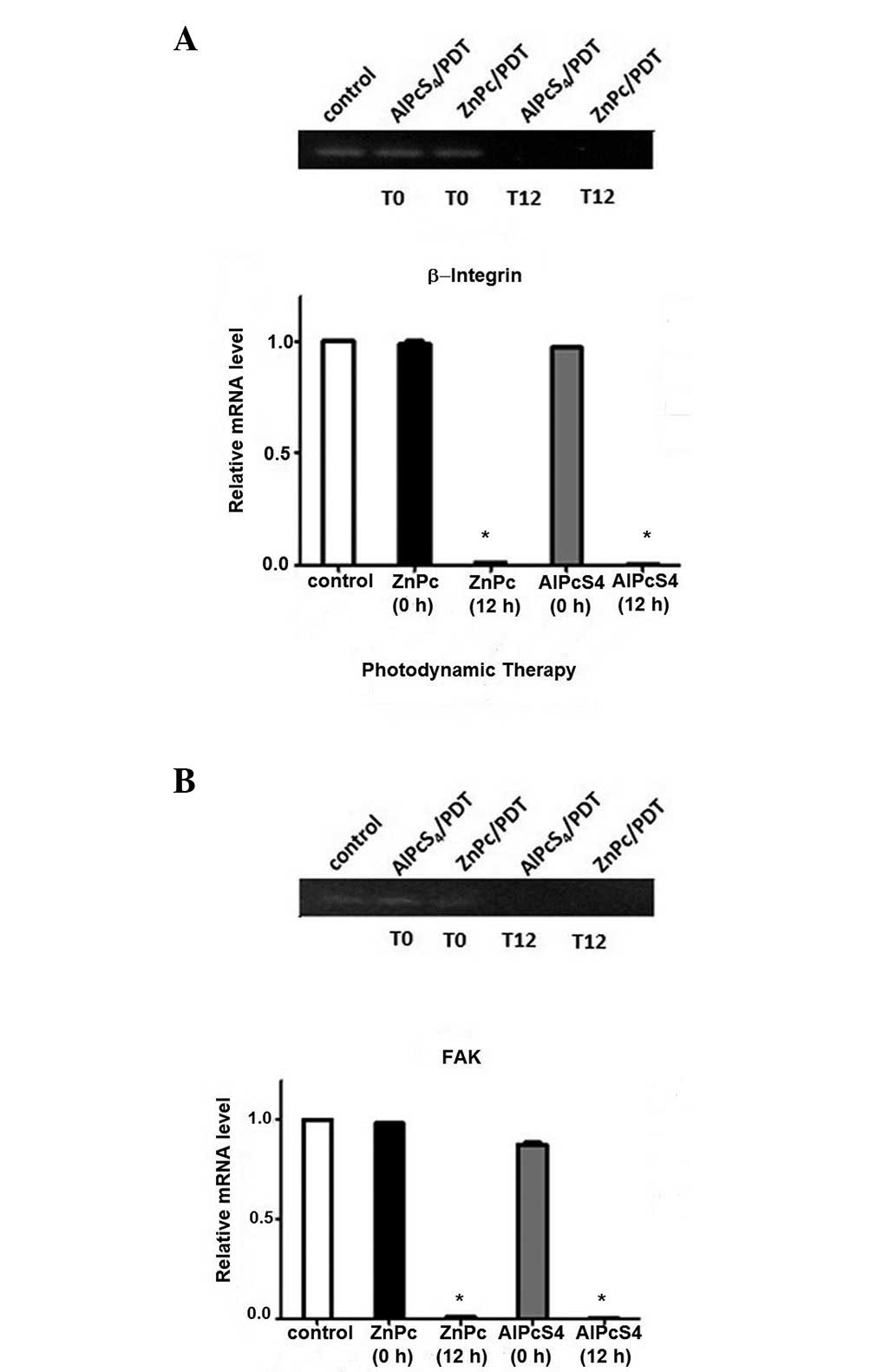
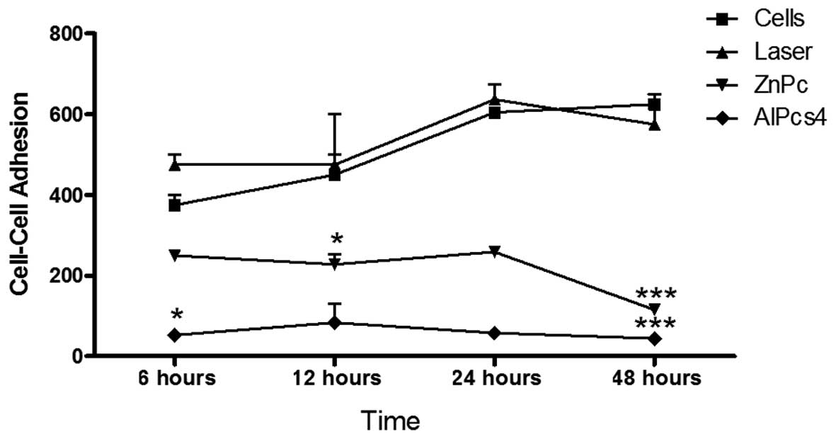
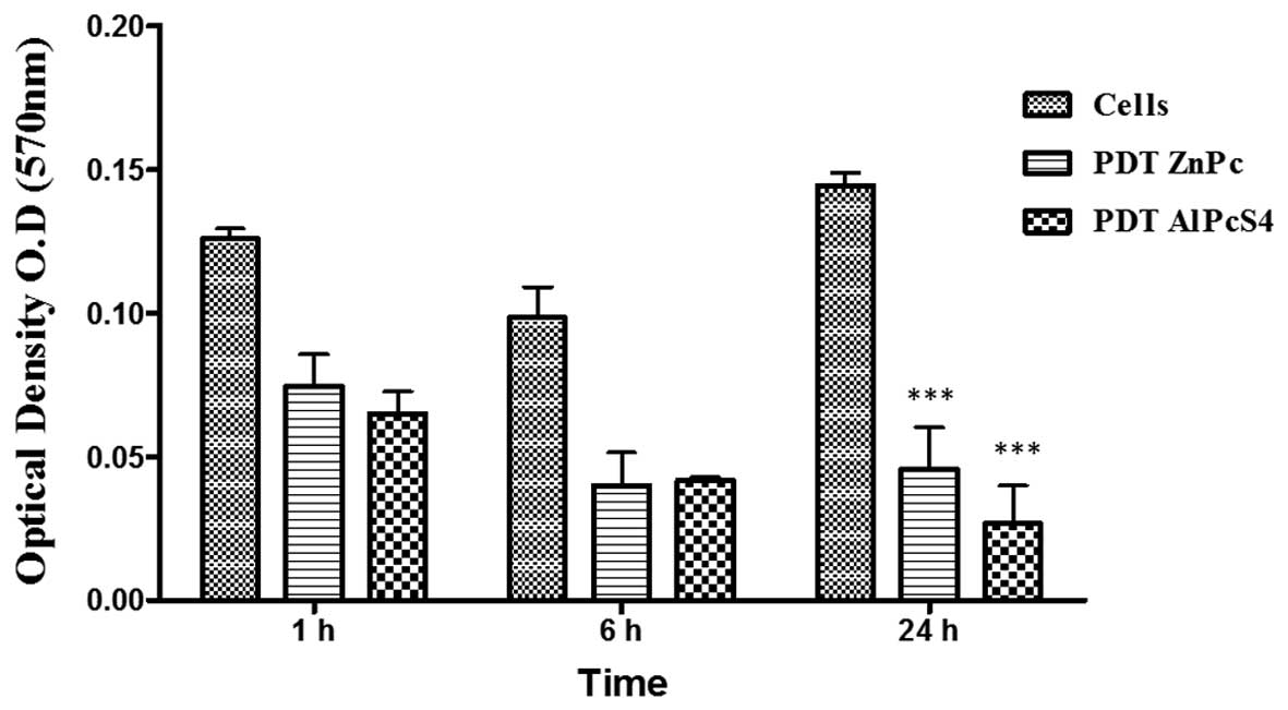
Photodynamic therapy (PDT) is a treatment modality that has clinical applications in both non‑neoplastic and neoplastic diseases. PDT involves a light‑sensitive compound (photosensitizer), light and molecular oxygen. This procedure may lead to several different cellular responses, including cell death. Alterations in the attachment of cancer cells to the substratum and to each other are important consequences of photodynamic treatment. PDT may lead to changes in the expression of cellular adhesion structure and cytoskeleton integrity, which are key factors in decreasing tumor metastatic potential. HEp‑2 cells were photosensitized with aluminum phthalocyanine tetrasulfonate and zinc phthalocyanine, and the proteins β1‑integrin and focal adhesion kinase (FAK) were assayed using fluorescence microscopy. The verification of expression changes in the genes for FAK and β1 integrin were performed by reverse transcription‑polymerase chain reaction (RT‑PCR). The results revealed that HEp‑2 cells do not express β‑integrin or FAK 12 h following PDT. It was concluded that the PDT reduces the adhesive ability of HEp‑2 cells, inhibiting their metastatic potential. The present study aimed to analyze the changes in the expression and organization of cellular adhesion elements and the subsequent metastatic potential of HEp‑2 cells following PDT treatment.