Laparoscopic liver biopsy in the diagnosis of hepatic epithelioid hemangioendothelioma: A case report
- Authors:
- Published online on: July 2, 2014 https://doi.org/10.3892/ol.2014.2308
- Pages: 1317-1319
Metrics: Total
Views: 0 (Spandidos Publications: | PMC Statistics: )
Total PDF Downloads: 0 (Spandidos Publications: | PMC Statistics: )
Abstract
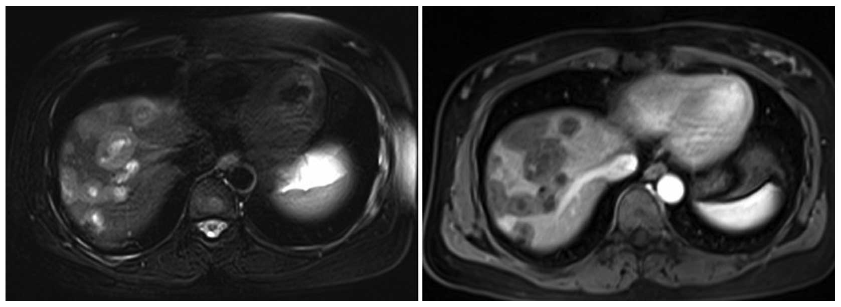
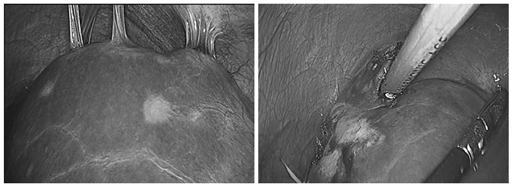
Hepatic epithelioid hemangioendothelioma (HEH) is a rare vascular tumor of the liver, and its definitive diagnosis is completely dependent on histopathological verification. In the present study, we report the case of a patient whose percutaneous liver biopsy failed to reveal a diagnosis of HEH, twice, and who was ultimately diagnosed by laparoscopic liver biopsy. The patient was a 42‑year‑old female with mild right upper quadrant discomfort. Ultrasonography and magnetic resonance imaging showed multiple mass lesions scattered throughout the liver, but no evidence of extrahepatic diseases. The initial laboratory tests included liver function tests and tumor markers were within normal limits. Subsequently, two, ultrasound (US)‑guided liver biopsies from the liver lesion were performed using an 18‑gauge needle, and both of these showed massive hepatocellular necrosis. To obtain adequate tissue samples for histological examination, the patient underwent laparoscopic liver biopsy. The overall immunohistochemical findings supported the diagnosis of HEH. In the present case, two, US‑guided percutaneous liver biopsies failed to diagnose HEH, and laparoscopic liver biopsy was safely performed to obtained adequate specimens for analysis. Although this method is not the preferred technique and has certain disadvantages, it is considered to be a useful and minimally invasive approach for liver lesions when other less‑invasive diagnostic modalities fail or are difficult to be performed.