Introduction
Gastric cancer is the fourth and fifth most
prevalent cancer diagnosed in males and females, respectively,
worldwide and the third and fifth most common cause of
cancer-related mortality in males and females, respectively
(1). Surgical resection remains the
mainstay of potentially curative treatment. However, local-regional
and distant recurrences are common, and the treatment outcome is
far from satisfactory (2). Thus,
surgery alone is insufficient for the treatment of the majority of
patients. In addition, gastric cancer is often diagnosed at an
advanced stage, which is unresectable. Therefore, chemotherapy and
chemoradiation are important for the treatment of gastric cancer,
particularly in patients with advanced and metastatic disease
(3,4).
Platinum-based drugs have been widely used and
extensively studied in anticancer therapy based on their ability to
covalently bind to DNA (5,6). At present, cisplatin remains the most
frequently used chemotherapy for different malignances. In gastric
cancer, regimens that contain platinum-based drugs have been
successfully administered in perioperative and postoperative
chemotherapy, as well as in chemotherapy for advanced/metastatic
disease, with significant efficacy (7–10).
However, despite the efficacy of platinum-based drugs against
gastric cancer, concerns exist regarding the use of these agents.
One concern is that cancer cells exhibit an inherent or acquired
refractory to platinum, which reduces its efficacy resulting in
disease relapse (11,12). An additional concern is their potent
toxicity and side effects, which significantly limit the tolerable
therapy dose.
Deguelin is a natural rotenoid isolated from several
plant species, including Mundulea sericea (Fig. 1). Deguelin has been found to exhibit
marked chemopreventive and antitumor activity against cancer, in
various model systems (13–15). Recently, it was reported that
deguelin induces DNA damage by reducing the expression of DNA
repair genes in human non-small cell lung cancer cells (16). The mechanism of platinum-refractory
or platinum-resistance in vivo is multi-factorial, including
increased tolerance to platinum-induced and enhanced repair of DNA
damage (17–22). Thus, to investigate whether deguelin
synergistically potentiates the antitumor effects of cisplatin, the
interactive effects of deguelin and cisplatin in vitro were
examined using the gastric carcinoma MGC-803 cell line. In
addition, the synergistic effects and possible mechanisms were
analyzed.
Materials and methods
Materials
3-(4,5)-dimethylthiazol(-2-yl)-2,5-diphenyltetrazolium
bromide (MTT), dimethyl sulfoxide (DMSO), propidium iodide (PI) and
cisplatin were purchased from Sigma-Aldrich (St. Louis, MO, USA).
The total protein extraction and comet assay kits were purchased
from Nanjing Keygen Biotech., Co., Ltd. (Nanjing, China). All the
chemicals used in this study were analytically pure and of culture
grade. The primary antibodies against BRCA1 (monoclonal rabbit
anti-human), ERCC1 (polyclonal rabbit anti-human), XRCC1
(polyclonal rabbit anti-human) and GAPDH (monoclonal rabbit
anti-human) were purchased from Cell Signaling Technology, Inc.
(Beverly, MA, USA), and the Bio-Rad protein assay kit I (cat. no.
500–0001) was purchased from Bio-Rad (Hercules, CA, USA).
Deguelin was purchased from Sigma-Aldrich, dissolved
in DMSO as a stock solution and stored at 4°C. The stock solution
was then diluted in cell culture medium to a final concentration of
0.05% DMSO (V/V).
Cell culture
The human gastric cancer MGC-803 cell line was
purchased from the Type Culture Collection of the Chinese Academy
of Sciences (Shanghai, China). The cells were cultured in RPMI-1640
medium (Life Technologies, Bedford, MA, USA) containing 10%
heat-inactivated fetal bovine serum, 100 U/ml penicillin and 100
U/ml streptomycin (all Life Technologies, Bedford, MA, USA) at 37°C
in a humidified atmosphere with 5% CO2.
Cell viability assay
The cell viability of the treated cancer cells was
determined using the MTT assay. Briefly, cells (4–5×10³)
were seeded in 96-well plates and cultured for 24 h, followed by
deguelin and cisplatin treatment. A volume of 10 μl of MTT (10
mg/ml) was added to each well and the cells were incubated for an
additional 4 h at 37°C. The supernatant fluid was then removed and
DMSO (150 μl/well) was added for 15–20 min. The optical densities
were measured at a wavelength of 570 nm using the SpectraMAX M5
microplate spectrophotometer (Molecular Devices, LLC, Sunnyvale,
CA, USA). All experiments were performed in triplicate. The effect
of deguelin on the cell proliferation was presented as the cell
growth inhibition, using the following formula: Inhibition rate (%)
= (A570 of control − A570 of treated
cells)/(A570 of control cells) × 100.
Isobologram analysis
The interactions of the combination treatment of
deguelin and cisplatin were analyzed by isobologram as previously
described (23). The dose-dependent
effects were determined for each compound and for one compound with
fixed concentrations of the other. The combination index (CI) was
calculated according to the following formula: CI =
(d1/Dx1) + (d2/Dx2), where Dx1
is the concentration of drug 1 (deguelin) required to produce ×
percentage effect alone, and d1 is the concentration of drug
1 required to produce the same × percentage effect in combination
with d2. Similarly, Dx2 is the concentration of drug
2 (cisplatin) required to produce × percentage effect alone, and
d2 is the concentration of drug 2 required to produce the
same × percentage effect in combination with d1. The CI
values were defined as follows: <1, synergism; 1, additive; and
>1, antagonism.
Morphological analysis
Following culture and drug treatment as previously
described, the morphological changes of the cells were observed.
The cells were fixed in 70% ethanol following washing with
phosphate-buffered saline (PBS). After examination for
morphological changes with an inverted microscope (XDS-800C; Ahghai
Caikon Optical Instrument Co., Ltd., Shanghai, China), the cells
were stained with PI (1 μg/ml in PBS) and analyzed under a
fluorescence microscope (Axiovert 200; Carl Zeiss, Göttingen,
Germany).
Western blot analysis
Following treatment with deguelin and cisplatin,
5×10⁶ cells were harvested and lysed in 1 ml lysis
buffer (Nanjing Keygen Biotech., Co., Ltd.), and the protein
concentration was determined using the Bio-Rad protein assay
reagent (Bio-Rad). The samples were then denatured in sample buffer
and the proteins were separated by sodium dodecyl
sulfate-polyacrylamide gel electrophoresis. Next, the gels were
electroblotted onto a polyvinylidene difluoride membrane, rinsed
with Tris-buffered saline with Tween 20 [20 mM Tris, 500 mM NaCl
and 0.1% Tween-20 (pH 7.6)] and blocked with 5% non-fat milk in
blocking buffer. The membrane was incubated with the appropriate
primary antibody overnight at 4°C. The membrane was then incubated
with the appropriate peroxidase-conjugated secondary antibody and
the immunoreactive bands were visualized using the enhanced
chemiluminescence method.
Statistical analysis
Data are presented as the mean ± standard deviation
and statistical analyses were performed using the analysis of
variance test. All data were analyzed using SPSS version 13.0
(SPSS, Chicago, IL, USA) and P<0.05 was considered to indicate a
statistically significant difference.
Results
Effects of deguelin and cisplatin on cell
proliferation
The antiproliferative effects of deguelin and
cisplatin alone on MGC-803 cells were investigated using the MTT
assay. Deguelin and cisplatin treatment resulted in a dose- and
time-dependent decrease in cell viability. Furthermore, when cells
were treated with deguelin for 48 and 72 h, the IC50
values were 10.74 and 6.52 μg/ml, respectively. The IC50
values of cells treated with cisplatin for 24, 48 and 72 h were
22.90, 7.66 and 3.89 μg/ml, respectively (Fig. 2).
Combined effects of deguelin and
cisplatin
The effects of cisplatin combined with deguelin in a
series of concentrations were examined. When combined with
deguelin, the IC50 of cisplatin in MGC-803 cells was
found to decrease significantly, in a dose-dependent manner. For
example, when the concentration of deguelin increased from 1.56 to
6.25 μg/ml, the IC50 of cisplatin was found to decrease
from 4.69 to 1.45 μg/ml (Fig.
3).
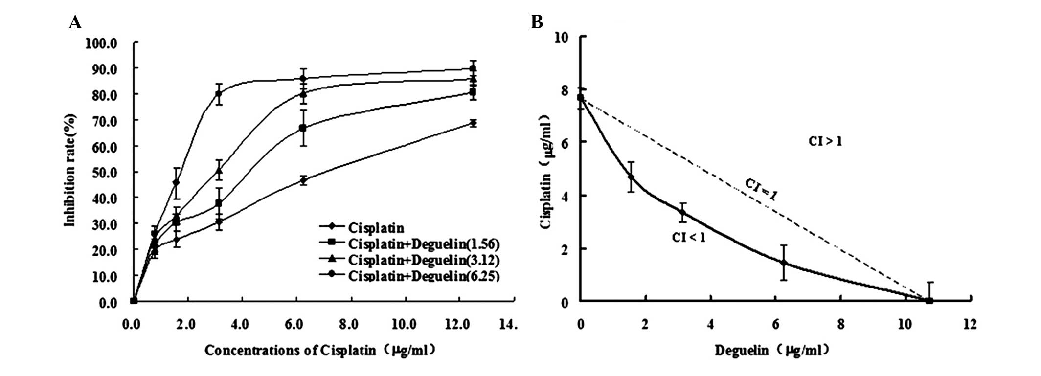 | Figure 3(A) Combined effects of deguelin and
cisplatin and (B) isobologram analysis. MGC-803 cells were treated
with cisplatin combined with deguelin at various concentrations (0,
1.56, 3.12 and 6.25 μg/ml). According to the data, when deguelin
(1.56, 3.12 and 6.25 μg/ml) was combined with cisplatin (4.49, 3.34
and 1.45 μg/ml, respectively), the CI was <1 (0.72, 0.75 and
0.76, respectively). Each point and vertical bar presents the mean
± standard deviation of three independent experiments. CI,
combination index. |
To evaluate the interaction between deguelin and
cisplatin, isobologram analysis was performed and the results
showed that the CI was considerably <1 when deguelin and
cisplatin were used in combination; CI=0.72 for 1.56 μg/mg deguelin
combined with 4.49 μg/mg cisplatin; CI=0.75 for 3.12 μg/mg deguelin
combined with 3.34 μg/mg cisplatin; and CI=0.76 for 6.25 μg/mg
deguelin combined with 1.45 μg/mg cisplatin). These results
indicated that deguelin and cisplatin exhibit synergistic effects
which inhibit the growth of MGC-803 cells (Fig. 3).
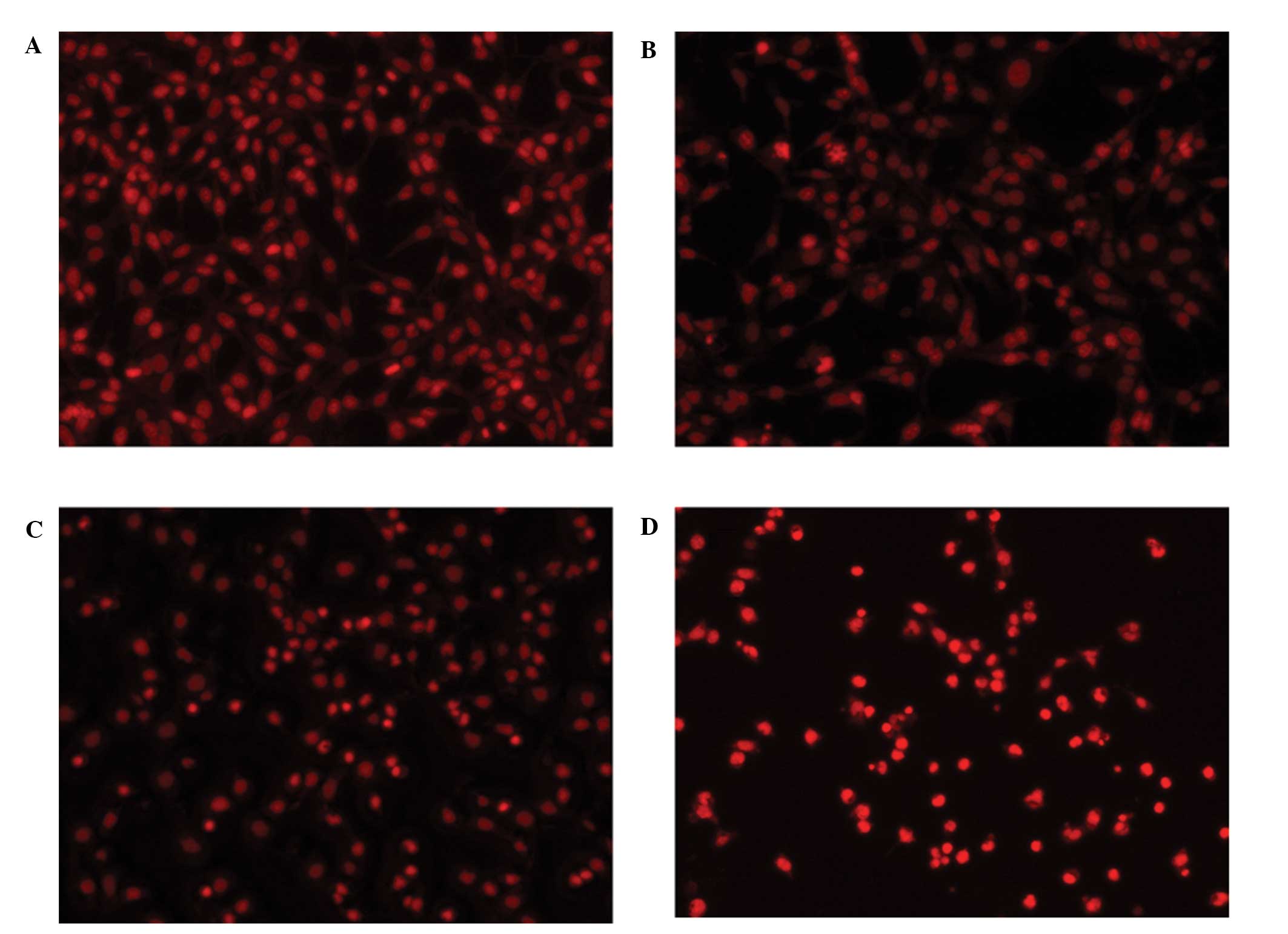
Furthermore, fluorescence microscopic examination of
PI-stained cells was performed to confirm the synergistic effects
between deguelin and cisplatin. Following treatment with 3 μg/ml
cisplatin and 6.25 μg/ml deguelin, the cell number was
significantly lower than that of the groups treated with cisplatin
or deguelin alone. However, the number of apoptotic cells had
increased (Fig. 4).
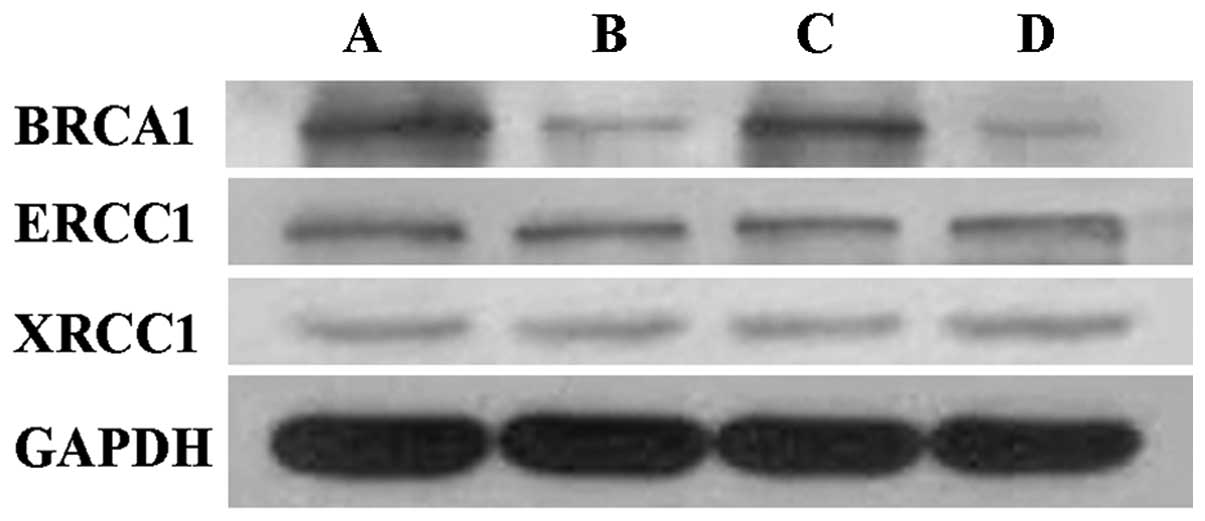
Mechanistic studies of deguelin-cisplatin
synergism
To identify the molecular mechanisms underlying the
synergism between deguelin and cisplatin, the expression of various
DNA damage repair genes was examined. The results revealed that
BRCA1 expression was significantly decreased following the
treatment of MGC-803 cells with deguelin alone or
deguelin-cisplatin in combination for 48 h, while no significant
differences were identified in the expression of ERCC1 and XRCC1.
However, treatment with cisplatin alone was not observed to alter
the expression of BRCA1, ERCC1 or XRCC1 (Fig. 5).
Discussion
Gastric cancer is prevalent in a number of countries
worldwide, accounting for 7.8% of all novel cancer cases and
~800,000 mortalities per year (1,24).
Despite the progress in recent years, the treatment outcome of
gastric cancer remains unsatisfactory with a five-year survival
rate of <30%. Therefore, novel drugs are urgently required to
improve the treatment of this malignancy.
Cisplatin, a representative platinum-based drug, is
one of the most commonly used drugs in the therapy of malignancies,
such as gastric cancer (25,26).
In cancer cells, platinum-based drugs efficiently bind to DNA to
form a variety of covalent adducts, which block replication and
transcription and eventually induce cell apoptosis or necrosis
(27). As DNA damage exerts
antitumor effects, increasing DNA repair may decrease the
sensitivity of cells or induce resistance to platinum-based drugs
(28,29). However, if DNA repair is inhibited,
the platinum-based drugs may exhibit more efficient antitumor
effects.
Deguelin, a naturally occurring rotenoid, is capable
of inhibiting phosphatidylinositide 3-kinase in premalignant and
malignant human bronchial epithelial cells (13). It has been reported that deguelin
markedly enhances the sensitivity of human U937 leukemia cells and
acute myeloid leukemia blasts to chemotherapeutic drugs via the
downregulation of Akt phosphorylation (30). However, the influences of deguelin
on platinum based drugs remain unclear. Therefore, in the present
study, the interaction between deguelin and cisplatin was examined
in the gastric cancer MGC-803 cell line.
In the present study, deguelin was found to inhibit
the proliferation of gastric cancer cells in a time- and
dose-dependent manner. Furthermore, DNA damage in cells was induced
by deguelin via the downregulation of DNA repair genes, consistent
with a previous report (16). When
combined with deguelin, the antitumor effect of cisplatin was
enhanced. Furthermore, the combination of deguelin with cisplatin
was found to increase the therapeutic efficacy of each drug,
resulting in a synergistic interaction (CI<1). In addition,
these results indicated that the inhibition of DNA damage repair
via the downregulation of BRCA1 underlies the synergistic effect of
deguelin and cisplatin.
In conclusion, deguelin in combination with
cisplatin exhibits a synergistic and significant antitumor effect
in gastric cancer cells. This may have promising therapeutic value
for gastric cancer and thus warrants further investigation.
Acknowledgements
This study was supported by the Technology Project
of Changzhou Social Development (grant no. CS20102016).
References
|
1
|
Jemal A, Bray F, Center MM, Ferlay J, Ward
E and Forman D: Global cancer statistics. CA Cancer J Clin.
61:69–90. 2011.
|
|
2
|
Songun I, Putter H, Kranenbarg EM, Sasako
M and van de Velde CJ: Surgical treatment of gastric cancer:
15-year follow-up results of the randomised nationwide Dutch D1D2
trial. Lancet Oncol. 11:439–449. 2010.
|
|
3
|
Jain VK, Cunningham D and Chau I:
Preoperative and postoperative chemotherapy for gastric cancer.
Surg Oncol Clin N Am. 21:99–112. 2012.
|
|
4
|
Pasini F, Fraccon AP and DE Manzoni G: The
role of chemotherapy in metastatic gastric cancer. Anticancer Res.
31:3543–3554. 2011.
|
|
5
|
Lebwohl D and Canetta R: Clinical
development of platinum complexes in cancer therapy: an historical
perspective and an update. Eur J Cancer. 34:1522–1534. 1998.
|
|
6
|
Monneret C: Platinum anticancer drugs.
From serendipity to rational design. Ann Pharm Fr. 69:286–295.
2011.
|
|
7
|
Kang YK, Kang WK, Shin DB, et al:
Capecitabine/cisplatin versus 5-fluorouracil/cisplatin as
first-line therapy in patients with advanced gastric cancer: a
randomised phase III noninferiority trial. Ann Oncol. 20:666–673.
2009.
|
|
8
|
Koizumi W, Tanabe S, Saigenji K, et al:
Phase I/II study of S-1 combined with cisplatin in patients with
advanced gastric cancer. Br J Cancer. 89:2207–2212. 2003.
|
|
9
|
Van Cutsem E, Moiseyenko VM, Tjulandin S,
et al; V325 Study Group. Phase III study of docetaxel and cisplatin
plus fluorouracil compared with cisplatin and fluorouracil as
first-line therapy for advanced gastric cancer: a report of the
V325 Study Group. J Clin Oncol. 24:4991–4997. 2006.
|
|
10
|
Cunningham D, Allum WH, Stenning SP, et
al: Perioperative chemotherapy versus surgery alone for resectable
gastroesophageal cancer. N Engl J Med. 355:11–20. 2006.
|
|
11
|
Rennicke A, Voigt W, Mueller T, et al:
Resistance mechanisms following cisplatin and oxaliplatin treatment
of the human teratocarcinoma cell line 2102EP. Anticancer Res.
25:1147–1155. 2005.
|
|
12
|
Timmer-Bosscha H, Mulder NH and de Vries
EG: Modulation of cis-diamminedichloroplatinum(II) resistance: a
review. Br J Cancer. 66:227–238. 1992.
|
|
13
|
Chun KH, Kosmeder JW II, Sun S, et al:
Effects of deguelin on the phosphatidylinositol 3-kinase/Akt
pathway and apoptosis in premalignant human bronchial epithelial
cells. J Natl Cancer Inst. 95:291–302. 2003.
|
|
14
|
Lee HY, Oh SH, Woo JK, et al:
Chemopreventive effects of deguelin, a novel Akt inhibitor, on
tobacco-induced lung tumorigenesis. J Natl Cancer Inst.
97:1695–1699. 2005.
|
|
15
|
Murillo G, Salti GI, Kosmeder JW II,
Pezzuto JM and Mehta RG: Deguelin inhibits the growth of colon
cancer cells through the induction of apoptosis and cell cycle
arrest. Eur J Cancer. 38:2446–2454. 2002.
|
|
16
|
Ji BC, Yu CC, Yang ST, et al: Induction of
DNA damage by deguelin is mediated through reducing DNA repair
genes in human non-small cell lung cancer NCI-H460 cells. Oncol
Rep. 27:959–964. 2012.
|
|
17
|
Altaha R, Liang X, Yu JJ and Reed E:
Excision repair cross complementing-group 1: gene expression and
platinum resistance. Int J Mol Med. 14:959–970. 2004.
|
|
18
|
Choi MK and Kim DD: Platinum transporters
and drug resistance. Arch Pharm Res. 29:1067–1073. 2006.
|
|
19
|
Howell SB, Safaei R, Larson CA and Sailor
MJ: Copper transporters and the cellular pharmacology of the
platinum-containing cancer drugs. Mol Pharmacol. 77:887–894.
2010.
|
|
20
|
Olaussen KA, Mountzios G and Soria JC:
ERCC1 as a risk stratifier in platinum-based chemotherapy for
nonsmall-cell lung cancer. Curr Opin Pulm Med. 13:284–289.
2007.
|
|
21
|
Saldivar JS, Wu X, Follen M and Gershenson
D: Nucleotide excision repair pathway review I: implications in
ovarian cancer and platinum sensitivity. Gynecol Oncol. 107(Suppl
1): S56–S71. 2007.
|
|
22
|
Wernyj RP and Morin PJ: Molecular
mechanisms of platinum resistance: still searching for the
Achilles’ heel. Drug Resist Updat. 7:227–232. 2004.
|
|
23
|
Hou W, Chen L, Yang G, et al: Synergistic
antitumor effects of liposomal honokiol combined with adriamycin in
breast cancer models. Phytother Res. 22:1125–1132. 2008.
|
|
24
|
Choi KS, Jun JK, Park EC, et al:
Performance of different gastric cancer screening methods in Korea:
a population-based study. PLoS One. 7:e500412012.
|
|
25
|
Hainsworth JD, Johnson DH and Greco FA:
Cisplatin-based combination chemotherapy in the treatment of poorly
differentiated carcinoma and poorly differentiated adenocarcinoma
of unknown primary site: results of a 12-year experience. J Clin
Oncol. 10:912–922. 1992.
|
|
26
|
Ohtsu A, Shimada Y, Yoshida S, et al:
Phase II study of protracted infusional 5-fluorouracil combined
with cisplatinum for advanced gastric cancer: report from the Japan
Clinical Oncology Group (JCOG). Eur J Cancer. 30A:2091–2093.
1994.
|
|
27
|
Fuertes MA, Castilla J, Alonso C and Pérez
JM: Cisplatin biochemical mechanism of action: from cytotoxicity to
induction of cell death through interconnections between apoptotic
and necrotic pathways. Curr Med Chem. 10:257–266. 2003.
|
|
28
|
Brabec V: DNA modifications by antitumor
platinum and ruthenium compounds: their recognition and repair.
Prog Nucleic Acid Res Mol Biol. 71:1–68. 2002.
|
|
29
|
Brabec V and Kasparkova J: Modifications
of DNA by platinum complexes. Relation to resistance of tumors to
platinum antitumor drugs. Drug Resist Updat. 8:131–146. 2005.
|
|
30
|
Bortul R, Tazzari PL, Billi AM, et al:
Deguelin, a PI3K/AKT inhibitor, enhances chemosensitivity of
leukaemia cells with an active PI3K/AKT pathway. Br J Haematol.
129:677–686. 2005.
|