A rare case of primary chondrosarcoma arising from the sternum: A case report
- Authors:
- Bin He
- Yuehan Huang
- Peng Li
- Xiaohe Ye
- Fuqiang Lin
- Lidong Huang
- Shengqiang Gao
- Hongqi Shi
- Yunfeng Shan
View Affiliations
Affiliations: Department of Hepatobiliary Surgery, The First Affiliated Hospital of Wenzhou Medical University, Wenzhou, Zhejiang 325000, P.R. China, Department of Pathology, The First Affiliated Hospital of Wenzhou Medical University, Wenzhou, Zhejiang 325000, P.R. China, Department of Radiology, The First Affiliated Hospital of Wenzhou Medical University, Wenzhou, Zhejiang 325000, P.R. China, Department of Cardiac and Thoracic Surgery, The First Affiliated Hospital of Wenzhou Medical University, Wenzhou, Zhejiang 325000, P.R. China
- Published online on: August 18, 2014 https://doi.org/10.3892/ol.2014.2453
-
Pages:
2233-2236
Metrics: Total
Views: 0 (Spandidos Publications: | PMC Statistics: )
Metrics: Total PDF Downloads: 0 (Spandidos Publications: | PMC Statistics: )
This article is mentioned in:
Abstract
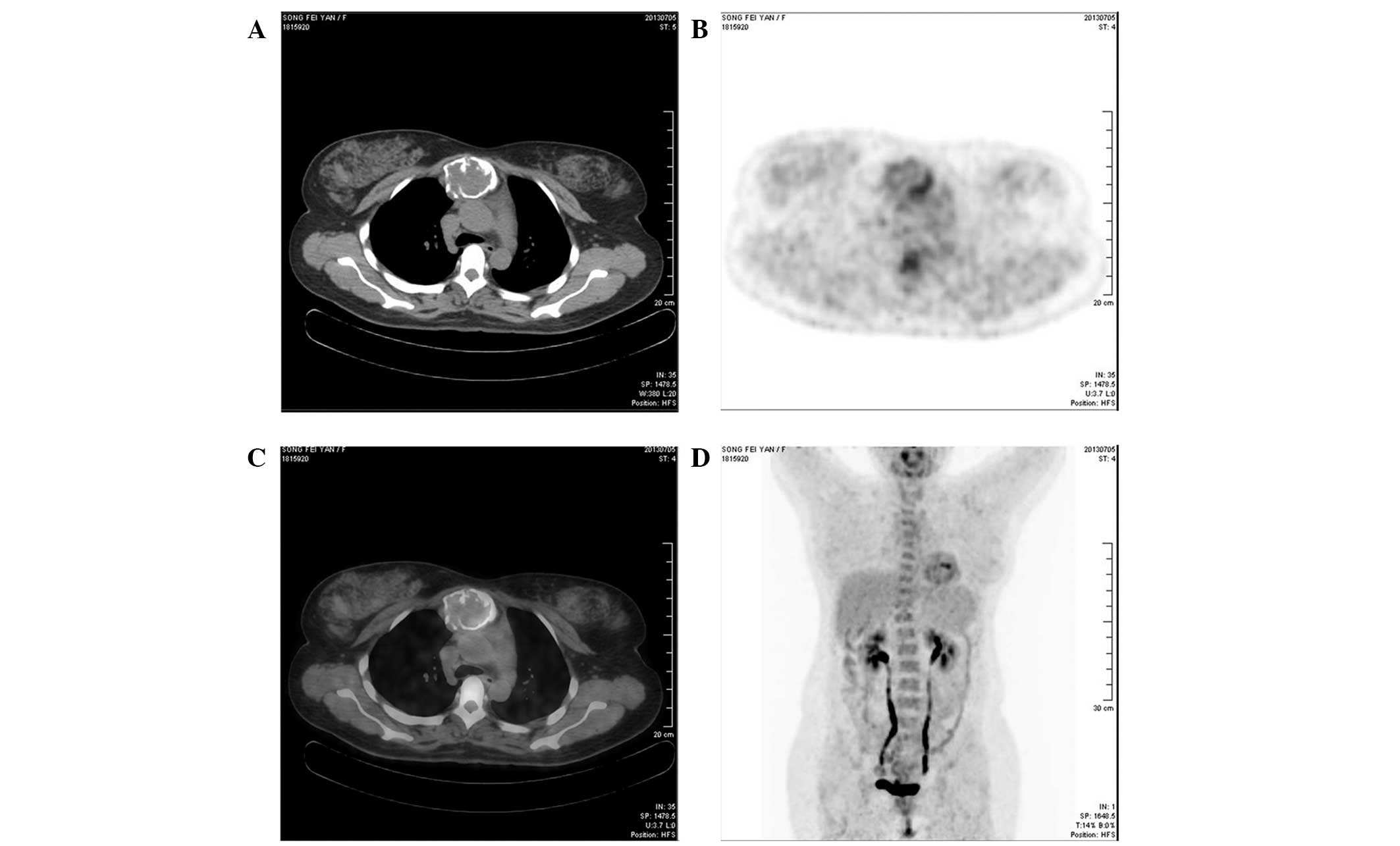
The current study reports a case of an extremely rare tumor that presented in an uncommon location, which was successfully treated via radical resection and reconstruction. A 37‑year‑old female, with no notable medical history, with the exception of a cesarean delivery, was admitted to The First Affiliated Hospital of Wenzhou Medical University (Wenzhou, China) due to pain and a lump of the anterior chest wall. The mass was identified on the manubrium sterni and was not tender on palpation. A chest computed tomography (CT) scan reconstruction identified the abnormal mass on the manubrium sterni (size, 5x4 cm in diameter) and positron emission tomography‑CT interpretation strongly indicated a type of well‑differentiated malignant tumor, such as a giant cell tumor. An aspiration needle biopsy was not conducted, however, the patient underwent tumor radical resection and sternal reconstruction using steel wire and titanium mesh. Histopathological examination of the surgical specimen determined the diagnosis of chondrosarcoma. A postoperative chest X‑ray revealed that the sternal defect had repaired well, therefore, this procedure may be highly beneficial in future for repairing defects in the sternum.
View References
1
|
Lequaglie C, Massone PB, Giudice G and
Conti B: Gold standard for sternectomies and plastic
reconstructions after resections for primary or secondary sternal
neoplasms. Ann Surg Oncol. 9:472–479. 2002.
|
2
|
Kużdżał J, Warmus J, Grochowski Z and
Gądek A: Reconstruction of the sternal manubrium. J Thorac
Cardiovasc Surg. 147:1986–1988. 2014.
|
3
|
Chapelier AR, Missana MC, Couturaud B, et
al: Sternal resection and reconstruction for primary malignant
tumors. Ann Thorac Surg. 77:1001–1006; discussion 1006–1007.
2004.
|
4
|
McCormack P, Bains MS, Beattie EJ Jr and
Martini N: New trends in skeletal reconstruction after resection of
chest wall tumors. Ann Thorac Surg. 31:45–52. 1981.
|
5
|
Liu ZC and Zhao H: Titanium internal
fixation system used for sternum reconstruction after resection of
chondrosarcoma. Chin Med J (Engl). 123:2621–2622. 2010.
|
6
|
Alpert JB, Nonaka D, Chachoua A, Pass HI
and Ko JP: Increasing dyspnea due to an anterior mediastinal mass.
Chest. 139:217–223. 2011.
|
7
|
Stanić V, Vulović T, Novaković M, et al:
Radical resection of giant chondrosarcoma of the anterior chest
wall. Vojnosanit Pregl. 65:64–68. 2008.
|
8
|
Waisberg DR, Abrão FC, Fernandez A, Terra
RM, Pêgo-Fernandes PM and Jatene FB: Surgically-challenging
chondrosarcomas of the chest wall: five-year follow-up at a single
institution. Clinics (Sao Paulo). 66:501–503. 2011.
|
9
|
Marulli G, Hamad AM, Cogliati E, Breda C,
Zuin A and Rea F: Allograft sternochondral replacement after
resection of large sternal chondrosarcoma. J Thorac Cardiovasc
Surg. 139:e69–e70. 2010.
|
10
|
Rocco G, Fazioli F, La Manna C, et al:
Omental flap and titanium plates provide structural stability and
protection of the mediastinum after extensive sternocostal
resection. Ann Thorac Surg. 90:e14–e16. 2010.
|