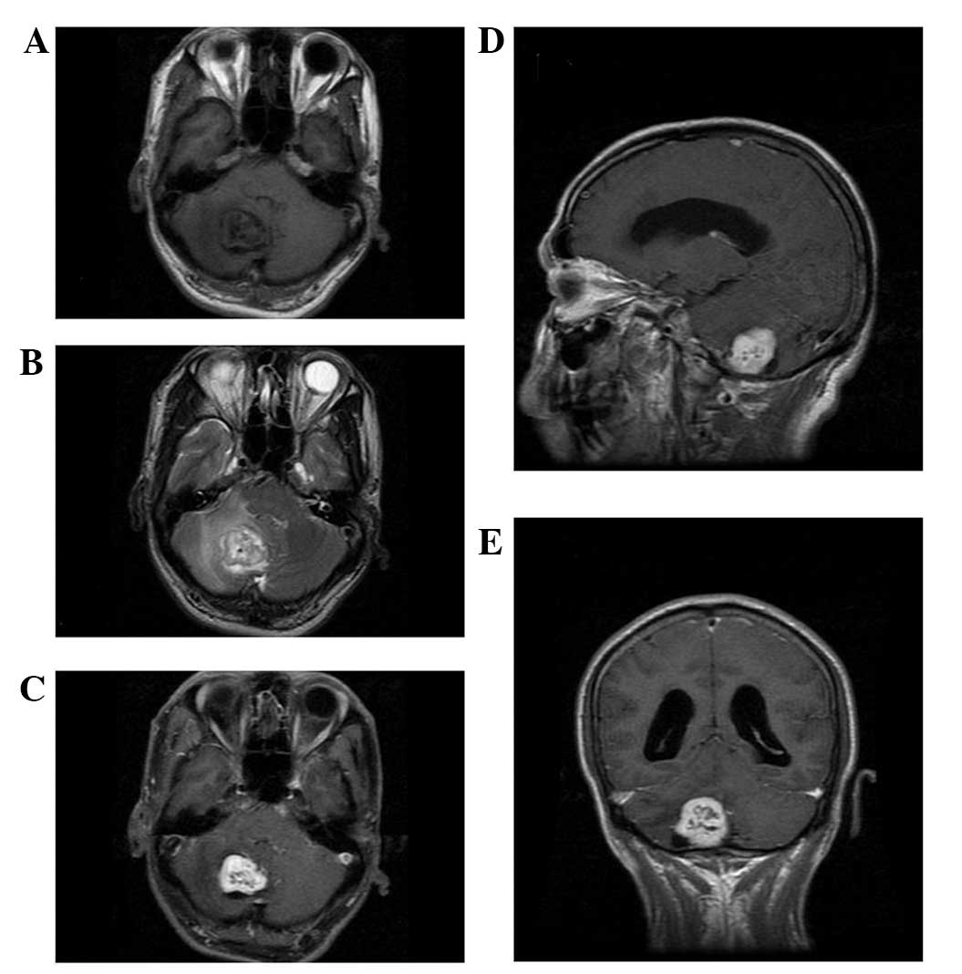
Magnetic resonance imaging of intracranial hemangiopericytoma and correlation with pathological findings

  • Authors:
    • Cong Ma
    • Feng Xu
    • Yu-Dong Xiao
    • Ramchandra Paudel
    • Yi Sun
    • En-Hua Xiao
  • View Affiliations

  • Published online on: September 4, 2014     https://doi.org/10.3892/ol.2014.2503
  • Pages: 2140-2144
Metrics: Total Views: 0 (Spandidos Publications: | PMC Statistics: )
Total PDF Downloads: 0 (Spandidos Publications: | PMC Statistics: )


Abstract

The present study aimed to evaluate the radiological and pathological features of intracranial hemangiopericytoma, and improve the understanding of this tumor. A retrospective analysis of radiological and pathological features of five cases of intracranial hemangiopericytoma was conducted between 2006 and 2012 in the Second Xiangya Hospital of Central South University. A total of five cases (three males and two females; aged 37‑60 years) were enrolled. Magnetic resonance imaging revealed that the lesions were lobulated with iso‑intensity T1‑weighted image signals and slightly long T2‑weighted image signals. Cystic degeneration, necrosis and flow void were observed. The case with the lesion located under the tentorium cerebelli exhibited compression of the fourth ventricle with lateral ventricle dilatation hydrocephalus. In all cases, the solid section of the lesion was markedly enhanced following injection of the contrast agent, and intratumoral vessels were observed. No case exhibited the dural tail sign. Immunohistochemical examination revealed positive expression of cluster of differentiation 34(CD34), vimentin and CD99, and negative expression of epithelial membrane antigen, S100 and glial fibrillary acidic protein. Proliferating cell nuclear antigen Ki‑67 immunohistochemical staining revealed that <5% of cells expressed Ki‑67 in two cases and 5‑10% of cells expressed Ki‑67 in three cases. In conclusion, intracranial hemangiopericytoma exhibits certain distinctive characteristics in radiological examination, allowing for improved diagnosis. However, pathological examination is required for confirmation.
View Figures
View References

Related Articles

Journal Cover

November-2014
Volume 8 Issue 5

Print ISSN: 1792-1074
Online ISSN:1792-1082

Sign up for eToc alerts

Recommend to Library

Copy and paste a formatted citation
x
Spandidos Publications style
Ma C, Xu F, Xiao Y, Paudel R, Sun Y and Xiao E: Magnetic resonance imaging of intracranial hemangiopericytoma and correlation with pathological findings. Oncol Lett 8: 2140-2144, 2014.
APA
Ma, C., Xu, F., Xiao, Y., Paudel, R., Sun, Y., & Xiao, E. (2014). Magnetic resonance imaging of intracranial hemangiopericytoma and correlation with pathological findings. Oncology Letters, 8, 2140-2144. https://doi.org/10.3892/ol.2014.2503
MLA
Ma, C., Xu, F., Xiao, Y., Paudel, R., Sun, Y., Xiao, E."Magnetic resonance imaging of intracranial hemangiopericytoma and correlation with pathological findings". Oncology Letters 8.5 (2014): 2140-2144.
Chicago
Ma, C., Xu, F., Xiao, Y., Paudel, R., Sun, Y., Xiao, E."Magnetic resonance imaging of intracranial hemangiopericytoma and correlation with pathological findings". Oncology Letters 8, no. 5 (2014): 2140-2144. https://doi.org/10.3892/ol.2014.2503