Introduction
Hepatocellular carcinoma (HCC) is the sixth most
common type of human malignancy worldwide (1) and one of the most common types of
malignant tumor in China. HCC is the third leading cause of
cancer-related mortality worldwide (2,3) and
the second leading cause of cancer-related mortality in China. The
annual incidence of HCC is increasing (4,5) and,
therefore, it presents a significant threat to human health.
Chemotherapy may be used to treat HCC; however, traditional
systemic chemotherapy exhibits a low curative rate for liver cancer
due to its high number of toxic effects, and is therefore not
widely accepted. Recently, treatments combining several
chemotherapeutic or chemopreventive agents have been used as they
not only enhance the treatment effect but also reduce drug
toxicity.
The compound 5-fluorouracil (5-FU) is one of the
most commonly used chemotherapeutic drugs for liver, colorectal and
gastric cancers in clinical practice (6). In addition, 5-FU is commonly used in
advanced-stage HCC chemotherapy, alone or in combination with other
drugs. Resistance to 5-FU is a key cause of chemotherapy failure in
advanced-stage HCC (7) and,
therefore, it is important to identify novel chemotherapeutic
agents with high therapeutic efficacy. The combination of novel
chemotherapeutic agents with existing drugs may enhance the
efficacy of liver cancer therapy.
Radix puerariae (the root of the kudzu plant,
Pueraria lobata) is one of the most popular traditional
Chinese medicines. Puerarin (daidzein-8-C-glucoside) and daidzein
(daidzein-7-O-glucoside) are the major isoflavones of the kudzu
root (8). Previous studies have
suggested that puerarin may exhibit anticarcinogenic effects
(9–16). In addition, one study has suggested
that puerarin may act as a sensitizer to enhance the inhibitory
effect of Bulbophyllum extract on HCC proliferation
(17).
The aim of the present study was to investigate the
effects of the combined treatment with puerarin and 5-FU on HCC
in vitro and in vivo.
Materials and methods
Chemicals and reagents
Puerarin (P5555) was obtained from Sigma-Aldrich
(St. Louis, MO, USA) with a purity of 98%, as assessed by
reverse-phase high-performance liquid chromatography. The puerarin
was stored as a 100-mM stock solution in dimethyl sulfoxide at
−20°C and diluted with serum-free culture medium for use in
experiments. The compound 5-FU was obtained from Sigma-Aldrich
(F6627) and diluted to various concentrations in serum-free culture
medium. The Cell Counting Kit-8 (CCK-8) was obtained from Dojindo
Molecular Technologies, Inc. (Kumamato, Japan).
The Annexin V-fluorescein isothiocyanate
(FITC)/propidium iodide (PI) kit was obtained from Beijing
MultiSciences Biotech Co., Ltd (Beijing, China) and the Hoechst
staining kit was obtained from the Beyotime Institute of
Biotechnology (Shanghai, China).
Cell line and cell culture
The HCC SMMC7721 cell line was obtained from the
China Center for Type Culture Collection (Wuhan, China). The cells
were cultured in Dulbecco’s modified Eagle’s medium (Gibco-BRL,
Gaithersburg, MD, USA) supplemented with 10% fetal bovine serum
(FBS; Gibco-BRL), 50 mg/ml streptomycin (Beyotime Institute of
Biotechnology), 50 IU/ml penicillin (Beyotime Institute of
Biotechnology), and 2 mM glutamine (Beyotime Institute of
Biotechnology) in a humidified atmosphere of 5% CO2 at
37°C.
Cell growth inhibition studies
The inhibitory effect of puerarin on SMMC7721 cell
growth in vitro was determined using CCK-8 dye, which is
only absorbed by living cells. Briefly, 5,000 cells/well were
seeded into 96-well microtiter plates. Following exposure to
puerarin (400, 800, 1,600, 3,200 or 6,400 μM), 5-FU (40, 80, 160,
320 or 640 μM) or puerarin and 5-FU (400:40; 800:80; 1600:60;
3200:320 μM, respectively) for 48 h, 10 μl CCK-8 solution was added
to each well, and the plates were incubated for an additional 2 h
(at 37°C with 5% CO2). The optical density was then
determined at a wavelength of 450 nm using a microplate reader
(iMark; Bio-Rad, Hercules, CA, USA). Each experiment was performed
in triplicate and the results were presented as the inhibition rate
(IR), which was calculated using the following formula: IR (%) =
[(A-B)/A] × 100, where A and B are the absorbance of the control
and sample groups following 48 h of incubation, respectively.
Evaluation of the combined effects of
puerarin and 5-FU
The following equation (18,19)
was used to evaluate the nature of the interaction between puerarin
and 5-FU: D = Dm[fa/(1 - fa)]1/m, where D is
the dose, Dm is the dose required to produce the median
effect (analogous to the IC50), fa is the fraction of
the system affected by D, and m is a Hill-type coefficient
signifying the sigmoidicity of the dose-effect curve. The
combination index (CI) values were obtained using Biosoft CalcuSyn
software (Biosoft, Cambridge, UK) written in BASIC for automatic
graphing of the CI with respect to fa, to determine whether the two
drugs were non-exclusive. Synergism was indicated by a CI value of
<1, summation by a CI value equal to 1 and antagonism by a CI
value of >1.
Cell apoptosis assay
Apoptotic cells were detected by Hoechst 33258
staining as follows. Following exposure to puerarin (1,600 μM),
5-FU (160 μM) or puerarin and 5-FU for 48 h, the SMMC7721 cells
were incubated with 20 μM Hoechst 33258 (Beyotime Institute of
Biotechnology) for 10 min at room temperature. The cells were then
washed twice with phosphate-buffered saline (PBS) and examined
under a fluorescence microscope (BX53F; Olympus, Tokoyo, Japan).
The apoptotic cells were identified by Hoechst 33258 staining of
the condensed chromatin and nuclear fragments. A total of 10 random
fields were counted for each sample.
Annexin V/PI staining
An Annexin V/PI apoptosis kit was used according to
the manufacturer’s instructions to quantify the percentage of cells
undergoing apoptosis. Firstly, SMMC7721 cells were incubated for 48
h with puerarin (1,600 μM) or 5-FU (160 μM) alone or in
combination. Next, the cells were washed twice with cold PBS and
resuspended in binding buffer at a concentration of
1×10⁶ cells/μl. Then, 5 μl of Annexin V-FITC and 10 μl
PI were added, and the cells were incubated for 5 min at room
temperature in the dark. Following incubation, 200 μl of binding
buffer was added and the cells were analyzed immediately by flow
cytometry (FACSAriaIII; BD Biosciences, Franklin Lakes, NJ, USA).
The flow cytometry analysis was performed using the Cell Quest
software (BD Biosciences). The Annexin V+/PI−
cells were identified as apoptotic cells, and the Annexin
V−/PI+ cells were identified as necrotic
cells (20). The entire procedure
was repeated three times for each sample.
Xenograft tumor model
All animal procedures, which complied with the
National Institutes of Health Guide for the Care and Use of
Laboratory Animals (21), were
approved by the Committee on Animal Experimentation of Wuhan
University (Wuhan, China). Four- to six-week-old male BALB/c-nu/nu
nude mice were purchased from the Center for Experimental Animals
of Wuhan University (Wuhan, China). All mice weighed 16–18 g and
were bred in autoclaved, filter-top, microisolator cages, which
were kept in an isolator unit with filtered air. The mice had
access to water and food ad libitum. The human HCC SMMC7721
cell line used for inoculation was cultured as previously
described. The mice were inoculated subcutaneously with
1×10⁷ SMMC7721 cells per mouse and the tumor sizes were
measured using micrometer calipers. The mice were randomly divided
into the following four groups, with six mice per group: Saline
tumor control, puerarin (50 mg/kg/day), 5-FU (12 mg/kg/day), and
puerarin (50 mg/kg/day) in combination with 5-FU (12
mg/kg/day).
Measuring tumor volume and weight
The tumor volume (TV) was calculated using the
following formula: TV (mm3) = d2x(D/2), where
d and D are the shortest and longest diameters, respectively. The
tumor size was measured with calipers every three days. The mice
were sacrificed by extracting the eyeball at the end of four weeks
and the tumor xenografts were removed, weighed and measured for
additional analyses.
Terminal deoxynucleotidyl transferase
dUTP nick end labeling assay
Sections of each tumor xenograft were fixed in 4%
formaldehyde, dehydrated with an ethanol gradient, embedded in
paraffin, dewaxed and rehydrated with a decreasing ethanol gradient
(100, 95, 90, 80 and 70%), according to standard instructions. An
in situ apoptosis detection kit (Roche Molecular Systems
Inc., Branchburg, NJ, USA) was used to detect apoptosis. All
procedures were performed according to the manufacturer’s
instructions. The specimens were incubated with proteinase K [15
μg/ml in 10 mM Tris/HCl (pH 7.5)] for 20 min at room temperature
after being dewaxed and rehydrated. Next, the specimens were rinsed
with 3% H2O2, and incubated with
equilibration buffer and terminal deoxynucleotidyl transferase
(Beyotime Institute of Biotechnology). The specimens were then
incubated with an anti-digoxigenin-peroxidase conjugate. Finally,
the 3,3′-diaminobenzidine substrate was added to react with the
peroxidase and the specimens were counterstained with hematoxylin,
mounted and analyzed using a light microscope (BX53F).
Evaluation of side effects
Following the completion of the study, blood was
collected from the nude mice by cardiac puncture using
heparin-rinsed 5-ml syringes and 22-gauge needles. Alanine
aminotransferase (ALT), aspartate aminotransferase (AST), blood
urea nitrogen (BUN) and serum creatinine (Cr) levels were measured
to evaluate liver and renal injury using an Olympus AU5400 Immuno
Analyzer (Olympus). All mice were sacrificed and dissected
following the completion of the study. In addition, metastasis,
hemorrhage and liver and kidney morphology were assessed by an
observer blinded to the treatment groups. This study was approved
by the Institutional Review Board of Renmin Hospital at Wuhan
University.
Statistical analysis
Statistical analyses were performed using SPSS 17.0
software (SPSS, Inc., Chicago, IL, USA). All data are presented as
the mean ± standard deviation (SD). The means of the different
groups were compared using non-parametric analysis (Mann-Whitney
rank-sum test) and P<0.05 was considered to indicate a
statistically significant difference.
Results
Effect of single drug exposure on the
growth of the HCC SMMC7721 cell line
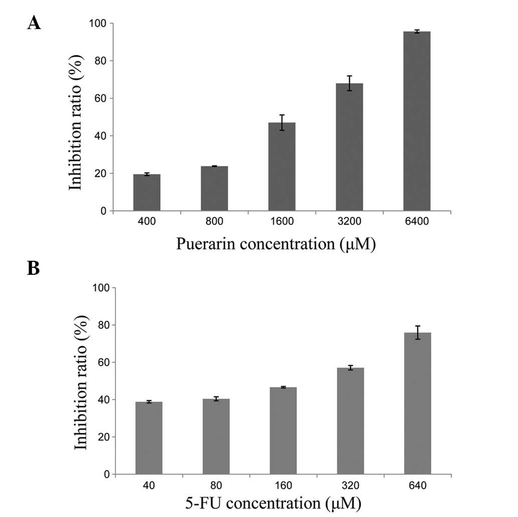
The inhibition of SMMC7721 proliferation by puerarin
and 5-FU was assessed following 48 h of drug exposure and 24 h of
culture in drug-free medium. After 48 h of treatment, in
vitro SMMC7721 cell growth was significantly inhibited in a
dose-dependent manner (P<0.01; Fig.
1). The mean ± SD rate of inhibition by puerarin was
19.51±0.72% at a concentration of 400 μM, and 95.56±0.81% at a
concentration of 6,400 μM. The rate of growth inhibition by 5-FU at
40 μM was 38.83±0.63%, while the rate was 75.91±3.54% at 640
μM.
Combined effect of puerarin and 5-FU on
SMMC7721 cell growth
The SMMC7721 cells were exposed to the two drugs in
combination at a fixed molar ratio (puerarin to 5-FU, 10:1) for 48
h. The effect on cell proliferation was then assessed by CCK-8
assay and the rate of growth inhibition was calculated according to
the method used by Chou and Talalay (18,19).
All experiments were repeated in triplicate. The CI values were
<1 when the fraction of affected cells was between 0.2555 and
0.7420 (Fig. 2).
Apoptosis induced by puerarin and
5-FU
Morphological changes in the SMMC7721 cells
indicating apoptosis were detected by Hoechst 33258 staining. The
classical characteristics of apoptotic cells are nuclear
fragmentation and chromatin condensation. Distinct chromatin
condensation and nuclear fragmentation were identified in the
treatment groups, while the nuclei of the control cells stained a
weak homogeneous blue (Fig. 3).
An Annexin V/PI apoptosis kit was used to quantify
the percentage of cells undergoing apoptosis. The proportion of
Annexin V-positive/PI-negative cells increased progressively over
48 h in the SMMC7721 cells incubated with low concentrations of
puerarin (1,600 μM) and 5-FU (160 μM) (Fig. 4). The cells treated with puerarin or
5-FU demonstrated a significantly higher rate of apoptosis when
compared with that of the control group (P<0.01; Fig. 4), and the rate of apoptosis was
significantly higher in the combined treatment group when compared
with that of the individual treatment groups (P<0.01; Fig. 4).
Effect of puerarin and 5-FU on tumor
development in vivo Xenograft tumor model and inhibition of
SMMC7721 tumor growth
The effect of puerarin and 5-FU on the growth of
primary tumor xenografts was investigated in nude mice. None of the
mice succumbed to the disease during the study, and 18 mice
successfully grew tumor xenografts. The mice were randomly divided
into three groups as previously described, and a saline-only
control group was included. No significant differences were
identified in the tumor sizes among the four groups at the start of
treatment. The mice were treated with puerarin, 5-FU, puerarin and
5-FU in combination, or saline only. Puerarin or 5-FU alone, as
well as the combined treatment with the two drugs, significantly
inhibited the SMMC7721 tumor growth rate (as determined by tumor
volume and weight) when compared with that of the control group
(P<0.05; Tables I and II).
 | Table IInhibitory effects of puerarin and
5-FU on SMMC7721 tumor volume in nude mice. |
Table I
Inhibitory effects of puerarin and
5-FU on SMMC7721 tumor volume in nude mice.
| Group | n | Volume,
mm3 (mean±SD) | Inhibition
rate,% |
|---|
| Puerarin | 6 | 134.89±45.71a,b | 70.58 |
| 5-FU | 6 | 108.83±12.93a,b | 76.26 |
| Puerarin + 5-FU | 6 | 31.58±8.83b | 93.11 |
| Control | 6 | 458.48±55.51 | |
| Table IIInhibitory effect of puerarin and 5-FU
on SMMC7721 tumor weight in nude mice. |
Table II
Inhibitory effect of puerarin and 5-FU
on SMMC7721 tumor weight in nude mice.
| Group | n | Weight, g
(mean±SD) | Inhibition
rate,% |
|---|
| Puerarin | 6 | 0.318±0.047)a,b | 46.20 |
| 5-FU | 6 | 0.297±0.068)a,b | 49.86 |
| Puerarin + 5-FU | 6 | 0.147±0.029)b | 75.21 |
| Control | 6 | 0.592±0.066) | |
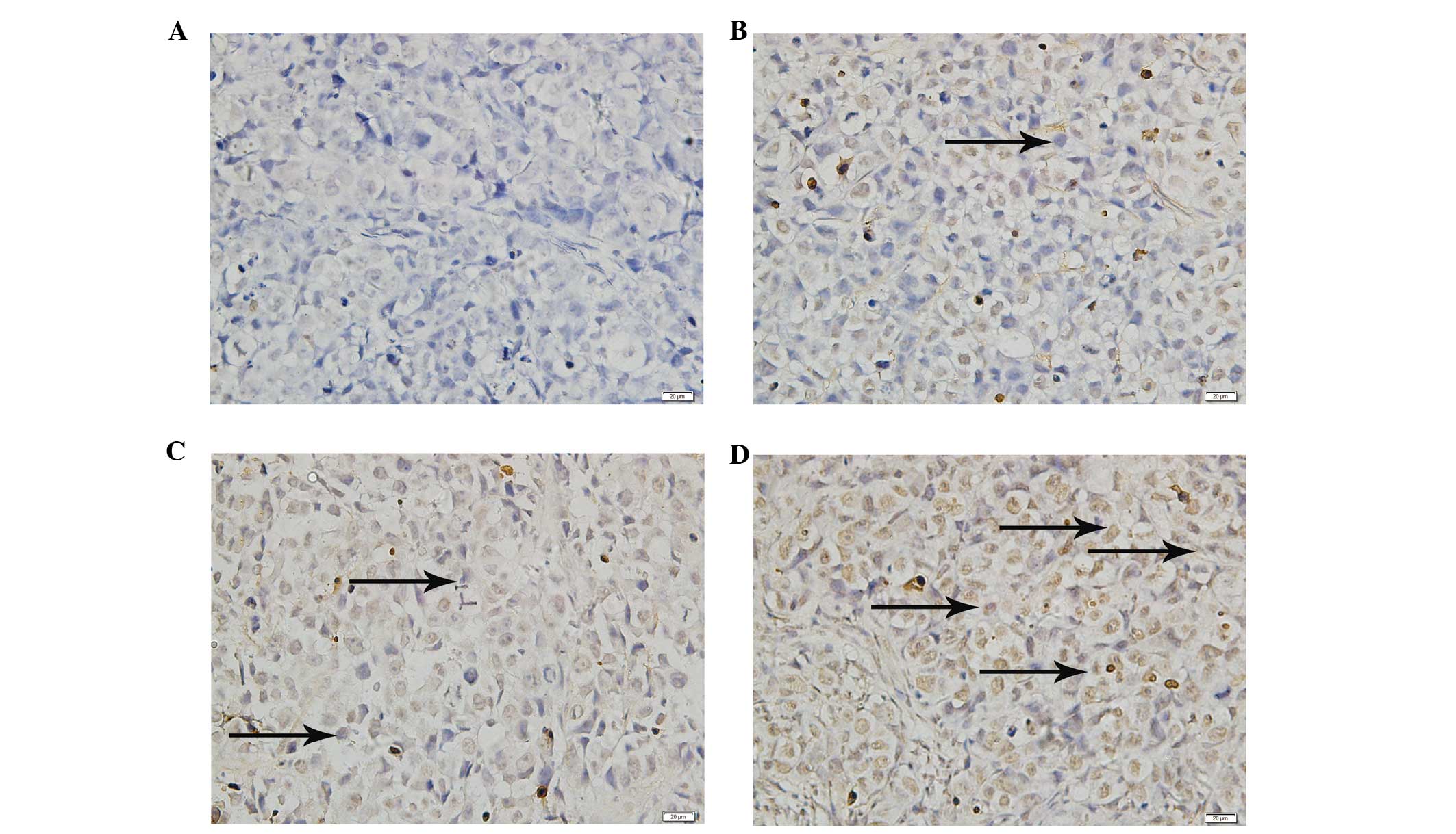
Detection of apoptotic cells in xenograft
tumor tissue
The TUNEL assay demonstrated that significant cell
death had occurred in the tumor masses from the treatment groups
when compared with that of the saline-only control group
(P<0.01; Fig. 5); however, the
degree of apoptosis differed in each group. The tumors from the
combined treatment group demonstrated a significantly increased
level of apoptosis when compared with that of the puerarin and 5-FU
groups (P<0.01; Fig. 5).
Evaluation of side effects
At the end of the study, blood was collected from
the mice by cardiac puncture using heparin-rinsed 5-ml syringes and
22-gauge needles. The ALT, AST, BUN and Cr levels were measured to
evaluate liver and renal injury (22). Serum ALT, AST, BUN and Cr levels
were not identified to be significantly elevated in the treated
mice when compared with those of the control group (P>0.05,
Table III), and no significant
difference in the levels of these biomarkers was identified between
the puerarin, 5-FU and combined treatment groups (P>0.05,
Table III). All mice were
sacrificed and dissected following completion of the study. In
addition, no liver or kidney injury, obvious metastasis or
hemorrhage was observed. Furthermore, no significant difference was
identified in liver or kidney weight between the treated groups and
the control group, and no evident lesions were observed by
histopathological examination.
| Table IIIEffect of puerarin combined with 5-FU
or alone on hepatic and renal function. |
Table III
Effect of puerarin combined with 5-FU
or alone on hepatic and renal function.
| Group | n | ALT, U/l
(mean±SD) | AST, U/l)
(mean±SD) | Urea, μmol/l)
(mean±SD) | Cr, μmol/l)
(mean±SD) |
|---|
| Puerarin | 6 | 46.17±6.55 | 143.33±28.93 | 7.98±2.15 | 16.64±5.61 |
| 5-FU | 6 | 46.00±3.16 | 144.83±21.27 | 7.96±0.96 | 15.86±5.29 |
| Puerarin + 5-FU | 6 | 45.33±8.16 | 146.67±25.84 | 8.29±1.59 | 16.24±3.54 |
| Tumor control | 6 | 46.33±4.08 | 145.33±14.85 | 8.05±0.51 | 16.57±4.54 |
| Normal control | 6 | 46.17±4.92 | 145.00±6.78 | 7.91±1.03 | 16.85±5.74 |
Discussion
HCC is a major public health threat that is
responsible for ~600,000 mortalities each year (2,3).
Recently, more effective treatments and earlier diagnosis have
increased the survival rate. However, the efficacy of surgical
techniques and radiotherapy is limited by the possibility of
metastasis. Therefore, chemotherapy is key for improving survival
in HCC patients.
5-FU is an important chemotherapeutic agent for the
treatment of hepatogastroenteric tumors (23). 5-FU is commonly used to treat HCC,
alone or in combination with other agents. Recently, more
traditional medicines have been used to prevent or treat tumors,
particularly in China. Puerarin has been used in traditional
Chinese medicine for many years. Certain studies have suggested
that puerarin exhibits anticancer activity and significant
antiproliferative and apoptotic effects (9–16).
In the present study, the synergistic effect of
puerarin and 5-FU treatment for hepatic carcinoma was investigated
in vitro and in vivo. Puerarin or 5-FU alone were
found to significantly inhibit the proliferation of SMMC7721 cells
in a dose-dependent manner (puerarin, 400–6,400 μM; 5-FU, 40–640
μM). Furthermore, combined treatment with puerarin and 5-FU
inhibited SMMC7721 cell proliferation at specific concentrations,
when the fraction of affected cells was between 0.2555 and 0.7420.
The mechanism of this synergistic effect was further investigated
and the results showed that puerarin or 5-FU alone induced
significant apoptosis when compared with that of the control group.
In addition, the combined treatment induced a greater degree of
apoptosis than puerarin or 5-FU alone in vitro and in
vivo. This suggested that combined treatment with puerarin and
5-FU is more effective in inhibiting the growth of human HCC
SMMC7721 cells.
A previous study suggested that puerarin may act as
a sensitizer to enhance the inhibition of hepatic carcinoma cell
proliferation by other chemotherapeutic agents (17). The results of the present study
indicated that puerarin enhances the efficacy of 5-FU in HCC
treatment in vitro and in vivo. Multiple factors have
been implicated in the increased resistance to chemotherapeutic
agents, including the reduction of intracellular drug accumulation
and DNA damage repair by the modulation of proliferative or
antiapoptotic proteins (24).
Puerarin most likely enhances HCC sensitivity to 5-FU by inhibiting
cell proliferation and inducing apoptosis.
Wang et al (11) showed that administration of puerarin
was able to reverse multidrug resistance in a nude mouse model of
human gastric carcinoma, and that decreased expression of
P-glycoprotein and multidrug resistance-associated protein may be
responsible for this effect. Crude pueraria extract and purified
puerarin induce apoptosis, which may correlate with the observed
inhibition of the cell cycle in the G0/G1 phase, as well as
increased Bax expression and decreased Bcl-2 expression (13). In addition, Tran et al
(15) revealed that puerarin
downregulates multidrug resistance protein 1 expression in
MCF-7/ADR cells via the upregulation of AMPK, which is dependent on
nuclear factor-κ-β and cAMP-responsive element transcriptional
activity. Further study is required to determine the molecular
mechanism by which combined treatment with puerarin and 5-FU
inhibits the growth of HCC. In conclusion, puerarin combined with
5-FU exhibited a significantly greater anti-liver cancer effect
than that of puerarin or 5-FU treatment alone, and thus combined
therapy may present a novel therapeutic regimen for HCC.
Acknowledgements
This study was performed at the Key Laboratory of
Hubei Province for Digestive System Disease with the assistance of
Mr. Hong Xia. The study was supported by the Fundamental Research
Funds for the Central Universities of China (grant no.
2012302020208).
References
|
1
|
Bosch FX, Ribes J, Díaz M and Cléries R:
Primary liver cancer: worldwide incidence and trends.
Gastroenterology. 127(Suppl 1): S5–S16. 2004.
|
|
2
|
Parkin DM, Bray F, Ferlay J and Pisani P:
Global cancer statistics, 2002. CA Cancer J Clin. 55:74–108.
2005.
|
|
3
|
Roberts LR: Sorafenib in liver cancer -
just the beginning. N Engl J Med. 359:420–422. 2008.
|
|
4
|
Hall AJ and Wild CP: Liver cancer in low
and middle income countries. BMJ. 326:994–995. 2003.
|
|
5
|
El-Serag HB and Rudolph KL: Hepatocellular
carcinoma: epidemiology and molecular carcinogenesis.
Gastroenterology. 132:2557–2576. 2007.
|
|
6
|
Diasio RB and Johnson MR: The role of
pharmacogenetics and pharmacogenomics in cancer chemotherapy with
5-fluorouracil. Pharmacology. 61:199–203. 2000.
|
|
7
|
Wang H, Jiang H, Zhou M, et al: Epidermal
growth factor receptor vIII enhances tumorigenicity and resistance
to 5-fluorouracil in human hepatocellular carcinoma. Cancer Lett.
279:30–38. 2009.
|
|
8
|
Lin YJ, Hou YC, Lin CH, Hsu YA, Sheu JJ,
Lai CH, Chen BH, Lee Chao PD, Wan L and Tsai FJ: Puerariae radix
isoflavones and their metabolites inhibit growth and induce
apoptosis in breast cancer cells. Biochem Biophys Res Commun.
378:683–688. 2009.
|
|
9
|
Yanagihara K, Ito A, Toge T and Numoto M:
Antiproliferative effects of isoflavones on human cancer cell lines
established from the gastrointestinal tract. Cancer Res.
53:5815–5821. 1993.
|
|
10
|
Jun M, Hong J, Jeong WS and Ho CT:
Suppression of arachidonic acid metabolism and nitric oxide
formation by kudzu isoflavones in murine macrophages. Mol Nutr Food
Res. 49:1154–1159. 2005.
|
|
11
|
Wang Li, Wei PK, Qin ZF, Xu YP, Lou LG, Li
YL and He Ji: Experimental study on puerarin injection reverse
multidrug resistance of nude mice of human gastric carcinoma
constructed using orthotopic transplantation. J Chendu Univ Tradit
Chin Med. 1:42–44; discussion 46, 2005 (In Chinese).
|
|
12
|
Yu Z and Li W: Induction of apoptosis by
puerarin in colon cancer HT-29 cells. Cancer Lett. 238:53–60.
2006.
|
|
13
|
Han P, Pei LY, Li J, Zhang L, Zhang HQ and
Xu D: Effect and mechanism of pueraria crude extract puerarin on
lung cancer H446 cell proliferation. Shandong Med J. 48:7–9.
2008.(In Chinese).
|
|
14
|
Hien TT, Kim HG, Han EH, Kang KW and Jeong
HG: Molecular mechanism of suppression of MDR1 by puerarin from
Pueraria lobata via NF-kappaB pathway and cAMP-responsive
element transcriptional activity-dependent up-regulation of
AMP-activated protein kinase in breast cancer MCF-7/adr cells. Mol
Nutr Food Res. 54:918–928. 2010.
|
|
15
|
Li XR, Zhang QQ, Cui YH, Hua QL and Yang
T: The growth influence on human esophageal cancer cells EC9706
induced by puerari. XIan Dai Zhang Liu Yi Xue. 10:1922–1924.
2010.
|
|
16
|
Jing XF, Li H and Li XM: Effect of
puerarin on AQP 1 mRNA expression level in esophageal cancer cell
line TE-1 and ECA109. Xinjiang J Med Univ. 34:258–260. 2011.(In
Chinese).
|
|
17
|
Tan GS, Sun L and Cao JG: Cytotoxic
constituents from Bulbuphyllum inconsipicum. You Ji Hua Xue.
3:372–374. 2006.(In Chinese).
|
|
18
|
Chou TC and Talalay P: Quantitative
analysis of dose-effect relationships: the combined effects of
multiple drugs or enzyme inhibitors. Adv Enzyme Regul. 22:27–55.
1984.
|
|
19
|
Chou TC, Motzer RJ, Tong Y and Bosl GJ:
Computerized quantitation of synergism and antagonism of taxol,
topotecan, and cisplatin against human teratocarcinoma cell growth:
a rational approach to clinical protocol design. J Natl Cancer
Inst. 86:1517–1524. 1994.
|
|
20
|
Kim BR, Shim JW, Sung DK, et al:
Granulocyte stimulating factor attenuates hypoxic-ischemic brain
injury by inhibiting apoptosis in neonatal rats. Yonsei Med J.
49:836–842. 2008.
|
|
21
|
Guide for the Care and Use of Laboratory
Animals. National Research Council (US) Committee for the Update of
the Guide for the Care and Use of Laboratory Animals. 8th edition.
National Academies Press; Washington (DC): 2011
|
|
22
|
Awad ME, Abdel-Rahman MS and Hassan SA:
Acrylamide toxicity in isolated rat hepatocytes. Toxicol In Vitro.
12:699–704. 1998.
|
|
23
|
Huang XW, Tang ZY, Lawrence TS and Zhang
M: 5-fluorouracil and hydroxyurea enhance adenovirus-mediated
transgene expression in colon and hepatocellular carcinoma cells. J
Cancer Res Clin Oncol. 131:184–190. 2005.
|
|
24
|
Siddik ZH: Cisplatin: mode of cytotoxic
action and molecular basis of resistance. Oncogene. 22:7265–7279.
2003.
|