Surgical resection of cerebellar hemangioblastoma with enhanced wall thickness: A report of two cases
- Authors:
- Zhenxing Sun
- Dan Yuan
- Yaxing Sun
- Pengxiang Yan
- Huancong Zuo
View Affiliations
Affiliations: Department of Spinal Cord and Spine Neurosurgery, Yuquan Hospital, Medical Center, Tsinghua University, Beijing 100049, P.R. China, Department of Nephrology, The Luhe Teaching Hospital of the Capital Medical University, Beijing 101149, P.R. China, Department of Psychiatry, The Second Municipal Hospital of Zaozhuang City, Zaozhuang, Shandong 277218, P.R. China, Department of Neurosurgery, Tiantan Hospital, Capital Medical University, Beijing 100050, P.R. China
- Published online on: February 10, 2015 https://doi.org/10.3892/ol.2015.2951
-
Pages:
1597-1599
-
Copyright: © Sun
et al. This is an open access article distributed under the
terms of Creative
Commons Attribution License.
Metrics: Total
Views: 0 (Spandidos Publications: | PMC Statistics: )
Metrics: Total PDF Downloads: 0 (Spandidos Publications: | PMC Statistics: )
This article is mentioned in:
Abstract
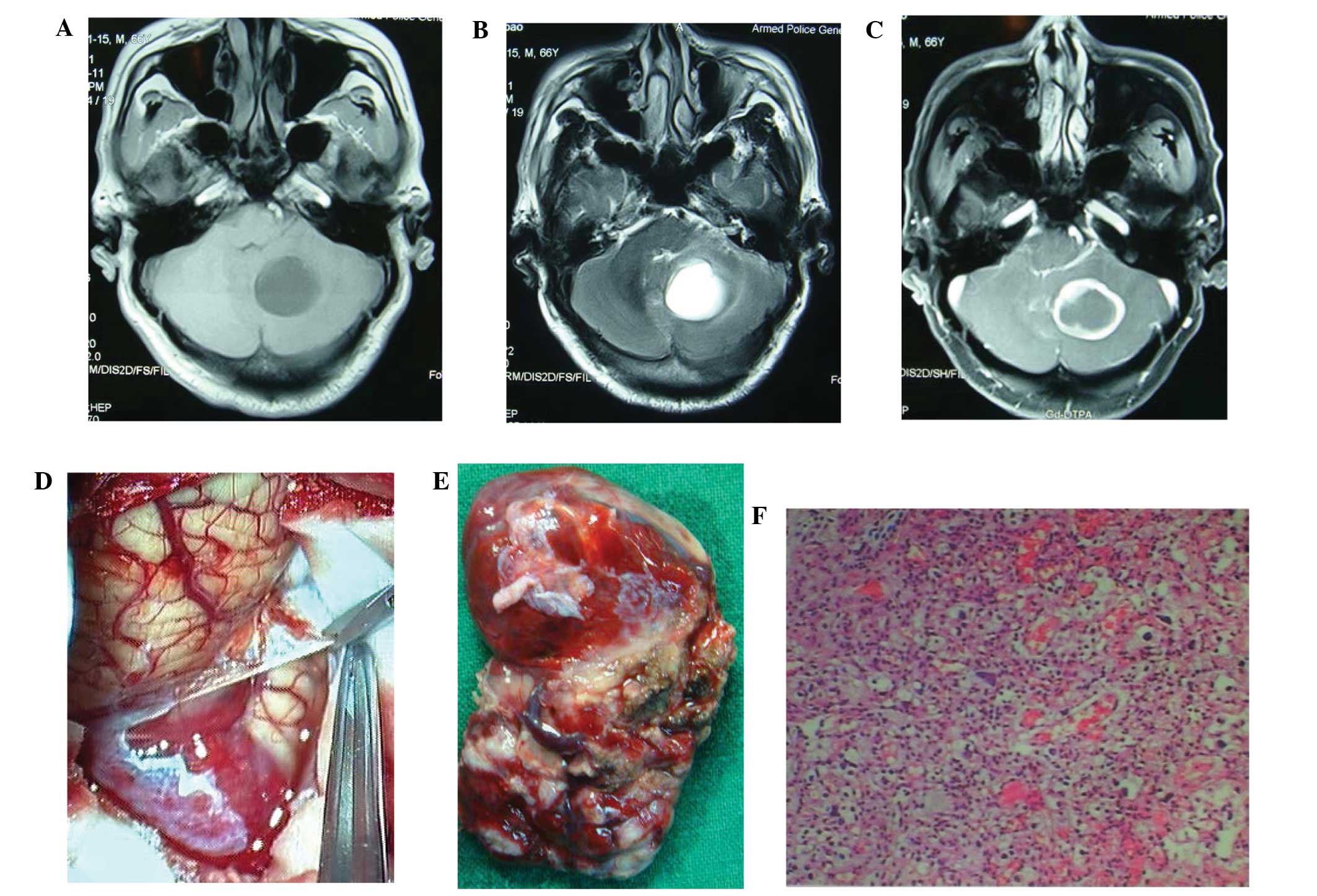
Hemangioblastomas are tumors of the central nervous system, and the cerebellum is the most common site of occurrence. Cerebellar hemangioblastoma with enhanced wall thickness is rare and often misdiagnosed preoperatively. At present, no unified radiological classification system based on magnetic resonance imaging (MRI) findings exists for cerebellar hemangioblastoma, and this tumor type can be solid or cystic mass, according to the MRI findings. The most common presentation of cerebellar hemangioblastoma observed radiologically is a large sac with small nodules, where the wall of the large cyst is not enhanced. A tumor with enhanced large cysts and tumor nodules is extremely rare. The most effective treatment is complete resection of the cyst and the solid growth. The present case reports the successful treatment of two cases of cerebellar hemangioblastoma with enhanced wall thickness, including the MRI findings for the differential diagnoses and the surgical experiences.
View References
1
|
Lee JY, Cho BM, Oh SM, et al: Delayed
diagnosis of cerebellar hemangioblastoma after intracerebellar
hemorrhage. Surg Neurol. 67:419–421. 2007. View Article : Google Scholar : PubMed/NCBI
|
2
|
Aldape KD, Plate KH, Vortmeyer AO, et al:
Haemangioblastoma. WHO Classification of Tumours of the Nervous
System. Louis DN, Ohgaki H, Wiestler OD and Cavenee WK: IARC Press;
Lyon: 2007
|
3
|
Simone CB II, Russell RR, Ondos J, et al:
Infratentorial craniospinal irradiation for von Hippel-Lindau: a
retrospective study supporting a new treatment for patients with
CNS hemangioblastomas. Neuro Oncol. 13:1030–1036. 2011. View Article : Google Scholar : PubMed/NCBI
|
4
|
Horvathy DB, Hauck EF, Ogilvy CS, et al:
Complete preoperative embolization of hemangioblastoma vessels with
Onyx 18. J Clin Neurosci. 18:401–403. 2011. View Article : Google Scholar : PubMed/NCBI
|
5
|
Gelabert González M: Posterior fossa
hemangioblastomas. Neurologia. 22:853–859. 2007.
|
6
|
Zhou LF and Du GH: Diagnosis and surgical
treatment of posterior fossa solid hemangilblastomas. Chin Med J.
113:129–132. 2000.
|
7
|
Wan JQ, Cui H and Wang Y: Surgical
management of large solid hemangioblastomas of the posterior fossa.
J Clin Neurosci. 18:39–42. 2011. View Article : Google Scholar
|
8
|
Neumann HP, Eggert HR, Weigel K, et al:
Hemangioblastomas of the central nervous system. A 10-year study
with special reference to von Hippel-Lindau syndrome. J Neurosurg.
70:24–30. 1989. View Article : Google Scholar : PubMed/NCBI
|
9
|
Jito J and Nozaki K: Treatment strategies
for cerebellar hemangioblastomas: simple or further studies? World
Neurosurg. 82:619–620. 2014. View Article : Google Scholar : PubMed/NCBI
|
10
|
Liao CC and Huang YH: Clinical features
and surgical outcomes of sporadic cerebellar hemangioblastomas.
Clin Neurol Neurosurg. 125:160–165. 2014. View Article : Google Scholar : PubMed/NCBI
|
11
|
Yan PX, Wang ZC, Yu CJ and Guan SS: The
diagnosis and microsurgical treatment of solid cerebellar
hemangioreticuloma. Zhonghua Wai Ke Za Zhi. 42:777–780. 2004.(In
Chinese). PubMed/NCBI
|