Introduction
Pancreatic cancer is a highly lethal disease that
affected ~38,000 individuals in the United States of America in
2013 (1). Early detection is
difficult, and it is often too late to provide clinical help to the
patient by the time the disease is identified. The poor prognosis
of pancreatic cancer arises due to its aggressive local invasion,
early metastasis and low responsiveness to conventional
chemotherapies. Pancreatic intraepithelial neoplasia (PanIN), which
is regarded as a cancer precursor, progresses slowly over a period
of years and eventually develops into invasive carcinoma (2). Therefore, preventive strategies to delay
the progression or cure this type of dormant pancreatic cancer are
currently of interest.
Rice is one of the major cereals, and is the most
consumed staple food worldwide, particularly in Asian countries.
Rice seeds and rice germ contain fiber and various types of
antioxidants, including phenolic acids, phytic acid, tocopherols
and γ-oryzanol (3). Numerous cellular
and preclinical studies support the hypothesis that antioxidants
protect against cancer (4–6). Among them, α-tocopherol has been
reported to prevent liver metastasis of chemically-induced
pancreatic cancer in animal models (7). Fermented brown rice by Aspergillus
oryzae (FBRA) is a processed food prepared by fermenting brown
rice and rice bran with A. oryzae. It has been demonstrated
that FBRA acts as a potent free-radical scavenger in vitro
(8). FBRA has also been shown to
exhibit chemopreventive effects against carcinogenesis in the colon
(9,10), liver (11), esophagus (12), stomach (13) and urinary bladder (14) of rodents. As FBRA has no systemic
toxicity in rodents, its potential chemopreventive effects against
chemically-induced pancreatic carcinogenesis are attractive.
The animal model of Syrian golden hamsters injected
with N-nitrosobis(2-oxopropyl)amine (BOP) provides pancreatic
lesions with similarities to the major forms of pancreatic cancer
in humans (15). PanIN lesions, such
as mild hyperplastic lesions (PanIN1) and papillary hyperplasia
(PanIN2), have been demonstrated to develop into carcinoma in
situ (PanIN3) and invasive ductal cancer following the
administration of BOP (16). For
example, Syrian golden hamsters treated with BOP demonstrate a
progression from mild hyperplasic lesions (PanIN1) to papillary
hyperplasia (PanIN2) at 8 weeks, to carcinoma in situ
(PanIN3) at 12 weeks and to invasive ductal adenocarcinoma at 24
weeks after the injection (17).
Furthermore, point mutations in codon 12 of the K-ras gene
frequently occur in this hamster pancreatic cancer model, as in
human pancreatic tumors (17,18). Similarly, in mouse lung neoplasms
induced by the tobacco-derived carcinogen
4-(methylnitrosamino)-1-(3-pyridyl)-1-butanone (NNK), mutations of
the K-ras gene are detected at a high frequency (19). Phutthaphadoong et al (20) revealed that FBRA treatement inhibited
NNK-induced lung carcinogenesis in mice. In the present study, the
chemopreventive activity of FBRA against BOP-induced pancreatic
tumorigenesis was examined in male Syrian golden hamsters by
evaluating the development of PanINs and invasive cancer.
Materials and methods
Animals, diet and chemicals
Male 4-week-old Syrian golden hamsters (n=172) were
purchased from Japan SLC, Inc. (Hamamatsu, Japan). After a 1-week
quarantine, the hamsters were housed between 3–5 animals per cage
on pulp-chip bedding in an air-conditioned animal room at 23±2°C
and 50±10% humidity. All hamsters were maintained under specific
pathogen-free conditions with a 12-h light/dark cycle, with free
access to water, and were maintained on a basal diet of MF
(Oriental Yeast Co., Ltd. Tokyo, Japan) until they reached 5 weeks
of age. The MF diet was used as the basal diet throughout the
study. The experimental diets were prepared by mixing 5 and 10%
FBRA, which were previously reported as effective concentrations
(9–14,20), with
the MF diet. FBRA was supplied by Genmai Koso Co., Ltd. (Sapporo,
Japan). Briefly, the manufacturing process for FBRA included
producing a fermentation base by steaming brown rice and rice bran.
A. oryzae was then seeded into the fermentation base and the
fermentation process was continued for 18–24 h. Subsequently, a
second fermentation was continued for an additional 12–24 h for
aging purposes. The fermented product was then dried and powdered.
BOP was obtained from Toronto Research Chemicals, Inc. (Ontario,
Canada).
Experimental procedure
Five-week-old male Syrian golden hamsters were
randomly divided into seven groups (group 1, n=34; groups 2 and 4,
n=30; groups 3 and 5, n=29; groups 6 and 7, n=10). The uneven group
distribution is due to one hamster in groups 1, 3 and 5 succumbing
during the early stages of the experiment. When they were 6 weeks
old, the hamsters in groups 1–5 received subcutaneous injections of
BOP 4 times (on days 1, 3, 5 and 7) at a dose of 10 mg/kg body
weight, whereas the 20 hamsters in groups 6 and 7 were treated with
saline as a vehicle control. Group 1 received a basal diet during
the experimental period, while the hamsters in groups 2 and 3 were
administered with 5 and 10% FBRA in the diet, respectively, during
the initiation stage (during and 1 week after BOP treatment) and
then were changed to the control diet until they were 27 weeks of
age. The hamsters in groups 4 and 5, which were maintained on the
basal diet during BOP treatment, received 5% and 10% FBRA in the
diet, respectively, during the post-initiation stage (1 week after
BOP treatment until 27 weeks of age). The hamsters in group 6
received 10% FBRA in their diet throughout the experimental period.
The animals in group 7 served as non-treatment controls. All
animals were weighed weekly using a dial scale (DH-3100N; Shinko
Denshi Co., Ltd., Tokyo, Japan). At the end of the experiment when
the hamsters were 27 weeks old, all surviving animals were
sacrificed under deep anesthesia by deep inhalation of isoflurane
and blood samples were collected from the aorta after overnight
fasting. Serum was obtained from the blood samples by
centrifugation at 1,000 × g for 30 min at 4°C and stored at −80°C
prior to analysis. The serum levels of triglyceride, total
cholesterol, high-density lipoprotein cholesterol and amylase, a
pancreatic exocrine enzyme, were measured by SRL, Inc. (Tokyo,
Japan). The effects of FBRA on a serum lipid profile possibly
associated with chemically-induced pancreatic cancer development in
rodents, as well as on amylase, were determined (21).
The animal husbandry and experimental protocols were
conducted in accordance with the guidelines for the use and care of
experimental animals, and were approved by the Committee for Animal
Research and Welfare of Gifu University (Gifu, Japan; project code
no. 25-1; approved on April 4, 2013).
Histopathological examination
At necropsy, the pancreas, kidneys, liver and bile
duct were carefully examined macroscopically. Four anatomical
sections of the pancreas (the gastric and splenic sections,
duodenal lobes, and the head portion) were fixed in 10%
phosphate-buffered formalin. All tissues were routinely processed,
embedded in paraffin, serially sectioned to a thickness of 4 µm,
and stained with hematoxylin and eosin (H&E) to assess the
histopathological features. Two independent investigators blinded
to the sample identity evaluated the sections of pancreas and
scored them according to the PanIN criteria within the following
categories: PanIN1, PanIN2, PanIN3 and invasive ductal
adenocarcinoma (17,22). The tumor incidence (percentage of
hamsters with pancreatic lesions) and multiplicity (number of
pancreatic lesions per hamster) were calculated on the basis of
these scores. In addition, the histopathological features of liver
tumors stained with H&E were assessed to distinguish
hepatocellular and cholangiocellular tumors, according to
previously established criteria (23). Histologically, hepatocellular adenoma
presents as single nodules, which compress the surrounding
parenchyma. By contrast to normal hepatocytes, those within
adenomas usually form solid aggregates or irregular sheets, which
vary in cellular thickness. Cholangiocellular adenoma is usually
composed of multilocular cystic structures lined by flattened
epithelium. Furthermore, cholangiocellular carcinoma, defined as a
malignant tumor arising from the intrahepatic ductular epithelial
cells, which range from columnar to cuboidal in shape, forms
distorted ductal structures.
Ki-67 labeling index of pancreatic
proliferative lesions
To assess cell proliferation, the avidin-biotin
peroxidase complex technique was used. The paraffin-embedded
sections (4 µm) were deparaffinized and rehydrated in
phosphate-buffered saline (PBS; pH 7.4, 0.01 mol/l; Wako Pure
Chemical Industries, Ltd., Osaka, Japan). The sections were then
placed in 10 mmol/l citrate buffer (pH 6.0) and heated in a Pascal
pressure cooker (S2800; Dako Japan KK, Tokyo, Japan) programmed for
1 min at 120°C for pretreatment. Endogenous peroxidase activity was
blocked by incubation with 3% H2O2 solution
for 20 min. After washing three times in PBS (pH 7.4; 0.01 mol/l),
the sections were pre-incubated with 2% bovine serum albumin (Wako
Pure Chemical Industries, Ltd.) for 40 min at room temperature to
block general protein. Subsequently, the sections were incubated
with a rabbit anti-hamster monoclonal Ki-67 antibody (cat no.
ab16667; 1:100 dilution; Abcam, Cambridge, MA, USA) overnight at
4°C. Sequential incubation with a biotinylated anti-rabbit
secondary antibody for 30 min and avidin-coupled peroxidase for 30
min (Vectastain Elite ABC kit, cat no. PK-6101; Vector Laboratory,
Burlingame, CA, USA) was performed, according to the manufacturer's
instructions. Binding was then visualized with
3,3′-diaminobenzidine tetrahydrochloride (Sigma-Aldrich, St. Louis,
MO, USA). Sections were counterstained with hematoxylin for
microscopic examination (BX40; Olympus Corporation, Tokyo, Japan).
The proliferating cells were quantified by counting the
Ki-67-positive cells at a magnification of ×400. The Ki-67 labeling
index of the pancreatic proliferative lesions was determined using
Ki-67-stained immunohistochemical sections of between 4–7 hamsters
per group, as certain PanINs were too small to evaluate and not all
hamsters developed adenocarcinoma. The Ki-67 status was determined
in >400 cells in each lesion and non-lesional epithelial sample,
and the Ki-67 labeling index was expressed as the percentage of
cells stained with the Ki-67 antibody.
Analysis of apoptosis
Apoptotic cells were detected by the terminal
deoxynucleotidyl transferase-mediated dUTP nick end labeling
(TUNEL) assay using an in situ Apoptosis Detection kit
(Takara Bio Inc., Otsu, Japan). The apoptosis rate was calculated
as the number of TUNEL-positive tumor cells as a proportion of the
total cell number. Cells were counted at a magnification of ×400
and the mean TUNEL-positive rate was calculated for three or four
randomly selected areas. At least 400 cells were counted per lesion
for evaluation, following counterstaining with hematoxylin, using a
BX40 microscope (Olympus Corporation).
Statistical analysis
The data are expressed as the mean ± standard
deviation, and significant differences among the 5 BOP-treated
groups were determined using one-way analysis of variance with the
Tukey-Kramer post-hoc test. The pathological grade distribution was
analyzed using the Kruskal-Wallis test with Dunn's post-hoc test.
All statistical analyses were performed using GraphPad Prism
software (version 6; GraphPad Software, Inc., La Jolla, CA, USA).
P<0.05 was considered to indicate a statistically significant
difference.
Results
Final body weights, and weights of the
liver and kidneys
All animals remained healthy throughout the
experimental period. The mean body, liver and kidney weights in all
groups at the end of the study are listed in Table I. The mean body and liver weights of
the hamsters in groups 4 (BOP→5% FBRA; P<0.0001 and P<0.0001,
respectively) and 5 (BOP→10% FBRA; P<0.0001 and P<0.0001,
respectively) were significantly higher compared with those of the
hamsters in group 1 (BOP alone). Furthermore, the mean relative
liver weight of group 4 (P=0.0024) was significantly greater than
that of group 1. No significant differences in kidney weight were
observed among the groups (P>0.05). There were no clear signs of
toxicity in the liver or kidneys of any of the hamsters as examined
by histological analysis.
 | Table I.Body, liver and kidney
weightsa of the hamsters
(age, 27 weeks). |
Table I.
Body, liver and kidney
weightsa of the hamsters
(age, 27 weeks).
| Group | Treatment (no. of
animals examined) | Body weight, g | Liver weight, g | Relative liver
weight, g/100 g body weight | Kidney weight, g |
|---|
| 1 | BOP (34) | 166.2±16.2 | 7.3±1.2 | 4.4±0.7 | 1.2±0.2 |
| 2 | BOP+5% FBRA (30) | 174.5±14.2 | 7.8±0.9 | 4.5±0.4 | 1.2±0.3 |
| 3 | BOP+10% FBRA
(29) | 167.6±18.2 | 7.5±1.3 | 4.5±0.5 | 1.2±0.2 |
| 4 | BOP→5% FBRA (30) |
186.0±19.1b | 9.4±2.0b | 5.0±0.7b | 1.2±0.2 |
| 5 | BOP→10% FBRA
(29) |
186.9±13.2b | 9.1±1.7b | 4.8±0.6 | 1.2±0.1 |
| 6 | 10% FBRA (10) | 191.0±20.3 | 8.7±1.6 | 4.5±0.6 | 1.3±0.2 |
| 7 | Control diet
(10) |
189.3±18.4b | 8.2±1.4 | 4.3±0.6 | 1.3±0.1 |
Serum triglyceride (TG), total
cholesterol (TC), high-density lipoprotein cholesterol (HDLC) and
amylase levels
The TG, TC, HDLC and amylase levels of the
27-week-old hamsters are listed in Table
II. A post-initiation diet of FBRA at a dose of 10%
significantly increased the TG level compared with group 1
(P=0.0343). The concentration of amylase in group 4 was
significantly higher compared with in group 1 (P=0.0209). However,
differences in TG and amylase levels between groups 6 and 7 were
not significant (P=0.3134 and P=0.9994, respectively). There were
no statistical differences in TC and HDLC levels (P>0.05).
| Table II.Serum levelsa of triglycerides, total
cholesterol, HDLC and amylase in hamsters (age, 27 weeks). |
Table II.
Serum levelsa of triglycerides, total
cholesterol, HDLC and amylase in hamsters (age, 27 weeks).
| Group | Treatment (no. of
animals examined) | Triglycerides,
mg/dl | Total cholesterol,
mg/dl | HDLC, mg/dl | Amylase, U/l |
|---|
| 1 | BOP (5) | 216.4±82.9 | 199.4±20.1 | 102.0±6.4 | 1080.4±124.8 |
| 2 | BOP+5% FBRA
(5) | 267.2±75.3 | 203.4±31.8 | 98.0±19.2 | 1087.0±183.8 |
| 3 | BOP+10% FBRA
(5) | 225.8±65.2 | 188.0±25.9 | 95.5±16.7 | 965.5±167.1 |
| 4 | BOP→5% FBRA
(5) | 319.8±44.8 | 195.2±12.4 | 103.2±10.5 |
1502.0±198.9b |
| 5 | BOP→10% FBRA
(5) |
392.0±65.6b | 212.6±8.6 | 112.8±7.0 | 1417.0±245.2 |
| 6 | 10% FBRA (5) | 245.4±111.9 | 188.2±25.4 | 103.3±21.5 | 1369.0±189.2 |
| 7 | Control diet
(5) | 362.6±113.2 | 190.2±17.6 | 91.8±27.1 | 1317.0±176.0 |
Incidence and multiplicity of
pancreatic proliferative lesions
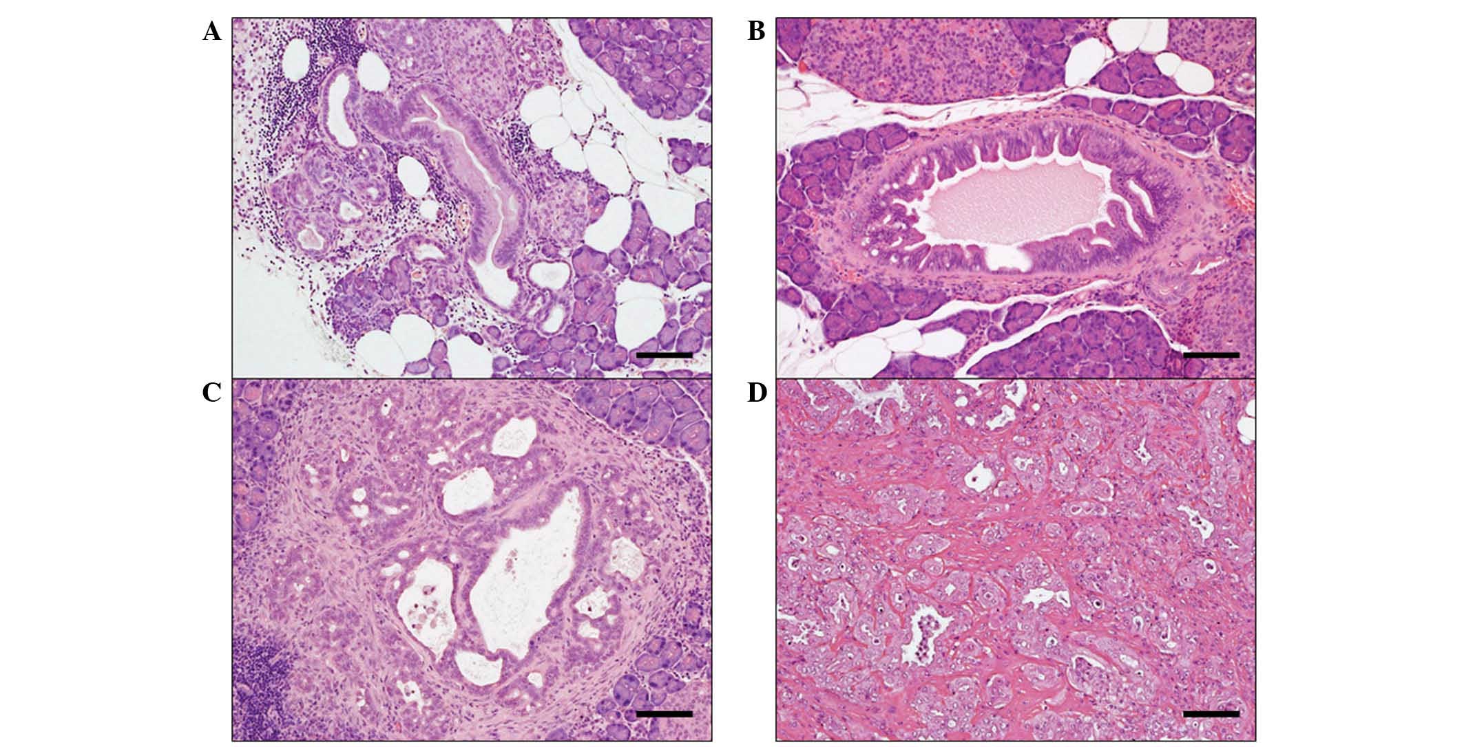
The administration of BOP induced pancreatic
proliferative lesions (Fig. 1A and D)
that were histopathologically diagnosed as PanIN1 (Fig. 1A), PanIN2 (Fig. 1B), PanIN3 (Fig. 1C) or invasive ductal adenocarcinoma
(Fig. 1D). PanIN1s (mild hyperplastic
lesions) are flat epithelial lesions composed of tall columnar
cells with basally located nuclei or a papillary pseudostratified
architecture (24). PanIN2s
(papillary hyperplasia) are flat or papillary mucinous lesions with
nuclear abnormalities, which may include loss of polarity, nuclear
crowding, enlarged nuclei and pseudostratification. PanIN3s
(carcinoma in situ) are identified as papillary or
micropapillary structures characterized with high-grade dysplasia
and loss of nuclear polarity, indicating the development of
pancreatic cancer. On the basis of the above criteria and on the
histopathological examination of H&E-stained pancreatic tissue,
the number and type of PanINs in the pancreatic tissue samples were
recorded.
The incidence of pancreatic proliferative lesions is
summarized in Table III. PanINs and
invasive ductal adenocarcinomas appeared in groups 1–5. There was
no evidence of these lesions in groups 6 and 7. The majority of the
animals in group 1 developed ductal adenocarcinomas (56%; including
32% PanIN3 and 24% invasive carcinoma), 41% had precancerous
lesions (12% PanIN1 and 29% PanIN2) and one hamster exhibited a
normal pancreas with no lesions (3%). Among the animals
administered 10% FBRA during the post-initiation phase, 3 hamsters
did not develop lesions in the pancreas (10%), 66% had precancerous
lesions (42% PanIN1 and 24% PanIN2), and 10 and 14% of the hamsters
exhibited PanIN3 and invasive carcinoma, respectively. Thus,
treatment with 10% FBRA was associated with a shift in the
pathological grade distribution toward normal/precancerous lesions,
and significantly inhibited the progression to pancreatic cancer
(P=0.0259).
| Table III.Effects of FBRA on the incidence and
distribution of pathological lesions in the pancreas. |
Table III.
Effects of FBRA on the incidence and
distribution of pathological lesions in the pancreas.
|
|
|
| Incidence of
highest-grade lesions in affected animals, n (%) |
|---|
|
|
|
|
|
|---|
| Group | Treatment (no. of
animals examined) | Animals with no
lesions, n (%) | PanIN1 | PanIN2 | PanIN3 | Invasive ca. |
|---|
| 1 | BOP (34) | 1 (3) | 4 (12) | 10 (29) | 11 (32) | 8 (24) |
| 2 | BOP+5% FBRA
(30) | 2 (7) | 3 (10) | 13 (43) | 5 (17) | 7 (23) |
| 3 | BOP+10% FBRA
(29) | 1 (4) | 9 (31) | 11 (38) | 3 (10) | 5 (17) |
| 4 | BOP→5% FBRA
(30) | 0 (0) | 2 (7) | 13 (43) | 7 (23) | 8 (27) |
| 5 | BOP→10% FBRA
(29) | 3
(10) | 12 (42) | 7 (24) | 3 (10) | 4 (14) |
| 6 | 10% FBRA (10) | 10
(100) | 0 (0) | 0 (0) | 0 (0) | 0 (0) |
| 7 | Control diet
(10) | 10
(100) | 0 (0) | 0 (0) | 0 (0) | 0 (0) |
The multiplicity of pancreatic proliferative lesions
are indicated in Table IV. The
multiplicity of total ductal adenocarcinomas in group 5 (10% FBRA
during the post-initiation phase) were significantly reduced
compared with group 1 that received BOP alone (0.24±0.44 vs.
0.71±0.72; P=0.0401). The multiplicity of PanIN2 in group 5 was
also significantly smaller than that of group 1 (0.45±0.69 vs.
1.26±1.24; P=0.0021). Conversely, the value of PanIN1 in group 5
was greater than the control group. Treatment with 10% FBRA during
the initiation phase (group 3) also significantly reduced the
multiplicity of PanIN2 compared with group 1 (P=0.0105).
| Table IV.Multiplicity of pancreatic
proliferative lesions. |
Table IV.
Multiplicity of pancreatic
proliferative lesions.
|
|
|
Lesions/animala, n |
|---|
|
|
|
|
|---|
|
|
|
|
| Ductal
adenocarcinoma |
|---|
|
|
|
|
|
|
|---|
| Group | Treatment (no. of
animals examined) | PanIN1 | PanIN2 | PanIN3 | Invasive ca. | Total |
|---|
| 1 | BOP (34) | 0.68±0.84 | 1.26±1.24 | 0.44±0.61 | 0.26±0.51 | 0.71±0.72 |
| 2 | BOP+5% FBRA
(30) | 0.70±0.79 | 0.87±0.82 | 0.23±0.50 | 0.23±0.43 | 0.47±0.63 |
| 3 | BOP+10% FBRA
(29) | 1.00±1.03 |
0.55±0.69b | 0.14±0.35 | 0.21±0.49 | 0.34±0.61 |
| 4 | BOP→5% FBRA
(30) | 1.13±0.94 | 0.97±0.61 | 0.37±0.56 | 0.27±0.45 | 0.63±0.76 |
| 5 | BOP→10% FBRA
(29) | 1.21±1.01 |
0.45±0.69c | 0.10±0.31 | 0.14±0.35 |
0.24±0.44b |
Cell proliferation and apoptosis in
pancreatic proliferative lesions
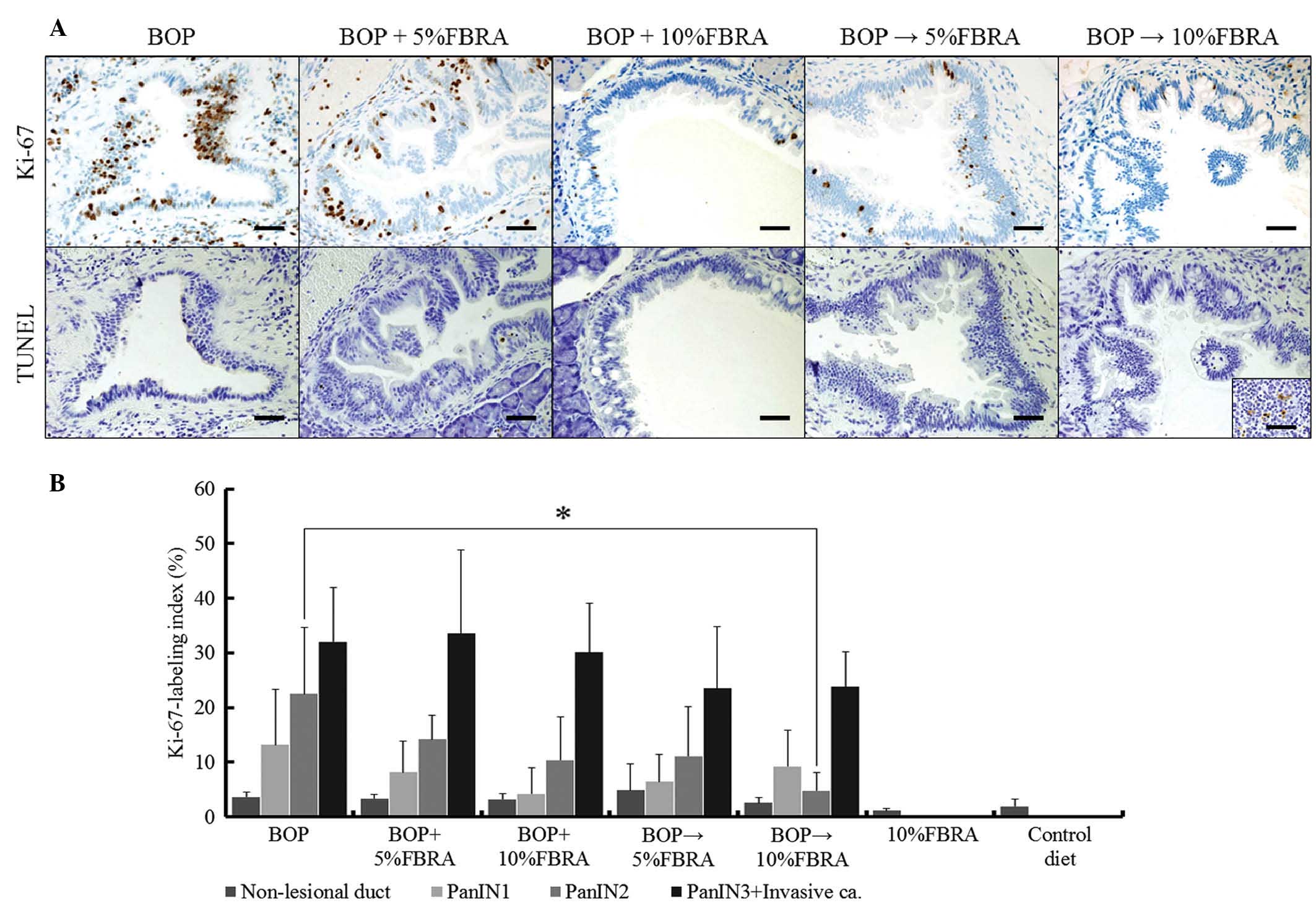
The proliferative potential of pancreatic
proliferative lesions were investigated, and the results of the
Ki-67-labeling index in the lesions of the hamsters in each group
are indicated in Fig. 2A and B.
Notably, the Ki-67-labeling index of the PanIN2 in group 5 was
significantly lower than that of group 1 (P=0.0054). Groups 6 and 7
were also analyzed; however PanINs and adenocarcinoma were not
observed due to a lack of BOP treatment. Furthermore, there was no
significant difference in the Ki-67 labeling index of non-lesional
ductal epithelia between the two groups (data not shown).
The apoptosis of pancreatic proliferative lesions
was also investigated. Expression of cells positive for the TUNEL
assay were frequently detected in the control section (lymph node;
Fig. 2A; inset of group 5). By
contrast, positive cells were rarely identified in BOP-induced
pancreatic tumors from the different groups, indicating that
cellular apoptosis rarely occurs in such tumors (data not
shown).
Hepatocellular and cholangiocellular
tumors
Liver tumors of hepatocellular or cholangiocellular
origin were sporadically observed, but no significant intergroup
variations were noted [P=0.5520, 0.5993, 0.5339 and 0.4724, for
hepatocellular adenoma, cholangiocellular adenoma,
cholangiocellular carcinoma, and cholangiocellular tumors (adenoma
+ adenocarcinoma), respectively; Table
V].
| Table V.Incidences of liver and bile duct
tumors induced by BOP. |
Table V.
Incidences of liver and bile duct
tumors induced by BOP.
|
|
| Animals with
tumors, n (%) |
|---|
|
|
|
|
|---|
|
|
|
| Cholangiocellular
tumors |
|---|
|
|
|
|
|
|---|
| Group | Treatment (no. of
animals examined) | Hepatocellular
adenoma | Adenoma | Adenocarcinoma | Total |
|---|
| 1 | BOP (34) | 2 (6) | 1 (3) | 0 (0) | 1 (3) |
| 2 | BOP+5% FBRA
(30) | 1 (3) | 0 (0) | 0 (0) | 0 (0) |
| 3 | BOP+10% FBRA
(29) | 0 (0) | 0 (0) | 1 (3) | 1 (3) |
| 4 | BOP→5% FBRA
(30) | 1 (3) | 1 (3) | 1 (3) | 2 (7) |
| 5 | BOP→10% FBRA
(29) | 0 (0) | 0 (0) | 0 (0) | 0 (0) |
Discussion
In the present study, dietary exposure to 10% FBRA
during the post-initiation phase of BOP-induced pancreatic
carcinogenesis significantly reduced the development of pancreatic
ductal adenocarcinoma (PanIN3 and invasive carcinoma). Furthermore,
post-initiation feeding of 10% FBRA also resulted in a significant
shift in the pathological grade distribution from pancreatic
cancerous lesions toward normal/precancerous lesions compared with
the predominance of cancerous lesions in group 1, where the animals
were treated with BOP alone. Thus, it appears that 10% FBRA was
effective for suppressing the progression of precancerous lesions
to pancreatic cancers. In BOP-induced pancreatic carcinogenesis,
FBRA appears to inhibit the occurrence of the various proliferative
lesions throughout tumorigenesis. Accordingly, it is suggested that
the suppressive effect of FBRA on pancreatic carcinogenesis is
associated with an arrest of the onset of carcinogenesis, and FBRA
blocks the transition from PanIN1 to PanIN2 and from PanIN2 to
PanIN3.
FBRA has previously been proven to inhibit
chemically-induced carcinogenesis in different organs, such as the
colon (9,10), liver (11), esophagus (12), stomach (13), urinary bladder (14) and lung (20) of rodents. Taken together, these
results and the results of the present study indicate that FBRA is
a candidate chemopreventive agent that may protect against
carcinogenesis in multiple organs, including the pancreas. Various
mechanisms by which chemopreventive agents exert their inhibitory
effects on tumorigenesis have been considered. For example, cell
proliferation is important in multistage carcinogenesis and
involves multiple genetic alterations (25). In the present study, FBRA inhibited
the cell proliferation of BOP-induced pancreatic intraductal
neoplasia. However, an association between FBRA intake and the
induction of apoptosis in the pancreatic lesions was not evident.
These results are in line with our previous studies using different
models of carcinogenesis (9–14,20). In
general, controlling cell proliferation has been proposed to be
relevant to carcinogenesis in various organs (26). Indeed, a high level of Ki-67
immunoreactivity is an indicator of poor prognosis of human
pancreatic ductal adenocarcinomas (27). In the present study, it was
demonstrated that dietary exposure to FBRA during the
post-initiation phase reduced tumor development in the pancreas.
Furthermore, the Ki-67 scores in PanIN2 lesions were reduced by
post-initiation treatment with FBRA. Similar results for FBRA were
previously obtained in precancerous lesions in the lungs of mice
(20). Thus, inhibiting the
proliferative activity in tumor cells may be one of the important
mechanisms underlying the chemopreventive effects of FBRA
administered at the post-initiation phase.
In the present study, simultaneous exposure to FBRA
and BOP decreased the multiplicity of PanIN2. These results are
consistent with our previous report, which identified that FBRA
suppresses NNK-induced lung carcinogenesis in A/J mice when
administered in the initiation phase (20). It has previously been proposed that
the chemopreventive effects of FBRA on lung carcinogenesis are
predominantly associated with inhibition of the metabolic
activation of carcinogens by cytochrome P450s (phase I enzymes).
Similarly, the intake of phenethyl isothiocyanate (PEITC) during
the initiation phase was reported to inhibit pulmonary and
pancreatic tumorigenesis in BOP-treated hamsters (28). Previous studies using mice also
demonstrated preventive effects of PEITC against NNK-induced lung
carcinogenesis, possibly through the inhibition of cytochrome P450s
(29,30). It may be important that BOP and NNK
share a metabolic pathway leading to active carcinogenic forms
(31). Accordingly, it is possible
that the mechanism underlying the anti-initiation effect of FBRA on
BOP-induced pancreatic carcinogenesis in hamsters is associated
with the inhibition of phase I enzymes.
FBRA is a processed food prepared by the solid-state
fermentation of brown rice and rice bran with A. oryzae.
Rice antioxidants are particularly abundant in the bran (3). The nutritional and sanitary advantages
of fermentation have long been recognized, although the details are
not well known. Solid-state fermentation of rice bran has been
employed to provide a higher bioavailable content of functional
compounds when compared with unfermented bran. Schmidt et al
(32) reported that the phenolic
compound content in rice bran is increased by fermentation with
Rhizopus oryzae. Solid-state fermentation of rice bran can
be applied for producing phospholipids, as well as for decreasing
the total fat and saturated fatty acids, with an increase of
unsaturated fatty acids (33). A
previous study reported that a diet high in certain unsaturated
fatty acids is associated with a reduced risk of developing
pancreatic cancer, whereas a diet high in saturated fatty acids may
be associated with an increased risk of human pancreatic cancer
(34). Although Takeuchi et al
(21) reported that hyperlipidemia
may be important for the development of pancreatic cancers in
hamsters, the association was not clarified in the current study,
which indicated that the inhibitory effects of FBRA on pancreatic
tumorigenesis were not associated with serum lipid levels.
Considering the aforementioned discussion and the absence of
studies in humans, the detailed mechanisms underlying the
inhibitory effects of FBRA on pancreatic tumorigenesis require
further investigation.
In conclusion, the present study clarified that the
administration of FBRA during the initiation and post-initiation
phases inhibited pancreatic tumorigenesis and progression induced
by BOP in male hamsters. Thus, FBRA is proposed to be a promising
agent for the prevention of human pancreatic cancer. In the future,
intervention experiments, which involve calorie-regulated high-fat
diet supplementation, should be performed to clarify the
association between BOP treatment and serum lipid profile, and to
investigate cancer chemoprevention by the intake of FBRA.
Acknowledgements
The authors would like to thank Miss Kyoko Takahashi
and Mr. Masayoshi Shimizu (Tumor Pathology, Graduate School of
Medicine, Gifu University), as well as Mr. Kouji Kato (Experimental
Pathology and Tumor Biology, Nagoya City University) for their
technical assistance and animal care.
References
|
1
|
Siegel R, Naishadham D and Jemal A: Cancer
statistics, 2013. CA Cancer J Clin. 63:11–30. 2013. View Article : Google Scholar : PubMed/NCBI
|
|
2
|
Yachida S, Jones S, Bozic I, Antal T,
Leary R, Fu B, Kamiyama M, Hruban RH, Eshleman JR, Nowak MA, et al:
Distant metastasis occurs late during the genetic evolution of
pancreatic cancer. Nature. 467:1114–1117. 2010. View Article : Google Scholar : PubMed/NCBI
|
|
3
|
Goufo P and Trindade H: Rice antioxidants:
Phenolic acids, flavonoids, anthocyanins, proanthocyanidins,
tocopherols, tocotrienols, y-oryzanol and phytic acid. Food Sci
Nutr. 2:75–104. 2014. View
Article : Google Scholar : PubMed/NCBI
|
|
4
|
Irani K, Xia Y, Zweier JL, Sollott SJ, Der
CJ, Fearon ER, Sundaresan M, Finkel T and Goldschmidt-Clermont PJ:
Mitogenic signaling mediated by oxidants in Ras-transformed
fibroblasts. Science. 275:1649–1652. 1997. View Article : Google Scholar : PubMed/NCBI
|
|
5
|
Sablina AA, Budanov AV, Ilyinskaya GV,
Agapova LS, Kravchenko JE and Chumakov PM: The antioxidant function
of the p53 tumor suppressor. Nat Med. 11:1306–1313. 2005.
View Article : Google Scholar : PubMed/NCBI
|
|
6
|
Vafa O, Wade M, Kern S, Beeche M, Pandita
TK, Hampton GM and Wahl GM: c-Myc can induce DNA damage, increase
reactive oxygen species and mitigate p53 function: A mechanism for
oncogene-induced genetic instability. Mol Cell. 9:1031–1044. 2002.
View Article : Google Scholar : PubMed/NCBI
|
|
7
|
Heukamp I, Kilian M, Gregor JI, Neumann A,
Jacobi CA, Guski H, Schimke I, Walz MK and Wenger FA: Effects of
the antioxidative vitamins A, C and E on liver metastasis and
intrametastatic lipid peroxidation in BOP-induced pancreatic cancer
in Syrian hamsters. Pancreatology. 5:403–409. 2005. View Article : Google Scholar : PubMed/NCBI
|
|
8
|
Tazawa K, Fuketa N and Hirohide N:
Superoxide scavenging effect of fermented brown rice determined by
ESR spin-trapping method. Food Style. 3:32–37. 1999.(In
Japanese).
|
|
9
|
Katyama M, Yoshimi N, Yamada Y, Sakata K,
Kuno T, Yoshida K, Qiao Z, Vihn PQ, Iwasaki T, Kobayashi H, et al:
Preventive effect of fermented brown rice and rice bran against
colon carcinogenesis in male F344 rats. Oncol Rep. 9:817–822.
2002.PubMed/NCBI
|
|
10
|
Phutthaphadoong S, Yamada Y, Hirata A,
Tomita H, Hara A, Limtrakul P, Iwasaki T, Kobayashi H and Mori H:
Chemopreventive effect of fermented brown rice and rice bran (FBRA)
on the inflammation-related colorectal carcinogenesis in
ApcMin/+mice. Oncol Rep. 23:53–59. 2010.PubMed/NCBI
|
|
11
|
Katayama M, Sugie S, Yoshimi N, Yamada Y,
Sakata K, Qiao Z, Iwasaki T, Kobayashi H and Mori H: Preventive
effect of fermented brown rice and rice bran on diethylnitrosoamine
and phenobarbital-induced hepatocarcinogenesis in male F344 rats.
Oncol Rep. 10:875–880. 2003.PubMed/NCBI
|
|
12
|
Kuno T, Hirose Y, Hata K, Kato K, Qiang
SH, Kitaori N, Hara A, Iwasaki T, Yoshimura T, Wada K, et al:
Preventive effect of fermented brown rice and rice bran on
N-nitrosomethylbenzylamine-induced esophageal tumorigenesis in
rats. Int J Oncol. 25:1809–1815. 2004.PubMed/NCBI
|
|
13
|
Tomita H, Kuno T, Yamada Y, Oyama T, Asano
N, Miyazaki Y, Baba S, Taguchi A, Hara A, Iwasaki T, et al:
Preventive effect of fermented brown rice and rice bran on
N-methyl-N'-nitro-N-nitrosoguanidine-induced gastric carcinogenesis
in rats. Oncol Rep. 19:11–15. 2008.PubMed/NCBI
|
|
14
|
Kuno T, Hirose Y, Yamada Y, Hata K, Qiang
SH, Asano N, Oyama T, Zhi H, Iwasaki T, Kobayashi H, et al:
Chemoprevention of mouse urinary bladder carcinogenesis by
fermented brown rice and rice bran. Oncol Rep. 15:533–538.
2006.PubMed/NCBI
|
|
15
|
Pour P, Althoff J, Krüger FW and Mohr U: A
potent pancreatic carcinogen in Syrian hamsters: N-nitrosobis
(2-oxopropyl) amine. J Natl Cancer Inst. 58:1449–1453.
1977.PubMed/NCBI
|
|
16
|
Grandhi BK, Thakkar A, Wang J and Prabhu
S: A novel combinatorial nanotechnology-based oral chemopreventive
regimen demonstrates significant suppression of pancreatic cancer
neoplastic lesions. Cancer Pre Res V (Phila). 6:1015–1025. 2013.
View Article : Google Scholar
|
|
17
|
Cerny WL, Mangold KA and Scarpelli DG:
K-ras mutation is an early event in pancreatic duct carcinogenesis
in the Syrian golden hamster. Cancer Res. 52:4507–4513.
1992.PubMed/NCBI
|
|
18
|
Fujii H, Egami H, Chaney W, Pour P and
Pelling J: Pancreatic ductal adenocarcinomas induced in Syrian
hamsters by N-nitrosobis (2-oxopropyl) amine contain a c-Ki-ras
oncogene with a point-mutated codon 12. Mol Carcinog. 3:296–301.
1990. View Article : Google Scholar : PubMed/NCBI
|
|
19
|
Yamakawa K, Kuno T, Hashimoto N, Yokohira
M, Suzuki S, Nakano Y, Saoo K and Imaida K: Molecular analysis of
carcinogen-induced rodent lung tumors: Involvement of microRNA
expression and Krαs or Egfr mutations. Mol Med Rep. 3:141–147.
2010.PubMed/NCBI
|
|
20
|
Phutthaphadoong S, Yamada Y, Hirata A,
Tomita H, Taguchi A, Hara A, Limtrakul PN, Iwasaki T, Kobayashi H
and Mori H: Chemopreventive effects of fermented brown rice and
rice bran against
4-(methylnitrosamino)-1-(3-pyridyl)-1-butanone-induced lung
tumorigenesis in female A/J mice. Oncol Rep. 21:321–327.
2009.PubMed/NCBI
|
|
21
|
Takeuchi Y, Takahashi M, Sakano K, Mutoh
M, Niho N, Yamamoto M, Sato H, Sugimura T and Wakabayashi K:
Suppression of N-nitrosobis(2-oxopropyl)amine-induced pancreatic
carcinogenesis in hamsters by pioglitazone, a ligand of peroxisome
proliferator-activated receptor gamma. Carcinogenesis.
28:1692–1696. 2007. View Article : Google Scholar : PubMed/NCBI
|
|
22
|
Hruban RH, Adsay NV, Albores-Saavedra J,
Compton C, Garrett ES, Goodman SN, Kern SE, Klimstra DS, Klöppel G,
Longnecker DS, et al: Pancreatic intraepithelial neoplasia: A new
nomenclature and classification system for pancreatic duct lesions.
Am J Surg Pathol. 25:579–586. 2001. View Article : Google Scholar : PubMed/NCBI
|
|
23
|
International Agency for Research on
Cancer: Pathology of Tumors in Laboratory Animals. III - Tumours of
the Hamster:Mohr U and Turusov VS: (2nd). (Lyon). IARC Press.
1996.
|
|
24
|
Löhr M, Klöppel G, Maisonneuve P,
Lowenfels AB and Lüttges J: Frequency of K-ras mutations in
pancreatic intraductal neoplasias associated with pancreatic ductal
adenocarcinoma and chronic pancreatitis: A meta-analysis.
Neoplasia. 7:17–23. 2005. View Article : Google Scholar : PubMed/NCBI
|
|
25
|
Perez-Sayans M, Somoza-Martin JM,
Barros-Angueira F, Reboiras-Lopez MD, Rey Gandara JM and
Garcia-Garcia A: Genetic and molecular alterations associated with
oral squamous cell cancer (Review). Oncology Reports. 22:1277–1282.
2009. View Article : Google Scholar : PubMed/NCBI
|
|
26
|
Mori H, Sugie S, Yoshimi N, Hara A and
Tanaka T: Control of cell proliferation in cancer prevention. Mutat
Res. 428:291–298. 1999. View Article : Google Scholar : PubMed/NCBI
|
|
27
|
Lundin J, Nordling S, von Boguslawsky K,
Roberts PJ and Haglund C: Prognostic value of Ki-67 expression,
ploidy and S-phase fraction in patients with pancreatic cancer.
Anticancer Res. 15:2659–2668. 1995.PubMed/NCBI
|
|
28
|
Nishikawa A, Furukawa F, Uneyama C,
Ikezaki S, Tanakamaru Z, Chung FL, Takahashi M and Hayashi Y:
Chemopreventive effects of phenethyl isothiocyanate on lung and
pancreatic tumorigenesis in N-nitrosobis (2-oxopropyl)
amine-treated hamsters. Carcinogenesis. 17:1381–1384. 1996.
View Article : Google Scholar : PubMed/NCBI
|
|
29
|
Morse MA, Amin SG, Hecht SS and Chung FL:
Effects of aromatic isothiocyanates on tumorigenicity,
O6-methylguanine formation, and metabolism of the tobacco-specific
nitrosamine 4-(methylnitrosamino)-1-(3-pyridyl)-1-butanone in A/J
mouse lung. Cancer Res. 49:2894–2897. 1989.PubMed/NCBI
|
|
30
|
Smith TJ, Guo Z, Li C, Ning SM, Thomas PE
and Yang CS: Mechanisms of inhibition of
4-(methylnitrosamino)-1-(3-pyridyl)-1-butanone bioactivation in
mouse by dietary phenethyl isothiocyanate. Cancer Res.
53:3276–3282. 1993.PubMed/NCBI
|
|
31
|
Nishikawa A, Lee IS, Uneyama C, Furukawa
F, Kim HC, Kasahara K, Huh N and Takahashi M: Mechanistic insights
into chemopreventive effects of phenethyl isothiocyanate in
N-nitrosobis (2-oxopropyl) amine-treated hamsters. Jpn J Cancer
Res. 88:1137–1142. 1997. View Article : Google Scholar : PubMed/NCBI
|
|
32
|
Schmidt CG, Gonçalves LM, Prietto L,
Hackbart HS and Furlong EB: Antioxidant activity and enzyme
inhibition of phenolic acids from fermented rice bran with fungus
Rizhopus oryzae. Food Chem. 146:371–377. 2014. View Article : Google Scholar : PubMed/NCBI
|
|
33
|
Mdos Oliveira S, Feddern V, Kupski L,
Cipolatti EP, Badiale-Furlong E and de Souza-Soares LA: Changes in
lipid, fatty acids and phospholipids composition of whole rice bran
after solid-state fungal fermentation. Bioresour Technol.
102:8335–8338. 2011. View Article : Google Scholar : PubMed/NCBI
|
|
34
|
Jansen RJ, Robinson DP, Frank RD, Anderson
KE, Bamlet WR, Oberg AL, Rabe KG, Olson JE, Sinha R, Petersen GM,
et al: Fatty acids found in dairy, protein and unsaturated fatty
acids are associated with risk of pancreatic cancer in a
case-control study. Int J Cancer. 134:1935–1946. 2014. View Article : Google Scholar : PubMed/NCBI
|