CT pelvimetry and clinicopathological parameters in evaluation of the technical difficulties in performing open rectal surgery for mid-low rectal cancer
- Authors:
- Published online on: October 26, 2015 https://doi.org/10.3892/ol.2015.3827
- Pages: 31-38
-
Copyright: © Zhou et al. This is an open access article distributed under the terms of Creative Commons Attribution License.
Metrics: Total
Views: 0 (Spandidos Publications: | PMC Statistics: )
Total PDF Downloads: 0 (Spandidos Publications: | PMC Statistics: )
Abstract
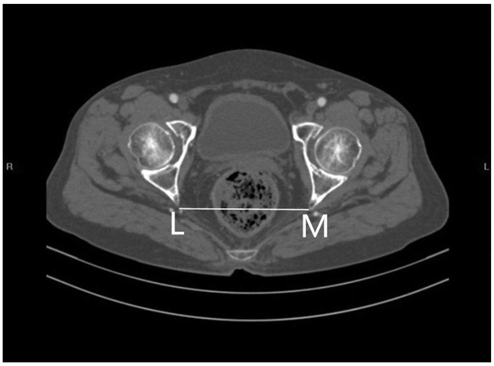
The present study aimed to evaluate the predictive value of pelvic anatomical and clinicopathological parameters for use in the estimation of the likely technical difficulties that may be encountered when performing open rectal surgery for mid‑low rectal cancer. Sixty consecutive patients, undergoing open rectal surgery for mid‑low rectal cancer were recruited between June 2009 and April 2014. All of the surgical procedures conducted, were low anterior resection (LAR) or abdominoperineal resection (APR). The operations were performed by the same surgeon and surgical team. Pelvic dimensions and angles were measured using three‑dimensional reconstruction of spiral computerized tomography (CT) images. Operative time and intraoperative blood loss were used as indicators of operative difficulty. The independent variables were pelvic anatomical and clinicopathological parameters, and the dependent variables were operative time and intraoperative blood loss. Univariate and multivariate analyses were performed in order to determine the predictive significance of these variables. The pelvis width was significantly wider in females than in males (P<0.05), while the sacrococcygeal bending degree was significantly greater in males than in females (P<0.05). No significant difference were detected between the pelvis depth of females and males (P>0.05). Multivariate analyses showed that body mass index (BMI), tumor height, lymph node metastasis, anteroposterior diameter of the pelvic inlet, anteroposterior diameter of the pelvic outlet, height of the pubic symphysis, the sacrococcygeal distance, sacrococcygeal‑pubic angle and diameter of the upper pubis to the coccyx were the main factors affecting the operative time (all P<0.05), while the maximum diameter of the tumor was the primary factor affecting intraoperative blood loss (P<0.05). Between the two procedures, the clinicopathological parameters appeared to be more valuable for predicting difficulty in LAR, in which operative time was associated with tumor height and tumor staging (RC2=0.312; P<0.001). By contrast, the pelvic anatomical parameters appeared to be more valuable predictors of variation in APR, in which intraoperative blood loss was associated with the anteroposterior diameter of the mid‑pelvis, the anteroposterior diameter of the pelvic outlet, the interspinous diameter, the depth of the sacral curvature and the sacropubic distance (RC2=0.608; P=0.002). BMI, tumor height and the maximum diameter of the tumor may be used to predict the operative difficulty in performing open rectal surgery for mid‑low rectal cancer. In addition to the associated clinicopathological parameters, wider, shallower and less curved pelvises may make the greatest contribution to reducing operative time and intraoperative blood loss. Operative difficulty is likely to be increased in deeper and narrower pelvises, or in those with greater sacrococcygeal curvature.