Pulmonary infiltration with eosinophilia complicated with mucosa-associated lymphoid tissue lymphoma: A case report
- Authors:
- Yin Liu
- Yinyan Tangsun
- Yonglong Xiao
- Deping Zhang
- Min Cao
View Affiliations
Affiliations: Department of Respiratory Medicine, Drum Tower Hospital, Nanjing University Medical School, Nanjing, Jiangsu 210008, P.R. China
- Published online on: July 11, 2016 https://doi.org/10.3892/ol.2016.4841
-
Pages:
1818-1820
-
Copyright: © Liu
et al. This is an open access article distributed under the
terms of Creative
Commons Attribution License.
Metrics: Total
Views: 0 (Spandidos Publications: | PMC Statistics: )
Metrics: Total PDF Downloads: 0 (Spandidos Publications: | PMC Statistics: )
This article is mentioned in:
Abstract
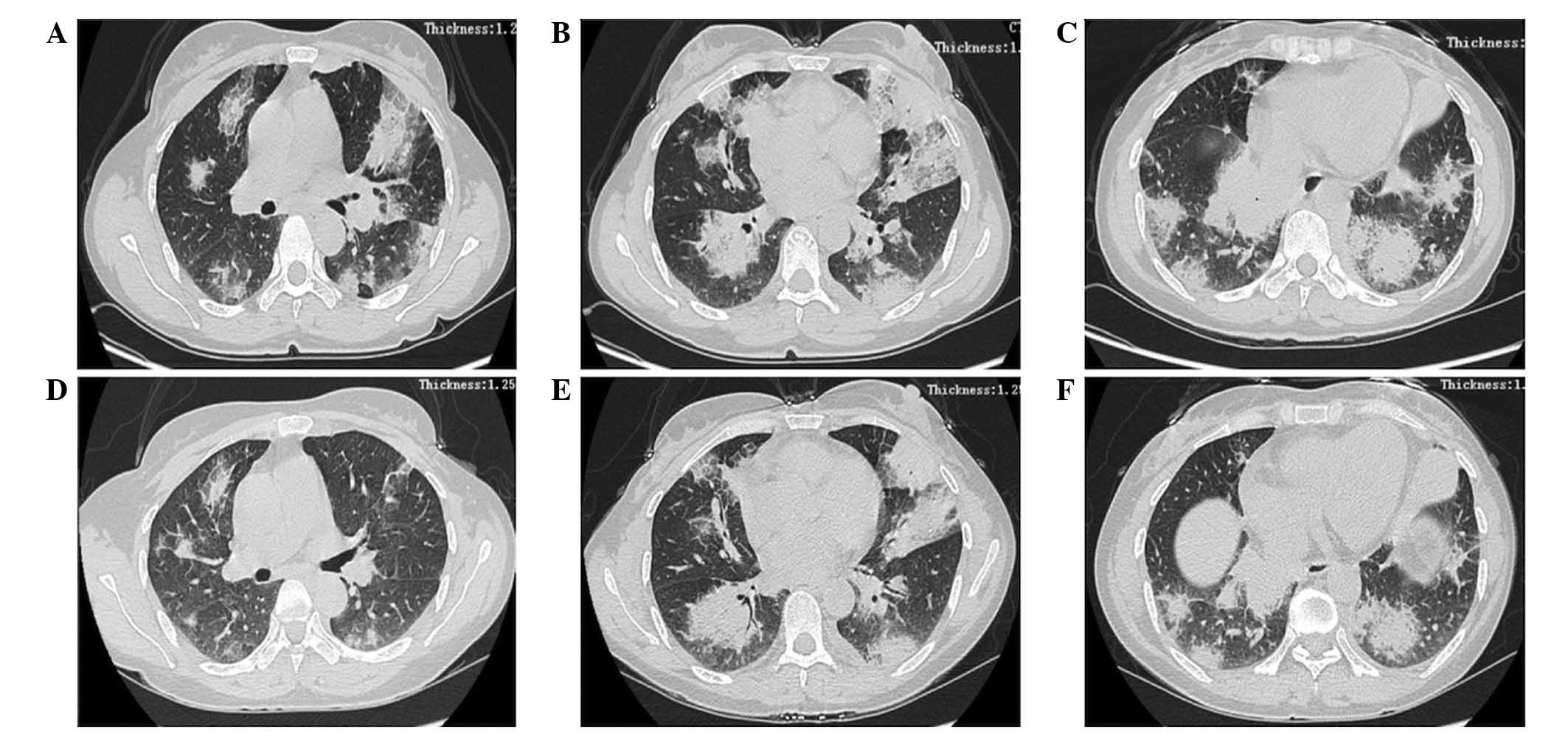
Tissue eosinophilia is rarely observed in cases of non-Hodgkin's lymphoma of B cell origin. The present study describes a rare case of mucosa-associated lymphoid tissue (MALT) lymphoma, which was initially misdiagnosed as eosinophilic pneumonia. The initial diagnosis was formed based on the results of chest radiography, peripheral eosinophilia tests and bronchoalveolar lavage, and the clinical course of the patient. Following administration of methylprednisolone (40 mg/day) for 4 days and oral administration of prednisolone (30 mg/day), the clinical course rapidly improved and the eosinophil count immediately decreased a to normal level. However, abnormal shadows observed on computed tomography (CT) scans of the chest did not diminish. At 6 months after the initiation of treatment, CT‑guided percutaneous lung biopsy was performed, and a final diagnosis of primary pulmonary mucosa‑associated lymphoid tissue lymphoma was made based on immunohistochemical examination. Primary lung MALT lymphoma remains a rare entity, with an indolent course and a reasonably favorable prognosis, whose diagnosis may be challenging.
View References
1
|
Bhatt NY and Allen JN: Update on
eosinophilic lung diseases. Semin Respir Crit Care Med. 33:555–571.
2012. View Article : Google Scholar : PubMed/NCBI
|
2
|
Mann B: Eosinophilic lung disease. Clin
Med Circ Respirat Pulm Med. 2:99–108. 2008.
|
3
|
Cadranel J, Wislez M and Antoine M:
Primary pulmonary lymphoma. Eur Respir J. 20:750–762. 2002.
View Article : Google Scholar : PubMed/NCBI
|
4
|
Jeong YJ, Kim KI, Seo IJ, Lee CH, Lee KN,
Kim KN, Kim JS and Kwon WJ: Eosinophilic lung diseases: A clinical,
radiologic, and pathologic overview. Radiographics. 27:617–639.
2007. View Article : Google Scholar : PubMed/NCBI
|
5
|
Thiruchelvam JK, Penfold CN and Akhtar S:
Chronic eosinophilic pneumonia associated with T-cell lymphoma. Int
J Oral Maxillofac Surg. 31:112–114. 2002. View Article : Google Scholar : PubMed/NCBI
|
6
|
Kunimasa K, Arita M, Arai Y, Uchino K,
Iwasaku M, Jo T, Konishi S and Ishida T: Peripheral T-cell lymphoma
not otherwise specified (PTCL-NOS) in a patient with
hypereosinophilic syndrome showing multiple nodules on chest
computed tomography. Intern Med. 50:2417–2421. 2011. View Article : Google Scholar : PubMed/NCBI
|
7
|
Watanabe K, Shinbo T, Kojima M, Naito M,
Tanahashi N and Nara M: B-cell lymphoma associated with
eosinophilia. Cancer. 64:1682–1685. 1989. View Article : Google Scholar : PubMed/NCBI
|
8
|
Fujii M, Tanimoto Y, Kiguchi T, Takehara
H, Fujimori Y, Teshima T, Kanehiro A, Shinagawa K, Tada S and
Kataoka M: Pulmonary infiltration with eosinophilia syndrome
complicated with non-Hodgkin's lymphoma of B cell lineage. Allergol
Int. 52:161–164. 2003. View Article : Google Scholar
|
9
|
Hare SS, Souza CA, Bain G, Seely JM Frcpc,
Gomes MM and Quigley M: The radiological spectrum of pulmonary
lymphoproliferative disease. Br J Radiol. 85:848–864. 2012.
View Article : Google Scholar : PubMed/NCBI
|
10
|
McCulloch GL, Sinnatamby R, Stewart S,
Goddard M and Flower CD: High-resolution computed tomographic
appearance of MALToma of the lung. Eur Radiol. 8:1669–1673. 1998.
View Article : Google Scholar : PubMed/NCBI
|
11
|
Bae YA, Lee KS, Han J, Ko YH, Kim BT,
Chung MJ and Kim TS: Marginal zone B-cell lymphoma of
bronchus-associated lymphoid tissue: Imaging findings in 21
patients. Chest. 133:433–440. 2008. View Article : Google Scholar : PubMed/NCBI
|