Introduction
Thyroid cancer is the most common type of endocrine
tumor; since 2000, overall thyroid cancer incidence rates have
increased by ~8% per year (1).
Papillary thyroid carcinoma (PTC) is the major histological type
and accounts for ~80% of all thyroid types of cancer (2,3). Highly
effective for the treatment of PTC involves surgery and radioactive
iodine; however, the traditional therapies are ineffective against
advanced radioactive iodine-resistant PTC. The majority of patients
with PTC demonstrate excellent clinical outcomes; however, distant
metastasis of PTC can be fatal (4).
Tyrosine kinase receptors represent targets of great
interest for cancer therapy. The tyrosine kinase-like orphan
receptor 2 (ROR2), is also a Wnt ligand receptor. ROR2 has been
revealed to specifically interact with Wnt5a. Aberrant activation
of Wnt signaling is involved in the development of various types of
tumors (4,5). In renal cell carcinoma, a high
expression level of ROR2 demonstrated a significant association
with higher clinical stage, nuclear grade and tumor stage (5). Recent data suggested that the Wnt
signaling pathway also altered PTC with RET/PTC mutations (6). To the best of our knowledge, the present
study revealed for the first time that ROR2 and Wnt5a expression
levels may also be altered in PTC tissues compared with normal
tissues.
The present study evaluated the protein expression
levels of ROR2 and Wnt5a in human PTC and adjacent normal tissues,
investigated the changes and clinical significance of ROR2 in PTC
and its association with Wnt5a expression.
Materials and methods
Patients and samples
The present study utilized 58 human PTC tissues and
paired adjacent noncancerous tissues excised from patients with
histologically confirmed PTC between January 2014 and July 2015 at
the Department of Oncological Surgery, Central Hospital of Cangzhou
(Cangzhou, China). The tissue samples were frozen immediately in
liquid nitrogen on removal from the patients. The present study was
approved by the Ethics Committee of the Central Hospital of
Cangzhou. Written informed consent was obtained from all patients
prior to enrollment in the present study.
Immunohistochemistry analysis
The rabbit anti-human ROR2 polyclonal antibody (cat.
no. SC-98486; 1:1,000) and the rabbit anti-human β-actin polyclonal
antibodies (cat. no. SC-130656; 1:1,000) were purchased from Santa
Cruz Biotechnology Inc. (Dallas, TX, USA). The rabbit anti-human
Wnt5a polyclonal antibody (cat. no. 55184-1-AP; 1:1,000) was
purchased from Abcam (Cambridge, UK). The tissues were fixed with
4% paraformaldehyde for 12 h and then embedded in paraffin and
sliced into 4-µm sections; these sections were then dewaxed in
xylene for 3 min three times, 100% ethanol for 2 min three times,
95% ethanol for 2 min, 80% ethanol for 2 min, 70% ethanol for 2 min
and PBS for 5 min. Endogenous peroxidase activity was blocked by
soaking slides in 3% H2O2 for 15 min at room
temperature (RT). The slides were then agitated and excess PBS
removed. All tumor sections were circled with a PAP pen. A total of
75 µl blocking buffer (Shanghai Xin Le Biological Technology Co.,
Ltd, Shanghai, China) was added to each section immediately. The
slides were then incubated for 1 h to overnight at RT in a
humidified chamber. The appropriate primary antibody was applied
overnight at 4°C. PBS-incubated slides were used as a negative
control. Subsequent to washing in PBS three times, the tissue
sections were incubated with biotin-conjugated secondary antibody
(Goat anti-rabbit antibody; cat. no. ab6720; Abcam; 1:1,000) at
room temperature for 1 h. Subsequently, the tissue sections were
visualized using 0.05% diaminobenzidine in PBS for 5 min at 37°C.
The tissue sections were counterstained with hematoxylin (10%) at
room temperature for 1 min, dehydrated using graded ethanol (70, 80
and 100%) and sealed and covered with glass coverslips. All slides
were processed by the same pathologist.
Two independent pathologists reviewed all
histological tissue sections and evaluated the immunohistochemistry
staining results according to the criteria of the World Health
Organization (7). The pathologists
randomly observed 10 high power fields of view with 100 cells in
each view using a light microscope at magnification, ×400 (Olympus
Corporation, Tokyo, Japan). Comprehensive evaluation was performed
according to the intensity and percentage of the stained tumor
cells. Staining intensity was graded according to the following
criteria: 0, no staining; 1, yellow; 2, deep yellow; and 3, brown.
The proportion of positive tumor cells was scored as follows: 0, no
positive tumor cells; 1, <10% positive tumor cells; 2, 10–50%
positive tumor cells; 3, 51–80% positive tumor cells; and 4,
>80% positive tumor cells. The immunohistochemical expression
level was based on the total scores. Total score = points of
staining intensity + points of percentage of positive cells. The
tissue samples were classified into two groups, as follows:
Negative expression, 0–1 points; and positive expression, 2–6
points.
Western blot analysis
Proteins were extracted from nitrogen-frozen tissue
fragments of tissue samples. The tissues were homogenized in 1 ml
radioimmunoprecipitation assay buffer (Beyotime Institute of
Biotechnology, Haimen, China) and subsequently with protease
inhibitor cocktail (Beyotime Institute of Biotechnology) at 0°C for
20 min, followed by incubation for 20 min on ice and centrifugation
at 12,000 × g for 15 min at 4°C. Following collection of the
supernatant fluid, the sample was boiled for 15 min then stored at
−80°C. Proteins (50 µg/well) were separated by 8% SDS-PAGE. The
absorbance of proteins at A562 nm was measured by a microplate
reader. The protein concentration was calculated from the standard
curve. Following electrophoresis, proteins were transferred to
polyvinylidene difluoride membranes and blocked for 1 h with 5%
fat-free milk at room temperature. The membranes were incubated
with primary antibodies, anti-ROR2 (SC-98486; 1:1,000) and
anti-Wnt5a (55184–1-AP; 1:1,000), overnight at 4°C. β-actin
antibody (SC-130656; 1:1,000) was used as the loading control.
Subsequently, the membranes were incubated with secondary antibody
(horseradish peroxidase-conjugated antibodies) at room temperature
for 1 h (goat anti-rabbit antibody; ab6721; Abcam, 1:1,000).
Following three washes with TBS for 15 min at room temperature, the
membranes were treated with a chemiluminescence detection kit
(Pierce; Thermo Fisher Scientific, Inc., Waltham, MA, USA). Protein
bands were quantified using densitometry (Gel-Doc Gel-It2 310
Imaging Analysis System, Bio-Rad Laboratories, Hercules, CA,
USA).
RNA extraction and cDNA synthesis and
reverse transcription-quantitative polymerase chain reaction
(RT-qPCR)
Total RNA isolation and cDNA synthesis were
performed as previously described (8). Total RNA of PTC tissues and paired
adjacent noncancerous tissues were isolated using
TRIzol® (Invitrogen; Thermo Fisher Scientific, Inc.),
according to manufacturer's instructions. RNA quality and
concentration were assessed on the NanoDrop 1000 (Thermo Fisher
Scientific, Inc.). A total of 1 µg RNA was reverse transcribed
using the First-Strand cDNA Synthesis kit (Thermo Fisher
Scientific, Inc.) with Oligo(dT) primer. PCR primers used were as
follows: ROR2 forward, 5′-GGCAGAACCCATCCTCGTG-3′ and reverse,
5′-CGACTGCGAATCCAGGACC-3′; Wnt5a forward,
5′-ACCACATGCAGTACATCGGAG-3′ and reverse,
5′-GAGGTGTTATCCACAGTGCTG-3′; and GAPDH forward,
5′-GAAGGTGAAGGTCGGAGTC-3′ and reverse, 5′-GAAGATGGTGATGGGATTTC-3′.
The PCR reaction was performed for 40 cycles of a 3-step program:
95°C for 15 sec, annealing temperature at 60°C for 15 sec, 72°C for
1 min. mRNA levels were quantified using the 2−ΔΔCq
method (9).
Statistical analysis
Statistical analysis was performed using SPSS
version 13.0 (SPSS, Inc., Chicago, IL, USA). The expression level
of mRNA was represented as fold change using the 2−ΔΔCq
method. The χ2 test was performed to determine the
association between protein ROR2 expression level and
clinicopathological features. P<0.05 was considered to indicate
a statistically significant difference.
Results
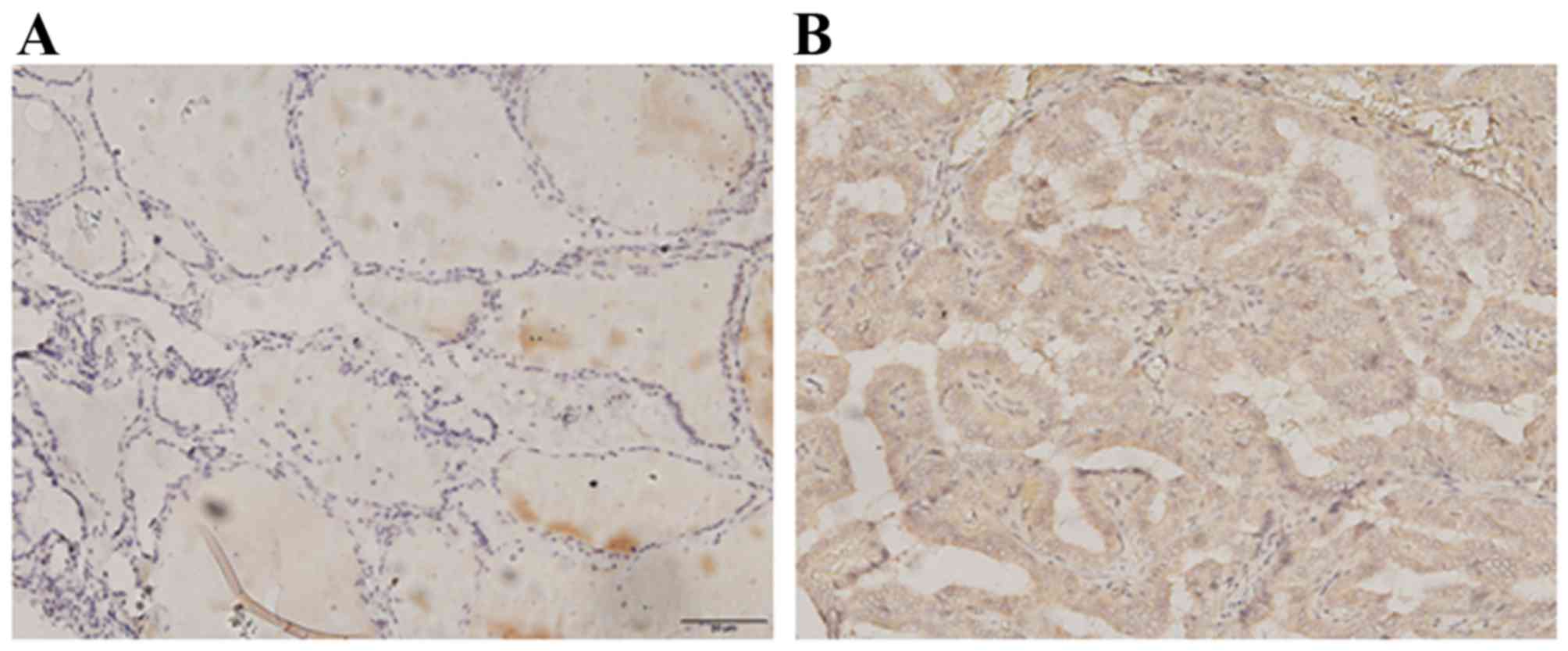
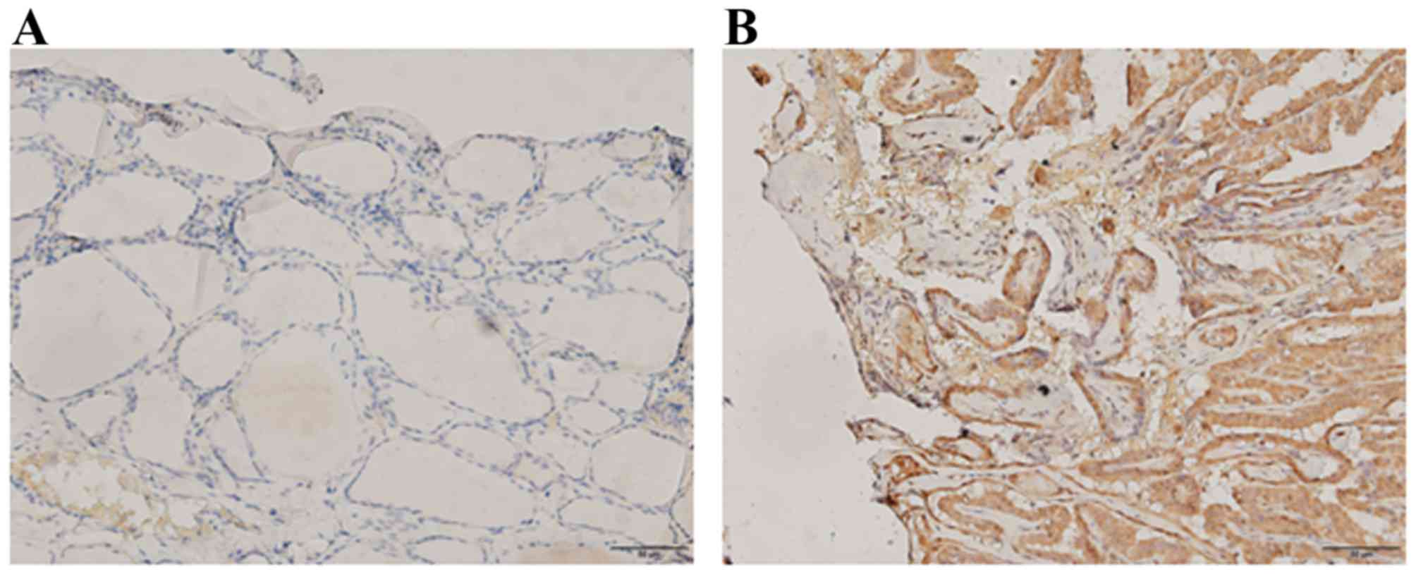
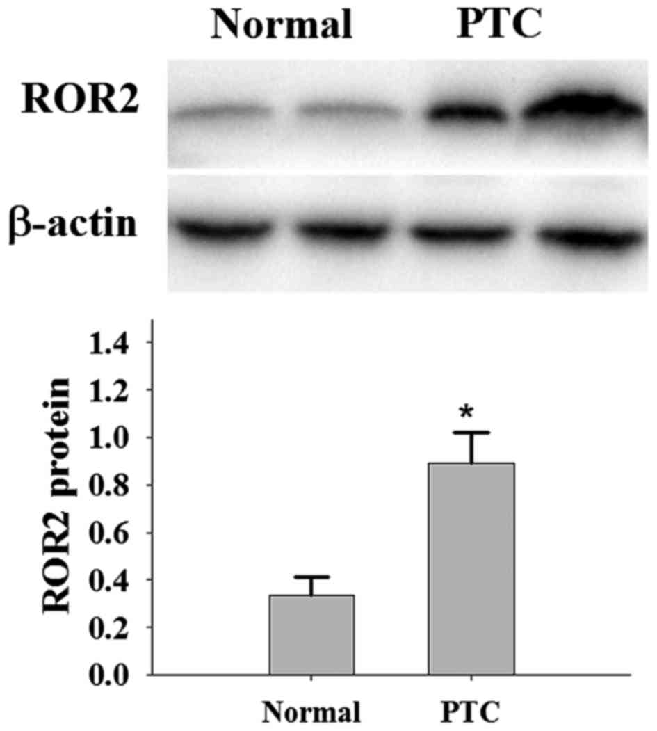
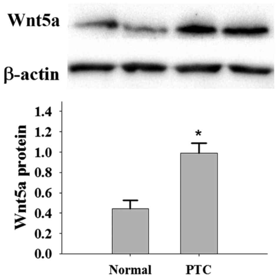
ROR2 protein and Wnt5a protein
expression is increased in PTC
The patients in this study comprised 43 females and
15 males with an age range of 16–57 years. The present study
selected the most representative PTC and adjacent normal tissues.
Immunohistochemistry demonstrated that 36 patients exhibited
positive expression of the ROR2 protein (P<0.05; Fig. 1; Table
I) and 39 patients exhibited positive expression of the Wnt5a
protein (P<0.05; Fig. 2; Table II). Western blot analysis
demonstrated that the protein expression levels of ROR2 and Wnt5a
in PTC were significantly increased compared with the expression in
normal thyroid tissue samples (Figs.
3 and 4).
 | Table I.Analysis of ROR2 expression level in
PTC and normal tissues. |
Table I.
Analysis of ROR2 expression level in
PTC and normal tissues.
|
|
| ROR2 expression,
n |
|
|
|---|
|
|
|
|
|
|
|---|
| Group | Cases, n | Negative | Positive | χ2 | P-value |
|---|
| PTC | 58 | 22 | 36 | 5.829 | 0.016 |
| Normal | 58 | 35 | 23 |
|
|
| Table II.Analysis of Wnt5a expression level in
PTC and normal tissues. |
Table II.
Analysis of Wnt5a expression level in
PTC and normal tissues.
|
|
| Wnt5a expression,
n |
|
|
|---|
|
|
|
|
|
|
|---|
| Group | Cases, n | Negative | Positive | χ2 | P-value |
|---|
| PTC | 58 | 19 | 39 | 15.211 | <0.001 |
| Normal | 58 | 40 | 18 |
|
|
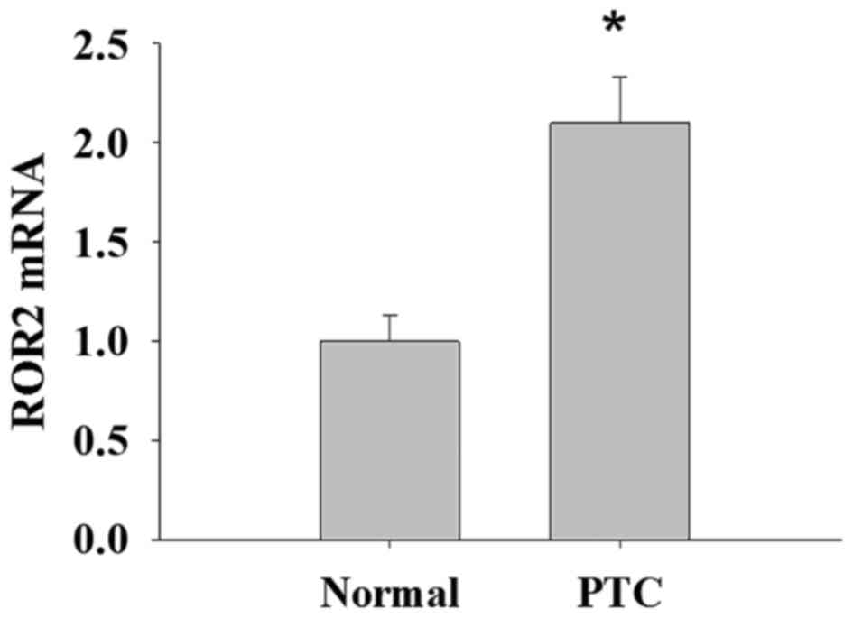
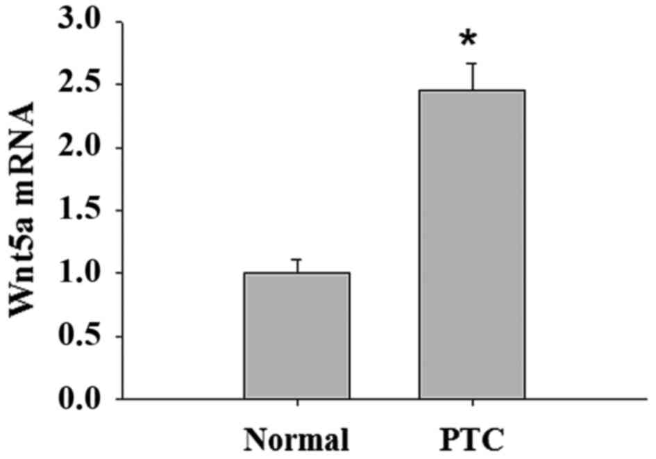
ROR2 gene and Wnt5a gene expression is
increased in PTC
ROR2 and Wnt5a mRNA expression levels were
significantly increased in PTC samples compared with in normal
thyroid tissue samples (Figs. 5 and
6).
ROR2 and Wnt5a expression with
pathological features
The expression level of ROR2 was associated with
tumor stage and lymph node metastasis in PTC; however, there was no
association with age, sex, tumor size and the number of tumors
(Table III). Wnt5a was also
associated with tumor stage and lymph node metastasis; no
significant differences were identified between Wnt5a expression
levels and other clinicopathological findings, including age, sex,
tumor size and the number of tumors (Table IV).
| Table III.Association between ROR2 expression
level and clinicopathological characteristics in patients with
PTC. |
Table III.
Association between ROR2 expression
level and clinicopathological characteristics in patients with
PTC.
|
|
| ROR2 expression,
n |
|
|
|---|
|
|
|
|
|
|
|---|
| Clinicopathological
characteristics | Cases, n | Negative | Positive | χ2 | P-value |
|---|
| Age |
|
|
| 0.648 | 0.421 |
| <45
years | 20 | 9 | 11 |
|
|
| ≥45
years | 38 | 13 | 25 |
|
|
| Sex |
|
|
| 2.039 | 0.153 |
| Male | 15 | 8 | 7 |
|
|
|
Female | 43 | 14 | 29 |
|
|
| Tumor Stage |
|
|
| 6.408 | 0.007 |
| I,
II | 40 | 20 | 20 |
|
|
| III,
IV | 18 | 2 | 16 |
|
|
| Tumor size |
|
|
| 0.741 | 0.389 |
| <1
cm | 41 | 17 | 24 |
|
|
| ≥1
cm | 17 | 5 | 12 |
|
|
| Lymph node
involvement |
|
|
| 5.084 | 0.028 |
|
Yes | 24 | 5 | 19 |
|
|
| No | 34 | 17 | 17 |
|
|
| Multifocal |
|
|
| 0.056 | 0.814 |
|
Yes | 20 | 8 | 12 |
|
|
| No | 38 | 14 | 24 |
|
|
| Table IV.Association between Wnt5a expression
level and clinicopathological characteristics in patients with
PTC. |
Table IV.
Association between Wnt5a expression
level and clinicopathological characteristics in patients with
PTC.
|
|
| Wnt5a expression,
n |
|
|
|---|
|
|
|
|
|
|
|---|
| Clinicopathological
characteristics | Cases, n | Negative | Positive | χ2 | P-value |
|---|
| Age |
|
|
| 0.727 | 0.394 |
| <45
years | 20 | 8 | 12 |
|
|
| ≥45
years | 38 | 11 | 27 |
| Sex |
|
|
| 0.482 | 0.488 |
|
Male | 15 | 6 | 9 |
|
|
|
Female | 43 | 13 | 30 |
|
|
| Tumor Stage |
|
|
| 7.069 | 0.003 |
| I,
II | 40 | 18 | 22 |
|
|
| III,
IV | 18 | 1 | 17 |
|
|
| Tumor size |
|
|
| 0.070 | 0.791 |
| <1
cm | 41 | 13 | 28 |
|
|
| ≥1
cm | 17 | 6 | 11 |
|
|
| Lymph node
involvement |
|
|
| 3.647 | 0.046 |
|
Yes | 24 | 4 | 20 |
|
|
| No | 34 | 15 | 19 |
|
|
| Multifocal |
|
|
| 0.070 | 0.792 |
|
Yes | 20 | 7 | 13 |
|
|
| No | 38 | 12 | 26 |
|
|
Correlation between ROR2 and Wnt5a in
PTC
There were significant positive associations between
ROR2 and Wnt5a (Table V).
| Table V.Association between ROR2 and Wnt5a
expression levels in PTC. |
Table V.
Association between ROR2 and Wnt5a
expression levels in PTC.
| Protein | r-value | P-value |
|---|
| ROR2 | 0.857 | 0.007 |
| Wnt5a |
|
|
Discussion
Thyroid cancer incidence increased, on average, 3.6%
per year during 1974–2013, this was primarily associated with
increases in PTC of 4.4% per year (10). The main treatment for PTC involves
total or subtotal thyroidectomy, radioactive iodine and thyroid
hormone inhibitory therapy; however, the traditional therapies are
ineffective against advanced radioactive iodine-resistant PTC
(11). The majority of patients with
PTC have a favorable prognosis and asymptomatic long-term survival;
however, invasion and metastasis is also one of the major causes of
mortality in patients with PTC (12).
The invasion and metastasis of PTC is an interactive effect
mediated by diverse genes and factors (13,14).
A previous study revealed that ROR2 has a role in
cell migration and invasion (15,16). ROR2
is a transmembrane protein and acts as a Wnt ligand receptor that
participates in Wnt signaling (17).
A previous study suggested that ROR2 mediates Wnt5a signaling in a
variety of tumor types, including in human metastatic melanoma,
leiomyosarcoma and gastrointestinal stromal tumors, renal cell
carcinoma and osteosarcoma (8,18–21). Although it is accepted that altered
Wnt signaling is a late event in thyroid cell transformation that
affects anaplastic thyroid tumors, previous data suggested that it
is also altered in PTC with RET/PTC mutations (22). The present study inferred that ROR2
and Wnt5a may also have an important role in PTC.
The present study demonstrated that ROR2 and Wnt5a
protein translation and gene transcription were upregulated in PTC
tissues in comparison with the matched adjacent normal thyroid
tissues. ROR2 is a member of a family of proteins known as receptor
protein kinases, which have a key role in cell growth,
differentiation and cell movement. Wnt5a binds to its receptor ROR2
and activates a serine/threonine-specific protein kinase, CamKII,
which negatively regulates the canonical Wnt/β-catenin signaling
via the MAPK signaling pathway. An association was revealed between
ROR2 expression level and tumor stage as well as lymph nodes
metastasis. Wnt5a protein expression level was observed to be
increased more significantly in patients with advanced stage and
lymph node metastases; however, the role of the Wnt5a/ROR2
signaling pathway in cancer remains unknown (23). Wnt5a was demonstrated to signal via
ROR2 to induce cellular migration and invasion in murine fibroblast
NIH3T3 cells (24). Wright et
al (21) demonstrated that ROR2
promotes tumor growth potential in renal cell carcinoma. McDonald
and Silver (23) revealed that in
cancer cells, the oncogenic potential of ROR2 may be conferred by
Wnt5a via the promotion of cancer cell invasion. In the present
study, compared with in the normal thyroid tissues, the expression
level of ROR2 and Wnt5a in PTC tissues was increased markedly;
therefore, ROR2 and Wnt5a may be involved in the progress of PTC or
may become additional tumor markers for the diagnosis of PTC.
However, it remains unclear how the Wnt5a/ROR2 signaling pathway
acts in PTC.
The present study investigated the clinical
significance of ROR2 and Wnt5a in patients with PTC for the first
time and the result demonstrated that ROR2 and Wnt5a were
significantly upregulated in PTC tissues compared with in the
matched adjacent normal thyroid tissues. High ROR2 and Wnt5a
expression levels were associated with tumor stage and lymph node
metastases. Furthermore, there was a significant positive
association between ROR2 and Wnt5a expression levels. These results
indicated the cancer-promoting activity of the Wnt5a/ROR2 signaling
pathway in PTC tissues. Rasmussen et al (6) revealed that in patients with renal cell
carcinoma a high expression level of ROR2 was associated with a
significantly lower overall survival rate, cancer specific survival
and recurrence free survival compared with renal cell carcinoma
patients with low expression levels of ROR2; these findings
suggested that ROR2 may be considered as a negative prognostic
biomarker and potential therapeutic target in this type of cancer.
A study investigating cervical cancer indicated that ROR2 was
significantly associated with cancer progression and poor prognosis
(25). Lara et al (26) demonstrated that epigenetic alteration
of ROR2 has a Wnt-mediated, pro-tumorigenic role in colon cancer.
In brief, ROR2 with Wnt5a may have an important role in the
infiltration and metastasis of PTC.
In summary, compared with normal thyroid tissues,
the present study revealed a high expression level of ROR2 and
Wnt5a in PTC tissues. Furthermore, the expression levels of ROR2
and Wnt5a were associated with tumor stage and lymph node
metastasis. There was a significant positive association between
ROR2 and Wnt5a expression levels. The results of the present study
indicated that the Wnt5a/ROR2 signaling pathway may have a critical
role in driving cell proliferation and migration. The expression
level of ROR2 and Wnt5a mRNA significantly increased with tumor
progression, which suggested that the ROR2 and Wnt5a genes have
potential as targets for cancer gene therapy. In conclusion, these
results indicated that ROR2 and Wnt5a may be promising biomarkers
and potential therapeutic targets for PTC in the future.
References
|
1
|
Haugen BR, Alexander EK, Bible KC, Doherty
GM, Mandel SJ, Nikiforov YE, Pacini F, Randolph GW, Sawka AM,
Schlumberger M, et al: 2015 American thyroid association management
guidelines for adult patients with thyroid nodules and
differentiated thyroid cancer: The American thyroid association
guidelines task force on thyroid nodules and differentiated.
Thyroid. 26:1–133. 2016. View Article : Google Scholar : PubMed/NCBI
|
|
2
|
La Vecchia C, Malvezzi M, Bosetti C,
Garavello W, Bertuccio P, Levi F and Negri E: Thyroid cancer
mortality and incidence: A global overview. Int J Cancer.
136:2187–2195. 2015. View Article : Google Scholar : PubMed/NCBI
|
|
3
|
Zimmermann MB and Galetti V: Iodine intake
as a risk factor for thyroid cancer: A comprehensive review of
animal and human studies. Thyroid Res. 8:82015. View Article : Google Scholar : PubMed/NCBI
|
|
4
|
Jeon MJ, Kim WG, Choi YM, Kwon H, Lee YM,
Sung TY, Yoon JH, Chung KW, Hong SJ, Kim TY, et al: Features
predictive of distant metastasis in papillary thyroid
microcarcinoma. Thyroid. 26:161–168. 2016. View Article : Google Scholar : PubMed/NCBI
|
|
5
|
Lara E, Calvanese V, Huidobro C, Fernández
AF, Moncada-Pazos A, Obaya AJ, Aguilera O, González-Sancho JM,
Sánchez L, Astudillo A, et al: Epigenetic repression of ROR2 has a
Wnt-mediated, pro-tumourigenic role in colon cancer. Mol Cancer.
9:1702010. View Article : Google Scholar : PubMed/NCBI
|
|
6
|
Rasmussen NR, Debebe Z, Wright TM, Brooks
SA, Sendor AB, Brannon AR, Hakimi AA, Hsieh JJ, Choueiri TK,
Tamboli P, et al: Expression of Ror2 mediates invasive phenotypes
in renal cell carcinoma. PLoS One. 9:e1161012014. View Article : Google Scholar : PubMed/NCBI
|
|
7
|
International Agency for Research on
Cancer: World Health Organization Classification of Tumours:
Pathology and Genetics of Tumours of Endocrine Organs. DeLellis RA,
Lloyd VR, Heitz PU and Eng C: IARC Press; Lyon, France: 8. 3rd. pp.
3202004
|
|
8
|
Sastre-Perona A and Santisteban P: Role of
the wnt pathway in thyroid cancer. Front Endocrinol (Lausanne).
3:312012.PubMed/NCBI
|
|
9
|
Livak KJ and Schmittgen TD: Analysis of
relative gene expression data using real-time quantitative PCR and
the 2(-Delta Delta C(T)) method. Methods. 25:402–408. 2001.
View Article : Google Scholar : PubMed/NCBI
|
|
10
|
Lim H, Devesa SS, Sosa JA, Check D and
Kitahara CM: Trends in thyroid cancer incidence and mortality in
the United States, 1974–2013. JAMA. 317:1338–1348. 2017. View Article : Google Scholar : PubMed/NCBI
|
|
11
|
La Greca A, Xu B, Ghossein R, Tuttle RM
and Sabra MM: Patients with multifocal macroscopic papillary
thyroid carcinoma have a low risk of recurrence at early follow-up
after total thyroidectomy and radioactive iodine treatment. Eur
Thyroid J. 6:31–39. 2017. View Article : Google Scholar : PubMed/NCBI
|
|
12
|
Carhill AA, Litofsky DR, Ross DS, Jonklaas
J, Cooper DS, Brierley JD, Ladenson PW, Ain KB, Fein HG, Haugen BR,
et al: Long-term outcomes following therapy in differentiated
thyroid carcinoma: NTCTCS registry analysis 1987–2012. J Clin
Endocrinol Metab. 100:3270–3279. 2015. View Article : Google Scholar : PubMed/NCBI
|
|
13
|
Cerutti JM, Latini FR, Nakabashi C,
Delcelo R, Andrade VP, Amadei MJ, Maciel RM, Hojaij FC, Hollis D,
Shoemaker J and Riggins GJ: Diagnosis of suspicious thyroid nodules
using four protein biomarkers. Clin Cancer Res. 12:3311–3318. 2006.
View Article : Google Scholar : PubMed/NCBI
|
|
14
|
Zou M, Baitei EY, Alzahrani AS, BinHumaid
FS, Alkhafaji D, Al-Rijjal RA, Meyer BF and Shi Y: Concomitant RAS,
RET/PTC, or BRAF mutations in advanced stage of papillary thyroid
carcinoma. Thyroid. 24:1256–1266. 2014. View Article : Google Scholar : PubMed/NCBI
|
|
15
|
Henry C, Llamosas E, Knipprath-Meszaros A,
Schoetzau A, Obermann E, Fuenfschilling M, Caduff R, Fink D, Hacker
N, Ward R, et al: Targeting the ROR1 and ROR2 receptors in
epithelial ovarian cancer inhibits cell migration and invasion.
Oncotarget. 6:40310–40326. 2015. View Article : Google Scholar : PubMed/NCBI
|
|
16
|
Kim YH, Choi YW, Han JH, Lee J, Soh EY,
Park SH, Kim JH and Park TJ: TSH signaling overcomes
B-RafV600E-induced senescence in papillary thyroid carcinogenesis
through regulation of DUSP6. Neoplasia. 16:1107–1120. 2014.
View Article : Google Scholar : PubMed/NCBI
|
|
17
|
Cheung R, Kelly J and Macleod RJ:
Regulation of villin by wnt5a/ror2 signaling in human intestinal
cells. Front Physiol. 2:582011. View Article : Google Scholar : PubMed/NCBI
|
|
18
|
O'Connell MP, Fiori JL, Xu M, Carter AD,
Frank BP, Camilli TC, French AD, Dissanayake SK, Indig FE, Bernier
M, et al: The orphan tyrosine kinase receptor, ROR2, mediates wnt5A
signaling in metastatic melanoma. Oncogene. 29:34–44. 2010.
View Article : Google Scholar : PubMed/NCBI
|
|
19
|
Edris B, Espinosa I, Mühlenberg T, Mikels
A, Lee CH, Steigen SE, Zhu S, Montgomery KD, Lazar AJ, Lev D, et
al: ROR2 is a novel prognostic biomarker and a potential
therapeutic target in leiomyosarcoma and gastrointestinal stromal
tumour. J Pathol. 227:223–233. 2012. View Article : Google Scholar : PubMed/NCBI
|
|
20
|
Wright TM and Rathmell WK: Identification
of Ror2 as a hypoxia-inducible factor target in von
hippel-lindau-associated renal cell carcinoma. J Biol Chem.
285:12916–12924. 2010. View Article : Google Scholar : PubMed/NCBI
|
|
21
|
Wright TM, Brannon AR, Gordan JD, Mikels
AJ, Mitchell C, Chen S, Espinosa I, van de Rijn M, Pruthi R, Wallen
E, et al: Ror2, a developmentally regulated kinase, promotes tumor
growth potential in renal cell carcinoma. Oncogene. 28:2513–2523.
2009. View Article : Google Scholar : PubMed/NCBI
|
|
22
|
Enomoto M, Hayakawa S, Itsukushima S, Ren
DY, Matsuo M, Tamada K, Oneyama C, Okada M, Takumi T, Nishita M and
Minami Y: Autonomous regulation of osteosarcoma cell invasiveness
by Wnt5a/Ror2 signaling. Oncogene. 28:3197–3208. 2009. View Article : Google Scholar : PubMed/NCBI
|
|
23
|
McDonald SL and Silver A: The opposing
roles of Wnt-5a in cancer. Br J Cancer. 101:209–214. 2009.
View Article : Google Scholar : PubMed/NCBI
|
|
24
|
Nomachi A, Nishita M, Inaba D, Enomoto M,
Hamasaki M and Minami Y: Receptor tyrosine kinase Ror2 mediates
Wnt5a-induced polarized cell migration by activating c-Jun
N-terminal kinase via actin-binding protein filamin A. J Biol Chem.
283:27973–27981. 2008. View Article : Google Scholar : PubMed/NCBI
|
|
25
|
Sun B, Ye X, Lin L, Shen M and Jiang T:
Up-regulation of ROR2 is associated with unfavorable prognosis and
tumor progression in cervical cancer. Int J Clin Exp Pathol.
8:856–861. 2015.PubMed/NCBI
|
|
26
|
Lara E, Calvanese V, Huidobro C, Fernández
AF, Moncada-Pazos A, Obaya AJ, Aguilera O, González-Sancho JM,
Sánchez L, Astudillo A, et al: Epigenetic repression of ROR2 has a
Wnt-mediated, pro-tumourigenic role in colon cancer. Mol Cancer.
9:1702010. View Article : Google Scholar : PubMed/NCBI
|