Introduction
With the improvement of high-throughput sequencing
technology, an increasing number of small non-coding (snc)RNAs have
been elucidated. These are RNAs that do not code for proteins and
include micro (mi)RNAs, circular RNAs, transfer (t)RNAs and
P-element-induced wimpy testis (PIWI)-interacting (pi)RNAs
(1). It has been reported that
sncRNAs serve vital roles in the regulation of transcription and
translation, as well as in the progression of multiple malignancies
(2). In recent years, increasing
evidence has demonstrated that tRNA-derived small (ts)RNAs are
involved in several metabolic pathways and immunological processes
(3), and are frequently
dysregulated in malignant tumors (4). tsRNAs may also act as biomarkers and
therapeutic targets for cancer prognosis and diagnosis, according
to several studies (5,6).
tsRNAs are a group of sncRNAs produced from tRNAs,
which are strongly associated with tRNA abundance and were
initially thought to be degradation fragments (7). However, there is evidence that tsRNAs
are byproducts of enzymatic digestion that specifically target
mature or precursor tRNAs under particular circumstances, such as
viral infection, stress induction (8,9).
tsRNAs are clusters of functional molecules with great stability
and inherently conserved features; they are not simply byproducts
of tRNA breakdown (10). The
present review describes the important characteristics and
biological purposes of tsRNAs. The dysregulation and roles of
tsRNAs in several malignancies are evaluated, and their potential
as biomarkers and therapeutic targets for cancer detection and
outlook is assessed.
Biogenesis and classification of tsRNAs
tsRNAs are classified into two groups based on
length and cleavage site (Fig. 1
and Table I): One group comprises
tRNA halves [tRNA-derived stress-induced (ti)RNAs], which have
lengths of 28–36 nucleotides (nt) and are produced by angiogenin
(ANG)-specific cleavage at the anticodon loop, including 3′ and 5′
tiRNAs (11); and the other group
comprises tRNA-derived fragments (tRFs), which are 14–30 nt in
length and are produced via cleavage of mature or precursor tRNAs
at specific sites. A total of five subclasses of tRFs have been
identified: tRF-1, tRF-2, tRF-3, tRF-5 and internal (i)-tRF
(12). Currently, it is unknown
exactly how tRF-2 and i-tRF are produced (13); however, tRF-5 and tRF-3 are reported
to be by-products of mature tRNA enzymatic digestion (14), whilst tRF-1 is a by-product of
precursor tRNA enzymatic shearing (15). The mature tRNA's D-loop (tRF-5a,
14–16 nt) or the sequences between the D-loop and the anticodon
loops (tRF-5b, 22–24 nt; and tRF-5c, 28–30 nt) are the sources of
tRF-5 (16). tRF-5 is typically
produced by Dicer and ANG cleavage to different termination sites
between the 5′ end and the anticodon loop. tRF-3, which is composed
of tRF-3a and tRF-3b, is generated by Dicer cleavage on the TψC
loop at the 3′ end of mature tRNAs and the tail of each tRF-3
contains a specific CCA structure at the 3′ end of the mature tRNA
(17). tiRNAs are present in the
cytoplasm, with tRF-5 common in the nucleus and tRF-3 and tRF-1
prominent in the cytoplasm (17).
The 3′ end of the tRNA precursor is where tRF-1 is produced, and it
finishes in U bases. tRF-1 is the product of ribonuclease Z,
shearing the 3′ end of precursor tRNAs (15).
 | Table I.tsRNA types. |
Table I.
tsRNA types.
| tsRNA | Length, nt | Subclass of
tsRNA | Generation
mechanism | Location |
|---|
| Trf | 14-30 | tRF-1 | Produced by
shearing the 3′ end of pre-tRNA by Rnase Z | Cytoplasm |
|
|
| tRF-3 | Generated by Dicer
cleavage on the TψC loop at the 3′ end of mature tRNAs | Cytoplasm |
|
|
| tRF-5 | Produced by Dicer
and ANG cleavage in the D-loop or the region between the D-loop and
the anticodon loop of mature tRNA | Nucleus |
|
|
| tRF-2 | Not clear | Not clear |
|
|
| i-tRF | Not clear | Not clear |
| TiRNA | 28-36 | 3′tiRNA | Produced by
ANG-specific cleavage at the anticodon loop of mature tRNA | Cytoplasm |
|
|
| 5′tiRNA | Produced by
ANG-specific cleavage at the anticodon loop of mature tRNA | Cytoplasm |
Biological functions of tsRNAs
The biological roles of tsRNAs have yet not been
fully elucidated; however, they serve a wide range of significant
biological functions, including in transcriptional regulation,
post-transcriptional modification and translational regulation
(18).
Transcriptional regulation
tsRNAs can regulate gene expression at the
transcriptional level. tsRNAs are similar to piRNAs in physical
structure and both are single-stranded RNAs. They are also similar
to piRNAs, which can interact with PIWI proteins to form complexes
and then interact with DNA methyltransferases to affect the
methylation of genes, thereby exerting transcriptional repression
(19,20). For example, Pekarsky et al
(21) assessed whether ts-3676 and
ts-4521 can interact with PIWI proteins as piRNAs by performing RNA
immunoprecipitation experiments. They reported that ts-3676 and
ts-4521 were notably enriched in complexes containing labeled
PIWI-like protein (PIWIL)2 compared with controls, suggesting that
ts-3676 and ts-4521 can interact with PIWIL2 as piRNAs. Zhang et
al (22) reported that the
tRNA-Glu-derived piRNA [td-piR(Glu)]/PIWIL4 complex recruits H3K9
methyltransferases (SETDB1, SUV39H1) and heterochromatin protein 1β
to the CD1A promoter region and promotes H3K9 methylation,
resulting in marked repression of CD1A transcription.
Post-transcriptional modification
tsRNAs serve important roles in the
post-transcriptional regulation of several biological processes by
regulating messenger (m)RNA stability. Similar to miRNAs, tsRNAs
can take part in the formation of RNA-induced silencing complexes
(RISCs) and regulate mRNA stability by RISC binding to the 3′
untranslated region (3′-UTR) of target genes. They may also
regulate gene expression through the post-transcriptional pathway
(23). tsRNAs can also control gene
expression by influencing the stability of targeted RNAs by
competitively binding to RNA-binding proteins (RBPs) (24). Competitively binding to the 3′-UTR
of RBP Y-box binding protein 1, tRFs displace oncogenic
transcripts, reducing their stability and resulting in tumor
suppressor and metastasis suppressor actions in breast cancer cells
(Fig. 2A) (25).
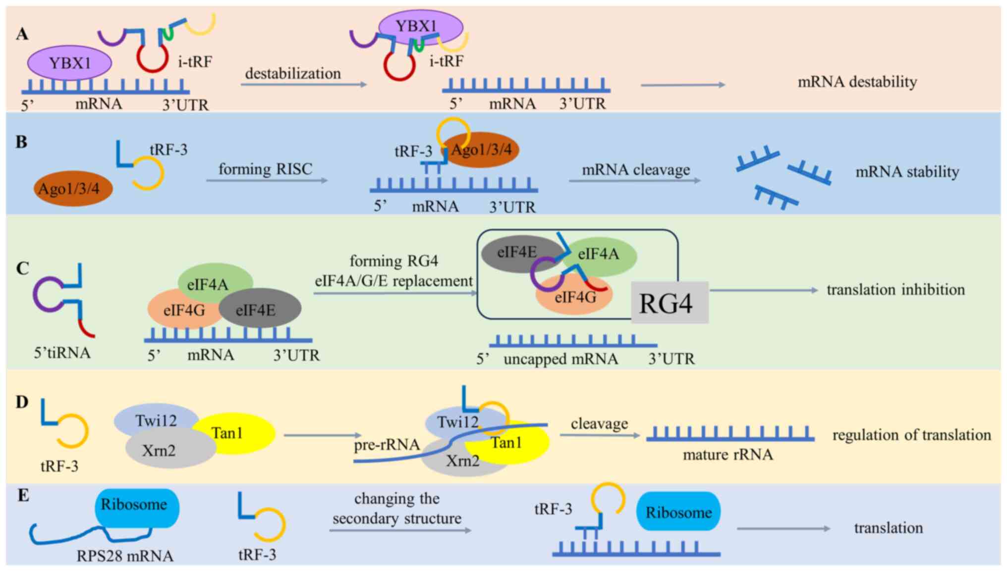 | Figure 2.Biological functions of tsRNAs. (A)
i-tRF bind to YBX1 to decrease oncogenic transcript stability. (B)
tRF-3 forms RISC with Ago proteins to silence mRNA. (C) 5′tiRNA
bind to the eIF4A/E/G complex, displacing mRNA without cap
structure to cause translational inhibition. (D) tRF-3 binds to
Twi12, Xrn2 and Tan1 to form a complex affecting the rRNA process.
(E) Regulation of ribosome biogenesis by changing RPS28 mRNA
secondary structure during translation. tRNA, transfer RNA; tsRNA,
tRNA-derived small RNA; tRF, tRNA-derived fragment; i-tRF, internal
tRF; YBX1, Y-box binding protein 1; RISC, RNA-induced silencing
complex; Ago, argonaute; tiRNA, tRNA-derived stress-induced RNA;
eIF4A/E/G, eukaryotic initiation factors 4A/E/G; Twi12, twinfilin
actin binding protein 2; Xrn2, 5′-3′ exoribonuclease 2; Tan-1,
translocation-associated notch protein 1; rRNA, ribosomal RNA;
RPS28, ribosomal protein S28; 3′ UTR, 3′ untranslated region; RG4,
RNA G-quadruplexes. |
Translational regulation
tsRNAs regulate gene expression at the translational
level in different ways, including the Argonaute
(AGO)-dependent/AGO-independent translational inhibition
approaches, ribosome-related translation inhibition and ribosomal
(r)RNA regulation. In the AGO-dependent translation inhibition
mechanism, tsRNAs bind to AGO proteins and regulate the
effectiveness of translation. tRFs preferentially bind to the AGO1,
AGO3 and AGO4 proteins to silence mRNA translation (Fig. 2B) (13). The primary effector protein of
miRNA-induced RISC is AGO2. In the AGO-independent translational
regulation approach, tsRNAs can form RNA G-quadruplexes (RG4),
whereas tiRNAs produced by ANG stress can form RG4 structures,
target and displace eukaryotic initiation factors (eIF)4A/E/G, and
can inhibit the initiation of mRNA translation (Fig. 2C) (26). Furthermore, it was reported that
tRFs are associated with the Tetrahymena Piwi protein 12 (Twi12),
which joins with 5′-3′ exoribonuclease 2 (Xrn2) and Twi-associated
novel 1 (Tan1) to form a complex that controls the regulation of
rRNA translation (Fig. 2D)
(27). In a study by Kim et
al (28), it was reported that
tsRNA binds to coding and noncoding 28′UTR sequences in ribosomal
protein S28 (RPS28) mRNA, altering its secondary structure and
enhancing its translation. Moreover, RPS28 mRNA translation was
reduced by LeuCAG3′tsRNA inhibition (Fig. 2E). In conclusion, tsRNAs have
several biological functions and important biological roles in
cells.
Roles of tsRNAs for diagnosis in several
malignant tumors
tsRNAs have a dual role in regulating cancers by
acting both as promoters and inhibitors; thus, tsRNAs are both
oncogenic and oncostatic molecules (24). This indicates that tsRNAs have
clinical diagnostic value (29).
The main cancers that have been investigated in detail include
breast cancer (BC) (30), gastric
cancer (GC) (31) and colorectal
cancer (CRC) (32). Furthermore,
other cancers have been studied, including urothelial bladder
carcinoma (UBC) (33), lung cancer
(34), gliomas (35), laryngeal squamous cell carcinoma
(LSCC) (36,37), epithelial ovarian cancer (EOC)
(38) and prostate cancer (PCa)
(39). In these studies, the
mechanisms by which tsRNAs regulate cancer are mainly characterized
by cell apoptosis, invasion, proliferation and mediation of
signaling pathways (40,41).
BC
tsRNAs serve crucial regulatory roles in BC
expression, and they are anticipated to be developed as biomarkers
for the diagnosis and monitoring of early-stage BC (42). Wang et al (43) reported that tRF-Glu-CTC-003,
tRF-Gly-CCC007, tRF-Gly-CCC-008, tRF-Leu-CAA-003, tRF-Ser-TGA-001
and tRF-Ser-TGA-002 were notably downregulated in the plasma and
tissues of a patient with early-stage BC. In addition, it was
reported that these six tRFs may distinguish between patients with
BC who had lymph node metastases and healthy individuals; however,
further research revealed no statistically significant results.
Zhang et al (44) reported
that tRF-Gly-CCC-046, tRF-Tyr-GTA-010 and tRF-Pro-TGG-001 were
downregulated in both the sera and tissues of patients with BC,
indicating that the three tRFs may function as circulating
biomarkers for the identification of BC. When these three tRFs were
combined with conventional biomarkers, the area under the curve
(AUC) and sensitivity increased, which markedly enhanced the
possibility of conventional biomarkers to diagnose early-stage BC.
Moreover, according to a study by Sun et al (45), exosomal tRF-16-K8J7K1B targets
tumor-necrosis factor related apoptosis-inducing ligand to induce
tamoxifen resistance in BC. tRF-16-K8J7K1B was reported to be
upregulated in the cells and sera of patients with
tamoxifen-resistant BC, and its overexpression boosted BC cell
proliferation, migration, invasion and apoptosis. To overcome
tamoxifen resistance, exosomal tRF-16-K8J7K1B may be a useful
forecasting marker and a treatment target. By targeting and
suppressing ribosomal protein-L27A, tRF-19-W4PU732S was observed to
increase the activity of BC cells in the study by Zhang et
al (46) tRF-19-W4PU732S was
notably highly expressed in BC tissues and cell lines (MCF-7 and
MDA-MB-231) and was associated with a poor prognosis for survival.
In a study by Mo et al (47)
it was reported that tRF-17-79 MP9PP was downregulated in BC
tissues and serum and inhibited BC cell invasion and migration via
the thrombospondin 1/transforming growth factor β1/Mothers against
decapentaplegic homolog 3 axis, whereas attenuated expression of
THBS 1 reversed tRF-17-79MP9PP-mediated inhibition of BC cells.
According to Ma and Liu (48),
tRF-20-S998LO9D was upregulated in BC tissues and may be a
cancer-promoting molecule in BC. Furthermore, the role of
5′-tRF-GlyGCC in the advancement of BC was assessed by Chen et
al (49) who reported that
5′-tRF-GlyGCC was upregulated in BC tissues and 5′-tRF-GlyGCC
restricted autophagy, enhanced fat mass and obesity-associated
protein demethylase activity, lowered eIF4G1 methylation, and
directly binded to proteins linked to adiposity and obesity. The
results point to 5′-tRF-GlyGCC as a possible option for BC therapy.
Additionally, research by Mo et al (50) assessed whether tiRNAs contribute to
the progression of BC. The expression of 5′-tiRNAVal was
markedly downregulated in BC tissues, and stage and lymph node
metastases were associated with serum 5′-tiRNAVal
downregulation. Moreover, 5′-tiRNAVal prevented the
Wnt/collagen signaling pathway that is regulated by frizzled class
receptor 3, which could be a potential therapeutic target in BC.
Wang et al (51) also
reported that miRNA-34 directly targets the
tRNAiMet precursor via AGO-mediated cleavage
to hinder the proliferation of BC cells.
6-phosphofructo-2-kinase/fructose 2, 6-bisphosphatase 3 (PFKFB 3)
has been reported to be a possible target of tRiMetF31 by
luciferase analysis. Whilst tRNAiMet and
PFKFB 3 were upregulated in BC and elevated PFKFB 3 was notably
associated with metastasis, miR-34a was downregulated in BC. Given
the research, tRiMetF31 offers an innovative target for therapeutic
intervention in BC by suppressing PFKFB 3 and inhibiting
angiogenesis. In conclusion, tsRNAs serve a vital regulatory role
in the biogenesis of BC, and investigations into their
dysregulation roles in BC and the possibility for them to serve as
novel diagnostic biomarkers are required.
GC
Studies have reported that tsRNAs can regulate GC
development and modulate GC progression in several ways. An
experimental investigation on the mechanism of tRF-Val-CAC-016 in
GC was performed by Xu et al (52), which revealed that tRF-Val-CAC-016
was markedly downregulated in GC tissues. tRF-Val-CAC-016
suppressed GC progression by controlling the calcium voltage-gated
channel α1 D-mediated MAPK signaling pathway. This finding
indicates that tRF-Val-CAC-016 may serve as a therapeutic target
for the early detection of GC. Zhang et al (53) reported that a high expression of
tRF-23-Q99P9P9NDD in GC serum could more effectively distinguish
between patients with GC, gastritis and healthy donors. A high
expression of tRF-23-Q99P9P9NDD was associated with a shorter
lifespan, according to Kaplan-Meier survival curve analysis.
Therefore, tRF-23-Q99P9P9NDD may be used for the potential
monitoring of patients with GC. According to Gu et al
(54), serum tRF-17-WS7K092
expression in patients with GC notably decreased compared with
healthy donors, and its high expression was associated with a poor
prognosis. After being combined with common biomarkers, the
sensitivity and AUC values of tRF-17-WS7K092 markedly improved,
indicating that it may be a GC diagnostic and prognostic biomarker.
Zheng et al (55) reported
that tRNA-Val-CAC-001 was downregulated in GC tissues and cells,
and could exert its effects by targeting LDL receptor-related
protein 6 through the Wnt/β-collagen signaling pathway in GC. Xu
et al (56) reported that
tRF-Glu-TTC-027 had a low expression of GC tissues compared with
normal tissues. In addition, tRF-Glu-TTC-027 could regulate the
progression of GC both in vivo and in vitro through
the MAPK signaling pathway. The aforementioned study reported that
tRF-Glu-TTC-027 could be exploited as a target for molecularly
targeted treatment in GC. Shen et al (57) revealed that tRF-33P4R8YP9LON4VDP was
downregulated in the preoperative serum of patients with GC, which
tended to be lower than that of healthy donors. The finding
predicted that tRF-33P4R8YP9LON4VDP may act as a tumor suppressor.
In the study by Li et al (58), the tRF-29-R9J8909NF5JP expression
levels in GC serum were associated with the degree of
differentiation, tumor stage, lymph node metastasis, tumor lymph
node metastasis stage and nerve/vascular infiltration. A high
expression of serum tRF-29-R9J8909NF5JP was associated with a
decreased rate of survival, based on the results of Kaplan-Meier
survival curve analysis. Wang et al (59) observed that tRF-41-YDLBRY73W0K5KKOVD
expression was decreased in GC cells and tissues. Functionally,
overexpression of tRF-41-YDLBRY73W0K5KKOVD diminished the cell
cycle, induced apoptosis and hindered cell proliferation and
migration. This implies that the protein tRF-41-YDLBRY73W0K5KKOVD
is a tumor suppressor and may potentially be used in the future to
treat GC. Cui et al (60)
reported that tRF-Val is highly expressed in GC tissues and cell
lines. Mechanistically, tRF-Val directly binds to the chaperone
molecule eukaryotic translation elongation factor 1 α1 specifically
and then translocates to the nucleus, increasing GC cell
proliferation and inhibiting GC cell apoptosis. These findings
indicate new molecular mechanisms for the development of GC.
Furthermore, according to Shen et al (61), there was low-level expression of
tRF-19-3L7L73JD in the preoperative plasma group compared to
post-operative plasma group and healthy donors. Moreover,
tRF-19-3L7L73JD had low expression in GC cell lines
(BGC-823/AGS/SGC-7901) compared with human epithelial cells
(GES-1). Functionally, tRF-19-3L7L73JD overexpression prevented GC
cells from proliferating and migrating, encouraged apoptosis and
preventing the cells from entering the G0/G1 phase. In a study by
Huang et al (62),
tRF-31-U5YKFN8DYDZDD was upregulated in the serum of patients with
GC compared with that of healthy donors. High expression of serum
tRF-31-U5YKFN8DYDZDD is associated with the tumor-node-metastasis
stage, tumor infiltration depth, lymph node metastasis and vascular
infiltration in GC. Receiver operating characteristic curve
analysis demonstrated that the detection efficiency of
tRF-31-U5YKFN8DYDZDD was greatly improved after combining it with a
conventional marker. Therefore, tRF-31-U5YKFN8DYDZDD may be a
therapeutic target in GC. Moreover, previous research by Wang et
al (63) revealed low
expression of tRF-24-V29K9UV3IU in GC tissues compared with that of
adjacent tissues. Knockdown of tRF-24-V29K9UV3IU expression
increased the expression of vimentin and N-cadherin, whilst
decreasing the expression of E-cadherin in mouse tumors. As a
downstream target gene of tRF-24-V29K9UV3IU, G protein-coupled
receptor 78 attenuated the inhibitory effects of overexpressed
tRF-24-V29K9UV3IU on GC cell proliferation, migration and invasion.
Additionally, Tong et al (64) reported the high expression of
tRF-3017 A in GC tissues and cell lines. tRF-3017 A regulated the
tumor suppressor gene neural EGFL like 2 (NELL2) by creating an
RISC with the AGO protein. The authors reported that tRF-3017 A
promoted GC cell invasion and migration by cutting off the tumor
inhibitor NELL2 and promoting GC cell migration and invasion. Zhu
et al (65) revealed that
tRF-5026a was upregulated in GC tissues, serum and cell lines, and
high expression of tRF-5026a was positively associated with
decreased survival time. Western blotting analyses demonstrated
that tRF-5026a diminished the proliferation, migration and cell
cycle progression of GC cells by controlling the PTEN/PI3K/AKT
signaling pathway. Finally, the impact of tRF-5026a on tumor growth
was assessed using a subcutaneous tumor model in nude mice and
animal tests revealed that upregulating tRF-5026a had a major
inhibitory impact on tumor growth. In summary, tsRNAs are crucial
to the development of GC and further studies are needed to explore
the molecular processes of tsRNAs in GC and discover more potential
therapeutic targets and biomarkers.
CRC
Wu et al (66) assessed the diagnostic value of
5′-tRF-GlyGCC in CRC and reported that the expression of
5′-tRF-GlyGCC was markedly upregulated in CRC tissues and plasma.
The sensitivity and AUC of 5′-tRF-GlyGCC were notably increased
when it was combined with the conventional biomarkers
carcinoembryonic antigen (CEA), carbohydrate antigen (CA)199 and
CA724. This suggests that 5′-tRF-GlyGCC has the potential to be a
novel biomarker for CRC treatment. Lu et al (67) identified a novel tRNA-derived
fragment, tRF-3022b, which was upregulated in CRC tissues and
plasma exosomes. The study revealed that tRF-3022b regulated
migration inhibitory factor (MIFs) in M2 macrophages by binding to
Galectin 1 and MIFs in CRC cells to reduce polarization.
Tsiakanikas et al (68)
reported that 5′-tiRNA-ProTGG was markedly upregulated in CRC
tissues. They assessed the value of 5′-tiRNA-ProTGG in CRC
prognosis. Kaplan-Meier survival curve analysis demonstrated that
high levels of 5′-tiRNA-ProTGG were associated with an unfavorable
prognostic value in patients with rectal cancer and/or moderately
differentiated CRC (grade II). As the underlying mechanisms of
epithelial-mesenchymal transition (EMT) in CRC are still unclear,
Chen et al (69) evaluated
the participation of tRF in EMT and its role in CRC progression.
tRF-phe-GAA-031 and tRF-VAL-TCA-002 were screened using
high-throughput sequencing and quantitative PCR, and it was
reported that their expression levels in CRC tissues were markedly
higher than that in paraneoplastic non-tumor tissues, and the
expression was associated with tumor metastasis and clinical stage.
tRF-phe-GAA-031 and tRF-VAL-TCA-002 may serve vital roles in the
metastasis of CRC. In conclusion, they could serve as probable
markers in the therapy of CRC. The study by Luan et al
(70) assessed the roles of key
tRFs in CRC progression and their associated mechanisms, and
reported that there was a low expression of tRF-20-M0NK5Y93 in CRC
cell lines (RKO/SW480). Functionally, tRF-20-M0NK5Y93 inhibited CRC
cell migration and invasion by targeting the EMT-associated
molecule Claudin-1. In a follow-up study, tRF-20-M0NK5Y93-induced
metastasis associated lung adenocarcinoma transcript 1 was reported
to promote CRC metastasis through selective splicing of structural
maintenance of chromosomes 1A. The two aforementioned findings
suggest that tRF-20-M0NK5Y93 may be a new potential therapeutic
target (71). In a study by Tao
et al (72), 5′tiRNA-His-GTG
was reported to be upregulated in CRC tissues. It was revealed to
be produced in response to the tumor hypoxic microenvironment and
was regulated through the hypoxia-inducible factor-1α/angiopoietin
axis. Large tumor suppressor kinase 2 (LATS2) was identified as an
important target of 5′tiRNA-His-GTG and it was reported that LATS2
‘switches off’ the 5′tiRNA-His-GTG through the regulation of the
Hippo signaling pathway and promotes anti-apoptosis-associated gene
expression. In summary, the potential mechanism by which tsRNAs
affect CRC development is not yet clear and needs to be further
explored to discover more potential tumor biomarkers and
therapeutic targets.
UBC
Qin et al (33) observed that tiRNA-Gly-GCC-1 was
markedly upregulated in UBC tissues and tiRNA-Gly-GCC-1 inhibited
toll-like receptor 4 (TLR4) expression by directly targeting the
3′UTR of TLR4.
Lung cancer
Hu et al (73) reported that tsRNA-5001A was markedly
elevated in lung adenocarcinoma tissues and high expression of
tsRNA-5001A was associated with decreased survival time. Cell
function assays indicated that tsRNA-5001A overexpression promoted
the proliferation of lung cancer cell lines (A549/PC9).
Glioma
Ren et al (74) proposed that tRFdb-3003a and
tRFdb-3003b serve key roles in glioma development. tRFdb-3003a and
tRFdb-3003b may bind directly to vav guanine nucleotide exchange
factor 2 to inhibit glioma progression. tRFdb-3003a/b expression is
notably reduced in glioma tissues. Xu et al (75) reported that in tsRNA derived from
tRNA-Leu-CAA, ts-26, tRFdb-3012 a/b expression was downregulated in
diffuse glioma tissues. The expression of tRFdb-3012 a/b in gliomas
was associated with isocitrate dehydrogenase mutation status and
O6-methylguanine-DNA methyltransferase promoter mutations. This
suggests that ts-26, tRFdb-3012 a/b may be used as diagnostic and
prognostic biomarkers for diffuse gliomas and that tRFdb-3012 a/b
and ts-26 may affect glioma progression by binding RNA-binding
motif protein 43 and homeobox A13, respectively.
LSCC
Deng et al (36) reported that tRF-33-Q1Q89P9L842205
expression was markedly downregulated in LSCC tissues. In addition,
tRF-33-Q1Q89P9L842205 expression was associated with lymph node
metastasis in LSCC. The results revealed that tRF-33-Q1Q89P9L842205
inhibits the proliferation, migration, invasion and induction of
apoptosis of LSCC cells through the direct silencing of the
catalytic subunit of phosphatidylinositol 3-kinase. It is
hypothesized that tRF-33-Q1Q89P9L842205 acts as a potential tumor
suppressor by directly targeting
phosphatidylinositol-4,5-bisphosphate 3-kinase catalytic subunit Δ
(PIK3D). In a study by Zhao et al (37), tRFTyr was reported to be
markedly elevated in LSCC tissues and cell lines
(AMC-HN8/TU212/TU686). Mechanistic studies showed that
tRFTyr interacted with lactate dehydrogenase A to
increase phosphorylation levels and activate lactate dehydrogenase
A to induce lactate accumulation in LSCC cells.
EOC
The clinical utility of tRF in EOC was first
assessed by Panoutsopoulou et al (76) who reported that serum i-tRF-GlyGCC
expression was upregulated in patients with EOC, indicating an
unfavorable prognostic value of highly expressed i-tRF-GlyGCC for
the therapy and survival of patients with EOC.
PCa
Wang et al (77) reported that the expression of
tRF-Glu-TTC-2 in PCa tissues and cell lines (PC3) was upregulated,
and overexpressed tRF-Glu-TTC-2 promoted the proliferation of PCa
cells. These results suggest that tRF-Glu-TTC-2 may be a novel
oncogene. Furthermore, according to Yang et al (78), tRF-315 expression was increased in
PCa cells (LNCaP, DU145 and PC3) compared with that in normal
prostate cells. In LNCaP and DU145 cells, tRF-315 not only
alleviated cisplatin induced apoptosis, but also regulated the
cisplatin-altered cell cycle by targeting the tumor suppressor
gene, growth arrest and DNA damage inducible α.
In summary, tsRNAs reflect the characteristics and
changes of tumor tissues and have potential tumor diagnostic value.
Analyzing tsRNAs in tumor tissues or body fluids could help predict
the treatment response and prognosis of patients.
Conclusion
The roles and mechanisms of tsRNAs in several
malignant tumors are summarized in Table II. The occurrence, progression and
prognosis of diseases have all been reported to be strongly
associated with anomalies in tsRNA expression in malignancies.
| Table II.Roles and mechanisms of tsRNAs in
malignant tumors. |
Table II.
Roles and mechanisms of tsRNAs in
malignant tumors.
| First author/s,
year | Cancer type | tsRNA | Expression of tsRNA
compared with normal tissue/healthy samples/normal cells | Effect | (Refs.) |
|---|
| Wang et al,
2020 | BC | tRF-Glu-CTC-003,
tRF-Gly-CCC007, tRF-Gly-CCC-008, tRF-Leu-CAA-003, tRF-Ser-TGA-001
and tRF-Ser-TGA-002 | Downregulated in BC
tissue/plasma | Inhibited BC cell
proliferation | (43) |
| Zhang et al,
2021 |
| tRF-Gly-CCC-046,
tRF-Tyr-GTA-010 and tRF-Pro-TGG-001 | Downregulated in BC
tissue/sera | Functioned as
circulating biomarkers for the identification of early-stage
BC | (44) |
| Sun et al,
2023 |
| tRF-16-K8J7K1B | Upregulated in BC
sera | Targeted TRAIL to
induce tamoxifen resistance in BC | (45) |
| Zhang et al,
2022 |
|
tRF-19-W4PU732S | Upregulated in BC
tissue | Targeted RPL27A to
increase the activity of BC cells | (46) |
| Mo et al,
2021 |
| tRF-17-79
MP9PP | Downregulated in BC
tissue/sera | Suppressed the
invasion and migration of BC cells through the THBS 1/TGF-β1/Smad 3
axis | (47) |
| Ma and Liu,
2022 |
|
tRF-20-S998LO9D | Upregulated in BC
tissue | Maybe an oncogene
in BC | (48) |
| Chen et al,
2023 |
| 5′-tRF-GlyGCC | Upregulated in BC
tissue | Promoted BC
metastasis by increasing fat mass and obesity-associated protein
demethylase activity | (49) |
| Mo et al, 2019 |
|
5′-tiRNAVal | Downregulated in BC
tissue/sera | Suppressed the
Wnt/β-catenin signaling pathway by targeting FZD3 in BC | (50) |
| Wang et al,
2022 |
| tRiMetF31 | Downregulated in BC
cell lines (MCF7, HCC1806, HCC1419 and ZR75-1) | Suppressed
migration and angiogenesis of BC cells via targeting PFKFB3 | (51) |
| Xu et al,
2022 | GC |
tRF-Val-CAC-016 | Downregulated in BC
tissue | Modulated the
transduction of the CACNA1d-mediated MAPK signaling pathway to
suppress GC cell proliferation | (52) |
| Zhang et al,
2022 |
|
tRF-23-Q99P9P9NDD | Upregulated in BC
tissue/sera | Promoted GC cell
proliferation, migration and invasion | (53) |
| Gu 0et al,
2022 |
| tRF-17-WS7K092 | Upregulated in BC
serum | Improved diagnostic
sensitivity and AUC when combined with CEA, CA19-9 and CA72-4 | (54) |
| Zheng et al,
2022 |
|
tRNA-Val-CAC-001 | Downregulated in BC
tissue | Prevented cell
proliferation by targeting LRP 6 through the Wnt/β-collagen
signaling pathway | (55) |
| Xu et al,
2021 |
| tRF-GluTTC-027 | Downregulated in BC
tissue | Regulated the
progression of GC through the MAPK signaling pathway | (56) |
| Shen et al,
2021 |
|
tRF-33P4R8YP9LON4VDP | Downregulated in BC
serum | Maybe a tumor
suppressor | (57) |
| Li et al,
2023 |
|
tRF-29-R9J8909NF5JP | Upregulated in BC
serum | High expression was
associated with a decreased rate of survival | (58) |
| Wang et al,
2023 |
|
tRF-41-YDLBRY73W0K5KKOVD | Downregulated in BC
tissue | Suppresses cell
cycle progression, induced apoptosis and hindered cell
proliferation and migration | (59) |
| Cui et al,
2022 |
| tRF-Val | Upregulated in BC
tissue | Promotes
proliferation and inhibited apoptosis by targeting EEF1A1 | (60) |
| Shen et al,
2021 |
|
tRF-19-3L7L73JD | Downregulated in BC
plasma | Inhibited cell
proliferation, migration and invasion; promoted apoptosis; and
inhibited the cells from entering the G0/G1 phase | (61) |
| Huang et al,
2021 |
|
tRF-31-U5YKFN8DYDZDD | Upregulated in BC
serum | Improved detection
efficiency after combining with conventional biomarkers | (62) |
| Wang et al,
2022 |
|
tRF-24-V29K9UV3IU | Downregulated in BC
tissue | Prevents GC
progression by inhibiting GPR78 expression | (63) |
| Tong et al,
2020 |
| tRF-3017 A | Upregulated in BC
tissue | Promoted metastasis
by inhibiting NELL2 | (64) |
| Zhu et al,
2021 |
| tRF-5026 a | Downregulated in BC
tissue/serum | Diminished cell
proliferation, migration and cell cycle progression by controlling
the PTEN/PI3K/AKT signaling pathway | (65) |
| Wu et al,
2021 | CRC | 5′-tRF-GlyGCC | Upregulated in CRC
tissue/plasma | May be a new
biomarker for CRC diagnosis | (66) |
| Lu et al,
2022 |
| tRF-3022b | Upregulated in CRC
tissue/plasma | Modulated cell
apoptosis and M2 macrophage polarization via binding to
cytokines | (67) |
| Tsiakanikas et
al, 2022 |
|
5′-tiRNA-ProTGG | Upregulated in CRC
tissue | High expression
levels were associated with poor prognosis | (68) |
| Chen et al,
2022 |
| tRF-phe-GAA-031 and
tRF-VAL-TCA-002 | Upregulated in CRC
tissue | Served as probable
markers in the therapy of CRC | (69) |
| Luan et al,
2021 and Luan et al, 2023 |
|
tRF-20-M0NK5Y93 | Downregulated in
CRC cell lines (RKO and SW480) under hypoxic conditions | Inhibited CRC cell
migration and invasion in part by targeting the EMT-associated
molecule Claudin-1 | (70,71) |
| Tao et al,
2021 |
| 5′ti
RNA-His-GTG | Upregulated in CRC
tissue | Responded to
hypoxia via the HIF-1α/ANG axis and promoted CRC progression by
regulating LATS2 | (72) |
| Qin et al,
2022 | UBC |
tiRNA-Gly-GCC-1 | Upregulated in UBC
tissue | Target TLR4 to
promote progression of UBC | (33) |
| Hu et al,
2021 | Lung Cancer | tsRNA-5001a | Upregulated in lung
cancer tissue | Promoted lung
cancer cell proliferation | (73) |
| Ren et al,
2022 | Glioma | tRFdb-3003a/b | Downregulated in
glioma tissue | Attached to VAV2 to
inhibit glioma progression | (74) |
| Xu et al,
2022 |
| ts-26, tRFdb-3012
a/b | Downregulated in
glioma tissue | Affected glioma
progression by binding RBM 43 and HOXA 15 | (76) |
| Deng et al,
2022 | LSCC |
tRF-33-Q1Q89P9L842205 | Downregulated in
LSCC tissue | A potential
diagnostic biomarker for LSCC. Acted as a tumor suppressor by
directly targeting PIK3CD | (36) |
| Zhao et al,
2023 |
|
tRFTyr | Upregulated in LSCC
tissue | Induced oncogenesis
and lactated accumulation in LSCC by interacting with LDHA | (37) |
| Panoutsopoulou
et al, 2021 | EOC | i-tRF-GlyGCC | Upregulated in EOC
serum | High levels of
i-tRF-GlyGCC expression associated with a lower survival | (76) |
| Wang et al,
2022 | PCa | tRF-Glu-TTC-3 | Upregulated in PCa
tissue | May be a novel
oncogene | (77) |
| Yang et al,
2021 |
| tRF-315 | Upregulated in PCa
cell lines (LNCaP, DU145 and PC3) | Prevented
cisplatin-induced apoptosis and alleviated cisplatin-induced
mitochondrial dysfunction in PCa cells | (78) |
With the continuous innovation and development of
technical means, tsRNAs have received attention (79). Several studies (80,81)
have reported that dysregulated tsRNAs serve a regulatory role in
several cancers, which is of great significance to the diagnosis,
treatment and prognosis of cancers, and thus tsRNAs have been
regarded as potential tumor biomarkers. However, there are still
limitations to the investigation of tsRNA as a potential biomarker.
The majority of existing studies are single longitudinal studies,
and research methods are not standardized. Furthermore, tsRNA
research is still in its infancy, the understanding of the
distribution and biological functions of tsRNAs is still
incomplete, and the mechanism by which tsRNAs regulate cancer has
not yet been fully elucidated. Therefore, tsRNAs currently cannot
be used for clinical diagnosis, and more in-depth studies are
required to improve and optimize them.
Finally, several studies have reported that the
properties of tsRNAs, such as high sensitivity, have a broad
application prospect in the early diagnosis of cancer. Currently,
there are fewer studies on tsRNA, and the potential of tsRNAs as
tumor biomarkers and therapeutic targets are being explored. In the
future, multi-longitudinal studies need to be performed to combine
multiple tsRNAs with traditional biomarkers such as CEA and CA199
to improve the diagnostic efficiency of cancer. Moreover, it is
necessary to expand the sample size to study whether tsRNAs can be
utilized for clinical analysis. Therefore, extensive further
research is still required to use tsRNA as a biomarker in clinical
practice.
Acknowledgements
Not applicable.
Funding
The present study was supported by the Key Medical Research
Projects of Jiangsu Provincial Health Commission (grant no.
ZD2022008).
Availability of data and materials
Not applicable.
Authors' contributions
CM contributed to writing the original manuscript.
CM, WY and RF provided the direction and guidance for design of
this manuscript. ZZ, YW and HC contributed to the
conceptualization. HC contributed to revising the manuscript. All
authors have read and approved the final manuscript. Data
authentication is not applicable.
Ethics approval and consent to
participate
Not applicable.
Patient consent for publication
Not applicable.
Competing interests
The authors declare that they have no competing
interests.
References
|
1
|
Xiong Q, Zhang Y, Li J and Zhu Q: Small
non-coding RNAs in human cancer. Genes (Basel). 13:20722022.
View Article : Google Scholar : PubMed/NCBI
|
|
2
|
Romano G, Veneziano D, Acunzo M and Croce
CM: Small non-coding RNA and cancer. Carcinogenesis. 38:485–491.
2017. View Article : Google Scholar : PubMed/NCBI
|
|
3
|
Gornalusse G, Spengler RM, Sandford E, Kim
Y, Levy C, Tewari M, Hladik F and Vojtech L: Men who inject opioids
exhibit altered tRNA-Gly-GCC isoforms in semen. Mol Hum Reprod.
29:gaad0032023. View Article : Google Scholar : PubMed/NCBI
|
|
4
|
Wang Y, Weng Q, Ge J, Zhang X, Guo J and
Ye G: tRNA-derived small RNAs: Mechanisms and potential roles in
cancers. Genes Dis. 9:1431–1442. 2022. View Article : Google Scholar : PubMed/NCBI
|
|
5
|
Jia Y, Tan W and Zhou Y: Transfer
RNA-derived small RNAs: Potential applications as novel biomarkers
for disease diagnosis and prognosis. Ann Transl Med. 8:10922020.
View Article : Google Scholar : PubMed/NCBI
|
|
6
|
Kim HK, Yeom JH and Kay MA: Transfer
RNA-derived small RNAs: Another layer of gene regulation and novel
targets for disease therapeutics. Mol Ther. 28:2340–2357. 2020.
View Article : Google Scholar : PubMed/NCBI
|
|
7
|
Qin C, Xu PP, Zhang X, Zhang C, Liu CB,
Yang DG, Gao F, Yang ML, Du LJ and Li JJ: Pathological significance
of tRNA-derived small RNAs in neurological disorders. Neural Regen
Res. 15:212–221. 2020. View Article : Google Scholar : PubMed/NCBI
|
|
8
|
Taxis TM, Bauermann FV, Ridpath JF and
Casas E: Analysis of tRNA halves (tsRNAs) in serum from cattle
challenged with bovine viral diarrhea virus. Genet Mol Biol.
42:374–379. 2019. View Article : Google Scholar : PubMed/NCBI
|
|
9
|
Drino A, Oberbauer V, Troger C, Janisiw E,
Anrather D, Hartl M, Kaiser S, Kellner S and Schaefer MR:
Production and purification of endogenously modified tRNA-derived
small RNAs. RNA Biol. 17:1104–1115. 2020. View Article : Google Scholar : PubMed/NCBI
|
|
10
|
Chen Q, Zhang X, Shi J, Yan M and Zhou T:
Origins and evolving functionalities of tRNA-derived small RNAs.
Trends Biochem Sci. 46:790–804. 2021. View Article : Google Scholar : PubMed/NCBI
|
|
11
|
Chu X, He C, Sang B, Yang C, Yin C, Ji M,
Qian A and Tian Y: Transfer RNAs-derived small RNAs and their
application potential in multiple diseases. Front Cell Dev Biol.
10:9544312022. View Article : Google Scholar : PubMed/NCBI
|
|
12
|
Shen Y, Yu X, Zhu L, Li T, Yan Z and Guo
J: Transfer RNA-derived fragments and tRNA halves: Biogenesis,
biological functions and their roles in diseases. J Mol Med (Berl).
96:1167–1176. 2018. View Article : Google Scholar : PubMed/NCBI
|
|
13
|
Yu X, Xie Y, Zhang S, Song X, Xiao B and
Yan Z: tRNA-derived fragments: Mechanisms underlying their
regulation of gene expression and potential applications as
therapeutic targets in cancers and virus infections. Theranostics.
11:461–469. 2021. View Article : Google Scholar : PubMed/NCBI
|
|
14
|
Yuan Y, Li J, He Z, Fan X, Mao X, Yang M
and Yang D: tRNA-derived fragments as new hallmarks of aging and
age-related diseases. Aging Dis. 12:1304–1322. 2021. View Article : Google Scholar : PubMed/NCBI
|
|
15
|
Lee YS, Shibata Y, Malhotra A and Dutta A:
A novel class of small RNAs: TRNA-derived RNA fragments (tRFs).
Genes Dev. 23:2639–2649. 2009. View Article : Google Scholar : PubMed/NCBI
|
|
16
|
Kumar P, Anaya J, Mudunuri SB and Dutta A:
Meta-analysis of tRNA derived RNA fragments reveals that they are
evolutionarily conserved and associate with AGO proteins to
recognize specific RNA targets. BMC Biol. 12:782014. View Article : Google Scholar : PubMed/NCBI
|
|
17
|
Kumar P, Mudunuri SB, Anaya J and Dutta A:
tRFdb: A database for transfer RNA fragments. Nucleic Acids Res.
43((Database Issue)): D141–145. 2015. View Article : Google Scholar : PubMed/NCBI
|
|
18
|
Zhao Y, Li X, Ye C, Huang C, Lv X and Li
J: The biogenesis, mechanism and function of the tRNA-derived small
RNA (tsRNA): A review compared with microRNA. Am J Cancer Res.
13:1656–1666. 2023.PubMed/NCBI
|
|
19
|
Siddiqi S and Matushansky I: Piwis and
piwi-interacting RNAs in the epigenetics of cancer. J Cell Biochem.
113:373–380. 2012. View Article : Google Scholar : PubMed/NCBI
|
|
20
|
Siddiqi S, Terry M and Matushansky I: Hiwi
mediated tumorigenesis is associated with DNA hypermethylation.
PLoS One. 7:e337112012. View Article : Google Scholar : PubMed/NCBI
|
|
21
|
Pekarsky Y, Balatti V, Palamarchuk A,
Rizzotto L, Veneziano D, Nigita G, Rassenti LZ, Pass HI, Kipps TJ,
Liu CG, et al: Dysregulation of a family of short noncoding RNAs,
tsRNAs, in human cancer. Proc Natl Acad Sci USA. 113:5071–5076.
2016. View Article : Google Scholar : PubMed/NCBI
|
|
22
|
Zhang X, He X, Liu C, Liu J, Hu Q, Pan T,
Duan X, Liu B, Zhang Y, Chen J, et al: IL-4 Inhibits the biogenesis
of an epigenetically suppressive pIWI-interacting RNA to upregulate
CD1a molecules on monocytes/dendritic cells. J Immunol.
196:1591–1603. 2016. View Article : Google Scholar : PubMed/NCBI
|
|
23
|
He L and Hannon GJ: MicroRNAs: Small RNAs
with a big role in gene regulation. Nat Rev Genet. 5:522–531. 2004.
View Article : Google Scholar : PubMed/NCBI
|
|
24
|
Li J, Zhu L, Cheng J and Peng Y: Transfer
RNA-derived small RNA: A rising star in oncology. Semin Cancer
Biol. 75:29–37. 2021. View Article : Google Scholar : PubMed/NCBI
|
|
25
|
Goodarzi H, Liu X, Nguyen HC, Zhang S,
Fish L and Tavazoie SF: Endogenous tRNA-derived fragments suppress
breast cancer progression via YBX1 displacement. Cell. 161:790–802.
2015. View Article : Google Scholar : PubMed/NCBI
|
|
26
|
Akiyama Y, Kharel P, Abe T, Anderson P and
Ivanov P: Isolation and initial structure-functional
characterization of endogenous tRNA-derived stress-induced RNAs.
RNA Biol. 17:1116–1124. 2020. View Article : Google Scholar : PubMed/NCBI
|
|
27
|
Couvillion MT, Bounova G, Purdom E, Speed
TP and Collins K: A Tetrahymena Piwi bound to mature tRNA 3′
fragments activates the exonuclease Xrn2 for RNA processing in the
nucleus. Mol Cell. 48:509–520. 2012. View Article : Google Scholar : PubMed/NCBI
|
|
28
|
Kim HK, Xu J, Chu K, Park H, Jang H, Li P,
Valdmanis PN, Zhang QC and Kay MA: A tRNA-derived small RNA
regulates ribosomal protein S28 protein levels after translation
initiation in humans and mice. Cell Rep. 29:3816–3824. 2019.
View Article : Google Scholar : PubMed/NCBI
|
|
29
|
Fu BF and Xu CY: Transfer RNA-derived
small RNAs: Novel regulators and biomarkers of cancers. Front
Oncol. 12:8435982022. View Article : Google Scholar : PubMed/NCBI
|
|
30
|
Wang J, Ma G, Ge H, Han X, Mao X, Wang X,
Veeramootoo JS, Xia T, Liu X and Wang S: Circulating tRNA-derived
small RNAs (tsRNAs) signature for the diagnosis and prognosis of
breast cancer. NPJ Breast Cancer. 7:42021. View Article : Google Scholar : PubMed/NCBI
|
|
31
|
Xu J, Wang Y, Li X, Zheng M, Li Y and
Zhang W: Clinical value assessment for serum hsa_tsr013526 in the
diagnosis of gastric carcinoma. Environ Toxicol. 39:2753–2767.
2024. View Article : Google Scholar : PubMed/NCBI
|
|
32
|
Huang T, Chen C, Du J, Zheng Z, Ye S, Fang
S and Liu K: A tRF-5a fragment that regulates radiation resistance
of colorectal cancer cells by targeting MKNK1. J Cell Mol Med.
27:4021–4033. 2023. View Article : Google Scholar : PubMed/NCBI
|
|
33
|
Qin C, Chen ZH, Cao R, Shi MJ and Tian Y:
A novel tiRNA-Gly-GCC-1 promotes progression of urothelial bladder
carcinoma and directly targets TLR4. Cancers (Basel). 14:45552022.
View Article : Google Scholar : PubMed/NCBI
|
|
34
|
Liang Y, Zhang X, Peng J, Liu J, Chen H
and Guo S: Vitamin D-mediated tsRNA-07804 triggers mitochondrial
dysfunction and suppresses non-small cell lung cancer progression
by targeting CRKL. J Cancer Res Clin Oncol. 150:512024. View Article : Google Scholar : PubMed/NCBI
|
|
35
|
Tu M, Zuo Z, Chen C, Zhang X, Wang S, Chen
C and Sun Y: Transfer RNA-derived small RNAs (tsRNAs) sequencing
revealed a differential expression landscape of tsRNAs between
glioblastoma and low-grade glioma. Gene. 855:1471142023. View Article : Google Scholar : PubMed/NCBI
|
|
36
|
Deng H, Wang J, Ye D, Chen J, Qiu S, Tang
M, Zhou C, Shen Y, Fang S, Shen Z, et al: A 5′-tiRNA fragment that
inhibits proliferation and migration of laryngeal squamous cell
carcinoma by targeting PIK3CD. Genomics. 114:1103922022. View Article : Google Scholar : PubMed/NCBI
|
|
37
|
Zhao R, Yang Z, Zhao B, Li W, Liu Y, Chen
X, Cao J, Zhang J, Guo Y, Xu L, et al: A novel tyrosine
tRNA-derived fragment, tRFTyr, induces oncogenesis and
lactate accumulation in LSCC by interacting with LDHA. Cell Mol
Biol Lett. 28:492023. View Article : Google Scholar : PubMed/NCBI
|
|
38
|
Panoutsopoulou K, Magkou P, Dreyer T, Dorn
J, Obermayr E, Mahner S, van Gorp T, Braicu I, Magdolen V,
Zeillinger R, et al: tRNA-derived small RNA
3′U-tRFValCAC promotes tumour migration and early
progression in ovarian cancer. Eur J Cancer. 180:134–145. 2023.
View Article : Google Scholar : PubMed/NCBI
|
|
39
|
Zhao C, Tolkach Y, Schmidt D, Muders M,
Kristiansen G, Müller SC and Ellinger J: tRNA-halves are prognostic
biomarkers for patients with prostate cancer. Urol Oncol.
36:503.e1–503.e7. 2018. View Article : Google Scholar : PubMed/NCBI
|
|
40
|
Di Fazio A and Gullerova M: An old friend
with a new face: TRNA-derived small RNAs with big regulatory
potential in cancer biology. Br J Cancer. 128:1625–1635. 2023.
View Article : Google Scholar : PubMed/NCBI
|
|
41
|
Gu X, Zhang Y, Qin X, Ma S, Huang Y and Ju
S: Transfer RNA-derived small RNA: An emerging small non-coding RNA
with key roles in cancer. Exp Hematol Oncol. 11:352022. View Article : Google Scholar : PubMed/NCBI
|
|
42
|
Pekarsky Y, Balatti V and Croce CM:
tRNA-derived fragments (tRFs) in cancer. J Cell Commun Signal.
17:47–54. 2023. View Article : Google Scholar : PubMed/NCBI
|
|
43
|
Wang J, Ma G, Li M, Han X, Xu J, Liang M,
Mao X, Chen X, Xia T, Liu X, et al: Plasma tRNA fragments derived
from 5′ ends as novel diagnostic biomarkers for early-stage breast
cancer. Mol Ther Nucleic Acids. 21:954–964. 2020. View Article : Google Scholar : PubMed/NCBI
|
|
44
|
Zhang Y, Bi Z, Dong X, Yu M, Wang K and
Song X, Xie L and Song X: tRNA-derived fragments: TRF-Gly-CCC-046,
tRF-Tyr-GTA-010 and tRF-Pro-TGG-001 as novel diagnostic biomarkers
for breast cancer. Thorac Cancer. 12:2314–2323. 2021. View Article : Google Scholar : PubMed/NCBI
|
|
45
|
Sun C, Huang X, Li J, Fu Z, Hua Y, Zeng T,
He Y, Duan N, Yang F, Liang Y, et al: Exosome-transmitted
tRF-16-K8J7K1B promotes tamoxifen resistance by reducing
drug-induced cell apoptosis in breast cancer. Cancers (Basel).
15:8992023. View Article : Google Scholar : PubMed/NCBI
|
|
46
|
Zhang Z, Liu Z, Zhao W, Zhao X and Tao Y:
tRF-19-W4PU732S promotes breast cancer cell malignant activity by
targeting inhibition of RPL27A (ribosomal protein-L27A).
Bioengineered. 13:2087–2098. 2022. View Article : Google Scholar : PubMed/NCBI
|
|
47
|
Mo D, He F, Zheng J, Chen H, Tang L and
Yan F: tRNA-derived fragment tRF-17-79MP9PP attenuates cell
invasion and migration via THBS1/TGF-β1/Smad3 axis in breast
cancer. Front Oncol. 11:6560782021. View Article : Google Scholar : PubMed/NCBI
|
|
48
|
Ma J and Liu F: Study of tRNA-derived
fragment tRF-20-S998LO9D in pan-cancer. Dis Markers.
2022:87993192022. View Article : Google Scholar : PubMed/NCBI
|
|
49
|
Chen F, Song C, Meng F, Zhu Y, Chen X,
Fang X, Ma D, Wang Y and Zhang C: 5′-tRF-GlyGCC promotes breast
cancer metastasis by increasing fat mass and obesity-associated
protein demethylase activity. Int J Biol Macromol. 226:397–409.
2023. View Article : Google Scholar : PubMed/NCBI
|
|
50
|
Mo D, Jiang P, Yang Y, Mao X, Tan X, Tang
X, Wei D, Li B, Wang X, Tang L, et al: A tRNA fragment,
5′-tiRNAVal, suppresses the Wnt/β-catenin signaling
pathway by targeting FZD3 in breast cancer. Cancer Lett. 457:60–73.
2019. View Article : Google Scholar : PubMed/NCBI
|
|
51
|
Wang B, Li D, Ilnytskyy Y, Kovalchuk I and
Kovalchuk O: A miR-34a-guided, tRNAiMet-derived, piR_019752-like
fragment (tRiMetF31) suppresses migration and angiogenesis of
breast cancer cells via targeting PFKFB3. Cell Death Discov.
8:3552022. View Article : Google Scholar : PubMed/NCBI
|
|
52
|
Xu W, Zheng J, Wang X, Zhou B, Chen H, Li
G and Yan F: tRF-Val-CAC-016 modulates the transduction of
CACNA1d-mediated MAPK signaling pathways to suppress the
proliferation of gastric carcinoma. Cell Commun Signal. 20:682022.
View Article : Google Scholar : PubMed/NCBI
|
|
53
|
Zhang Y, Gu X, Qin X, Huang Y and Ju S:
Evaluation of serum tRF-23-Q99P9P9NDD as a potential biomarker for
the clinical diagnosis of gastric cancer. Mol Med. 28:632022.
View Article : Google Scholar : PubMed/NCBI
|
|
54
|
Gu X, Zhang Y, Huang Y and Ju S:
Comprehensive evaluation of serum tRF-17-WS7K092 as a promising
biomarker for the diagnosis of gastric cancer. J Oncol.
2022:84387262022. View Article : Google Scholar : PubMed/NCBI
|
|
55
|
Zheng J, Li C, Zhu Z, Yang F, Wang X,
Jiang P and Yan F: A 5′-tRNA derived fragment
namedtiRNA-Val-CAC-001 works as a suppressor in gastric cancer.
Cancer Manag Res. 14:2323–2337. 2022. View Article : Google Scholar : PubMed/NCBI
|
|
56
|
Xu W, Zhou B, Wang J, Tang L, Hu Q, Wang J
and Chen H, Zheng J, Yan F and Chen H: tRNA-derived fragment
tRF-Glu-TTC-027 regulates the progression of gastric carcinoma via
MAPK signaling pathway. Front Oncol. 11:7337632021. View Article : Google Scholar : PubMed/NCBI
|
|
57
|
Shen Y, Yu X, Ruan Y, Li Z, Xie Y, Yan Z
and Guo J: Global profile of tRNA-derived small RNAs in gastric
cancer patient plasma and identification of tRF-33-P4R8YP9LON4VDP
as a new tumor suppressor. Int J Med Sci. 18:1570–1579. 2021.
View Article : Google Scholar : PubMed/NCBI
|
|
58
|
Li X, Zhang Y, Li Y, Gu X and Ju S: A
comprehensive evaluation of serum tRF-29-R9J8909NF5JP as a novel
diagnostic and prognostic biomarker for gastric cancer. Mol
Carcinog. 62:1504–1517. 2023. View Article : Google Scholar : PubMed/NCBI
|
|
59
|
Wang Y, Li Z, Weng Q, Zheng Y, Lin Y, Guo
J and Ye G: Clinical diagnostic values of transfer RNA-derived
fragment tRF-41-YDLBRY73W0K5KKOVD and its effects on the growth of
gastric cancer cells. DNA Cell Biol. 42:176–187. 2023. View Article : Google Scholar : PubMed/NCBI
|
|
60
|
Cui H, Li H, Wu H, Du F, Xie X, Zeng S,
Zhang Z, Dong K, Shang L, Jing C, et al: A novel 3′tRNA-derived
fragment tRF-Val promotes proliferation and inhibits apoptosis by
targeting EEF1A1 in gastric cancer. Cell Death Dis. 13:4712022.
View Article : Google Scholar : PubMed/NCBI
|
|
61
|
Shen Y, Xie Y, Yu X, Zhang S, Wen Q, Ye G
and Guo J: Clinical diagnostic values of transfer RNA-derived
fragment tRF-19-3L7L73JD and its effects on the growth of gastric
cancer cells. J Cancer. 12:3230–3238. 2021. View Article : Google Scholar : PubMed/NCBI
|
|
62
|
Huang Y, Zhang H, Gu X, Qin S, Zheng M,
Shi X, Peng C and Ju S: Elucidating the role of serum
tRF-31-U5YKFN8DYDZDD as a novel diagnostic biomarker in gastric
cancer (GC). Front Oncol. 11:7237532021. View Article : Google Scholar : PubMed/NCBI
|
|
63
|
Wang H, Huang W, Fan X, He X, Chen S, Yu S
and Zhang Y: The tRNA-derived fragment tRF-24-V29K9UV3IU functions
as a miRNA-like RNA to prevent gastric cancer progression by
inhibiting GPR78 expression. J Oncol. 2022:87776972022.PubMed/NCBI
|
|
64
|
Tong L, Zhang W, Qu B, Zhang F, Wu Z, Shi
J, Chen X, Song Y and Wang Z: The tRNA-derived fragment-3017A
promotes metastasis by inhibiting NELL2 in human gastric cancer.
Front Oncol. 10:5709162020. View Article : Google Scholar : PubMed/NCBI
|
|
65
|
Zhu L, Li Z, Yu X, Ruan Y, Shen Y, Shao Y,
Zhang X, Ye G and Guo J: The tRNA-derived fragment 5026a inhibits
the proliferation of gastric cancer cells by regulating the
PTEN/PI3K/AKT signaling pathway. Stem Cell Res Ther. 12:4182021.
View Article : Google Scholar : PubMed/NCBI
|
|
66
|
Wu Y, Yang X, Jiang G, Zhang H, Ge L, Chen
F, Li J, Liu H and Wang H: 5′-tRF-GlyGCC: A tRNA-derived small RNA
as a novel biomarker for colorectal cancer diagnosis. Genome Med.
13:202021. View Article : Google Scholar : PubMed/NCBI
|
|
67
|
Lu S, Wei X, Tao L, Dong D, Hu W, Zhang Q,
Tao Y, Yu C, Sun D and Cheng H: A novel tRNA-derived fragment
tRF-3022b modulates cell apoptosis and M2 macrophage polarization
via binding to cytokines in colorectal cancer. J Hematol Oncol.
15:1762022. View Article : Google Scholar : PubMed/NCBI
|
|
68
|
Tsiakanikas P, Adamopoulos PG, Tsirba D,
Artemaki PI, Papadopoulos IN, Kontos CK and Scorilas A: High
expression of a tRNAPro derivative associates with poor
survival and independently predicts colorectal cancer recurrence.
Biomedicines. 10:11202022. View Article : Google Scholar : PubMed/NCBI
|
|
69
|
Chen H, Xu Z, Cai H, Peng Y, Yang L and
Wang Z: Identifying differentially expressed tRNA-derived small
fragments as a biomarker for the progression and metastasis of
colorectal cancer. Dis Markers. 2022:26461732022.PubMed/NCBI
|
|
70
|
Luan N, Chen Y, Li Q, Mu Y, Zhou Q, Ye X,
Deng Q, Ling L and Wang J and Wang J: TRF-20-M0NK5Y93 suppresses
the metastasis of colon cancer cells by impairing the
epithelial-to-mesenchymal transition through targeting Claudin-1.
Am J Transl Res. 13:124–142. 2021.PubMed/NCBI
|
|
71
|
Luan N and Wang J, Sheng B, Zhou Q, Ye X,
Zhu X, Sun J, Tang Z and Wang J: tRF-20-M0NK5Y93-induced MALAT1
promotes colon cancer metastasis through alternative splicing of
SMC1A. Am J Cancer Res. 13:852–871. 2023.PubMed/NCBI
|
|
72
|
Tao EW, Wang HL, Cheng WY, Liu QQ, Chen YX
and Gao QY: A specific tRNA half, 5′tiRNA-His-GTG, responds to
hypoxia via the HIF1α/ANG axis and promotes colorectal cancer
progression by regulating LATS2. J Exp Clin Cancer Res. 40:672021.
View Article : Google Scholar : PubMed/NCBI
|
|
73
|
Hu F, Niu Y, Mao X, Cui J, Wu X, Simone CB
II, Kang HS, Qin W and Jiang L: tsRNA-5001a promotes proliferation
of lung adenocarcinoma cells and is associated with postoperative
recurrence in lung adenocarcinoma patients. Transl Lung Cancer Res.
10:3957–3972. 2021. View Article : Google Scholar : PubMed/NCBI
|
|
74
|
Ren J, Wu X, Shang FF, Qi Y, Tang Z, Wen
C, Cao W, Cheng Q, Tan L, Chen H, et al: The tRNA-Cys-GCA derived
tsRNAs suppress tumor progression of gliomas via regulating VAV2.
Dis Markers. 2022:87083122022. View Article : Google Scholar : PubMed/NCBI
|
|
75
|
Xu B, Liang J, Zou H, Wang J, Xiong Y and
Pei J: Identification of Novel tRNA-Leu-CAA-Derived tsRNAs for the
diagnosis and prognosis of diffuse gliomas. Cancer Manag Res.
14:2609–2623. 2022. View Article : Google Scholar : PubMed/NCBI
|
|
76
|
Panoutsopoulou K, Dreyer T, Dorn J,
Obermayr E, Mahner S, Gorp TV, Braicu I, Zeillinger R, Magdolen V,
Avgeris M, et al: tRNA(GlyGCC)-derived internal fragment
(i-tRF-GlyGCC) in ovarian cancer treatment outcome and progression.
Cancers (Basel). 14:242021. View Article : Google Scholar : PubMed/NCBI
|
|
77
|
Wang L, Liu Y, Yan W, Huang C, Ding Z,
Yang J, Jiang S and Sun L: Clinical significance of high expression
of tRF-Glu-TTC-2 in prostate carcinoma and its effect on growth. Am
J Mens Health. 16:155798832211359702022. View Article : Google Scholar : PubMed/NCBI
|
|
78
|
Yang C, Lee M, Song G and Lim W:
tRNALys-derived fragment alleviates cisplatin-induced
apoptosis in prostate cancer cells. Pharmaceutics. 13:552021.
View Article : Google Scholar : PubMed/NCBI
|
|
79
|
Yang N, Li R, Liu R, Yang S, Zhao Y, Xiong
W and Qiu L: The emerging function and promise of tRNA-Derived
small RNAs in cancer. J Cancer. 15:1642–1656. 2024. View Article : Google Scholar : PubMed/NCBI
|
|
80
|
Chen Q, Li D, Jiang L, Wu Y, Yuan H, Shi
G, Liu F, Wu P and Jiang K: Biological functions and clinical
significance of tRNA-derived small fragment (tsRNA) in tumors:
Current state and future perspectives. Cancer Lett. 587:2167012024.
View Article : Google Scholar : PubMed/NCBI
|
|
81
|
Salehi M, Kamali MJ, Rajabzadeh A, Minoo
S, Mosharafi H, Saeedi F and Daraei A: tRNA-derived fragments: Key
determinants of cancer metastasis with emerging therapeutic and
diagnostic potentials. Arch Biochem Biophys. 753:1099302024.
View Article : Google Scholar : PubMed/NCBI
|


