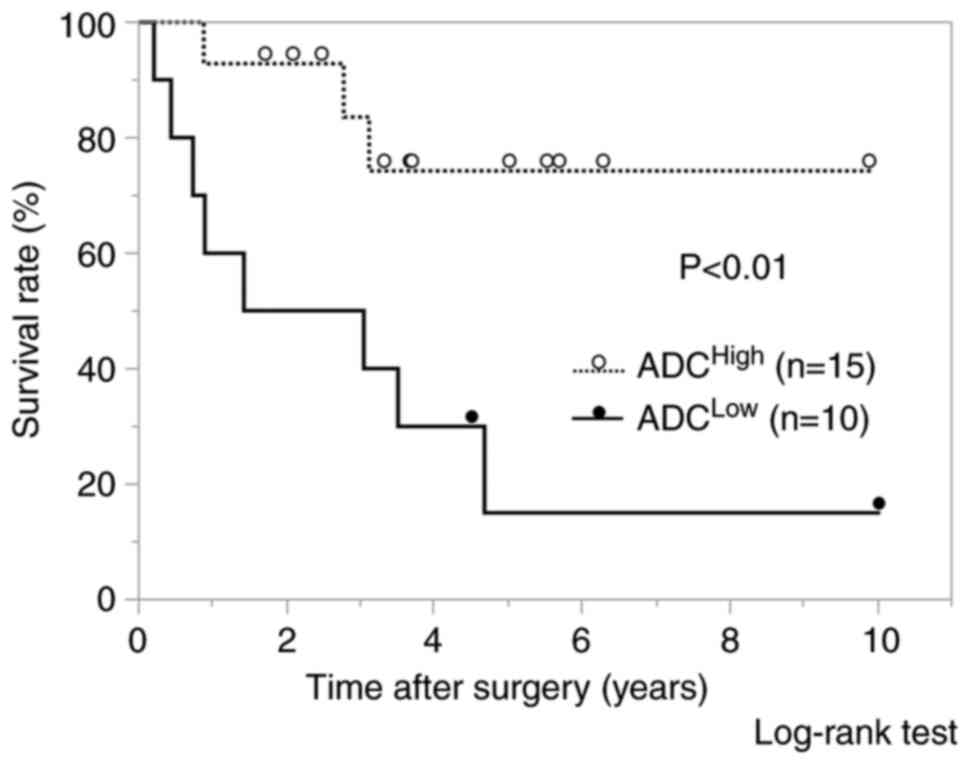
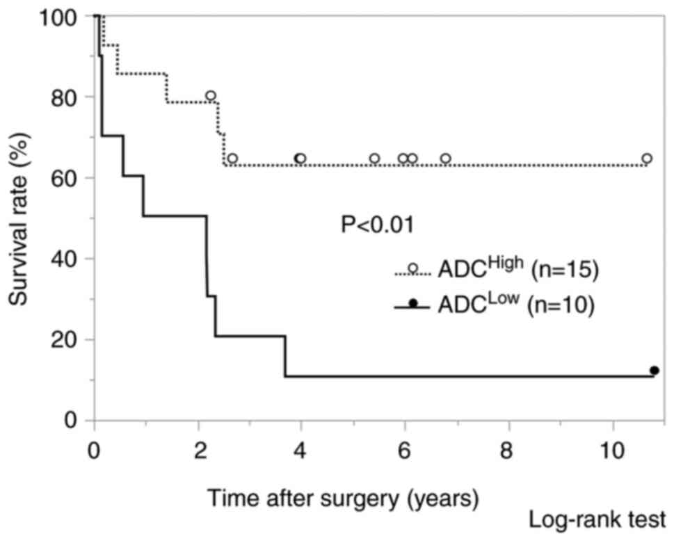
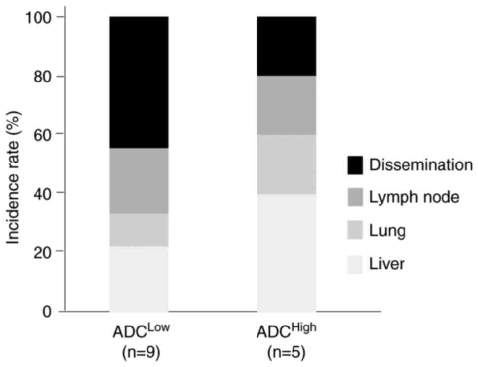
|
1
|
Hickman L and Contreras C: Gallbladder
cancer: Diagnosis, surgical management, and adjuvant therapies.
Surg Clin North Am. 99:337–355. 2019. View Article : Google Scholar : PubMed/NCBI
|
|
2
|
Cubertafond P, Gainant A and Cucchiaro G:
Surgical treatment of 724 carcinomas of the gallbladder. Results of
the French Surgical Association Survey. Ann Surg. 219:275–280.
1994. View Article : Google Scholar : PubMed/NCBI
|
|
3
|
Lim H, Seo DW, Park DH, Lee SS, Lee SK,
Kim MH and Hwang S: Prognostic factors in patients with gallbladder
cancer after surgical resection: Analysis of 279 operated patients.
J Clin Gastroenterol. 47:443–448. 2013. View Article : Google Scholar : PubMed/NCBI
|
|
4
|
Alizadeh AA, Aranda V, Bardelli A,
Blanpain C, Bock C, Borowski C, Caldas C, Califano A, Doherty M,
Elsner M, et al: Toward understanding and exploiting tumor
heterogeneity. Nat Med. 21:846–853. 2015. View Article : Google Scholar : PubMed/NCBI
|
|
5
|
Scara S, Bottoni P and Scatena R: CA 19-9:
Biochemical and clinical aspects. Adv Exp Med Biol. 867:247–260.
2015. View Article : Google Scholar : PubMed/NCBI
|
|
6
|
Ratanaprasatporn L, Uyeda JW, Wortman JR,
Richardson I and Sodickson AD: Multimodality imaging, including
dual-energy CT, in the evaluation of gallbladder disease.
Radiographics. 38:75–89. 2018. View Article : Google Scholar : PubMed/NCBI
|
|
7
|
Kitazume Y, Taura S, Nakaminato S, Noguchi
O, Masaki Y, Kasahara I, Kishino M and Tateishi U:
Diffusion-weighted magnetic resonance imaging to differentiate
malignant from benign gallbladder disorders. Eur J Radiol.
85:864–873. 2016. View Article : Google Scholar : PubMed/NCBI
|
|
8
|
Parikh T, Drew SJ, Lee VS, Wong S, Hecht
EM, Babb JS and Taouli B: Focal liver lesion detection and
characterization with diffusion-weighted MR imaging: Comparison
with standard breath-hold T2-weighted imaging. Radiology.
246:812–822. 2008. View Article : Google Scholar : PubMed/NCBI
|
|
9
|
Jiang T, Xu JH, Zou Y, Chen R, Peng LR,
Zhou ZD and Yang M: Diffusion-weighted imaging (DWI) of
hepatocellular carcinomas: A retrospective analysis of the
correlation between qualitative and quantitative DWI and tumour
grade. Clin Radiol. 72:465–472. 2017. View Article : Google Scholar : PubMed/NCBI
|
|
10
|
Kurosawa J, Tawada K, Mikata R, Ishihara
T, Tsuyuguchi T, Saito M, Shimofusa R, Yoshitomi H, Ohtsuka M,
Miyazaki M and Yokosuka O: Prognostic relevance of apparent
diffusion coefficient obtained by Diffusion-Weighted MRI in
pancreatic cancer. J Magn Reson Imaging. 42:1532–1537. 2015.
View Article : Google Scholar : PubMed/NCBI
|
|
11
|
Parsian S, Giannakopoulos NV, Rahbar H,
Rendi MH, Chai X and Partridge SC: Diffusion-weighted imaging
reflects variable cellularity and stromal density present in breast
fibroadenomas. Clin Imaging. 40:1047–1054. 2016. View Article : Google Scholar : PubMed/NCBI
|
|
12
|
Yamada S..Morine Y, Imura S, Ikemoto T,
Arakawa Y, Saito Y, Yoshikawa M, Miyazaki K and Shimada M:
Prognostic prediction of apparent diffusion coefficient obtained by
diffusion-weighted MRI in mass-forming intrahepatic
cholangiocarcinoma. J Hepatobiliary Pancreat Sci. 27:388–395. 2020.
View Article : Google Scholar : PubMed/NCBI
|
|
13
|
Lee NK, Kim S, Moon JI, Shin N, Kim DU,
Seo HI, Kim HS, Han GJ, Kim JY and Lee JW: Diffusion-weighted
magnetic resonance imaging of gallbladder adenocarcinoma: Analysis
with emphasis on histologic grade. Clin Imaging. 40:345–351. 2016.
View Article : Google Scholar : PubMed/NCBI
|
|
14
|
Japanese Society of Biliary Surgery, .
Classification of biliary tract carcinoma, second English edition.
Tokyo: Kanehara & Co., Ltd.; 2004
|
|
15
|
Min JH, Kang TW, Cha DI, Kim SH, Shin KS,
Lee JE, Jang KT and Ahn SH: Apparent diffusion coefficient as a
potential marker for tumour differentiation, staging and long-term
clinical outcomes in gallbladder cancer. Eur Radiol. 29:411–421.
2019. View Article : Google Scholar : PubMed/NCBI
|
|
16
|
Yun EJ, Cho SG, Park SW, Kim WH, Kim HJ
and Suh CH: Gallbladder carcinoma and chronic cholecystitis:
Differentiation with two-phase spiral CT. Abdom Imaging.
29:102–108. 2004. View Article : Google Scholar : PubMed/NCBI
|
|
17
|
Yoshimitsu K, Honda H, Kaneko K, Kuroiwa
T, Irie H, Ueki T, Chijiwa K, Takenaka K and Masuda K: Dynamic MRI
of the gallbladder lesions: Differentiation of benign from
malignant. J Magn Reson Imaging. 7:696–701. 1997. View Article : Google Scholar : PubMed/NCBI
|
|
18
|
Demachi H, Matsui O, Hoshiba K, Kimura M,
Miyata S, Kuroda Y, Konishi K, Tsuji M and Miwa A: Dynamic MRI
using a surface coil in chronic cholecystitis and gallbladder
carcinoma: Radiologic and histopathologic correlation. J Comput
Assist Tomogr. 21:643–651. 1997. View Article : Google Scholar : PubMed/NCBI
|
|
19
|
Levy AD, Murakata LA and Rohrmann CA Jr:
Gallbladder carcinoma: Radiologicepathologic correlation.
RadioGraphics. 21:295–314, questionnaire, 549–555. 2001. View Article : Google Scholar : PubMed/NCBI
|
|
20
|
Levy AD, Murakata LA, Abbott RM and
Rohrmann CA Jr: From the archives of the AFIP. Benign tumors and
tumorlike lesions of the gallbladder and extrahepatic bile ducts:
Radiologic-pathologic correlation. Armed Forces Institute of
Pathology. Radiographics. 22:387–413. 2002. View Article : Google Scholar : PubMed/NCBI
|
|
21
|
Piccolo G and Piozzi GN: Laparoscopic
radical cholecystectomy for primary or incidental early gallbladder
cancer: The new rules governing the treatment of gallbladder
cancer. Gastroenterol Res Pract. 2017:85705022017. View Article : Google Scholar : PubMed/NCBI
|
|
22
|
Sugita R, Yamazaki T, Furuta A, Itoh K,
Fujita N and Takahashi S: High b-value diffusion-weighted MRI for
detecting gallbladder carcinoma: Preliminary study and results. Eur
Radiol. 19:17942009. View Article : Google Scholar : PubMed/NCBI
|
|
23
|
Irie H, Kamochi N, Nojiri J, Egashira Y,
Sasaguri K and Kudo S: High b-value diffusion-weighted MRI in
differentiation between benign and malignant polypoid gallbladder
lesions. Acta Radiol. 52:236–240. 2011. View Article : Google Scholar : PubMed/NCBI
|
|
24
|
Ogawa T, Horaguchi J, Fujita N, Noda Y,
Kobayashi G, Ito K, Koshita S, Kanno Y, Masu K and Sugita R: High
b-value diffusion-weighted magnetic resonance imaging for
gallbladder lesions: Differentiation between benignity and
malignancy. J Gastroenterol. 47:1352–1360. 2012. View Article : Google Scholar : PubMed/NCBI
|
|
25
|
Solak A, Solak I, Genc¸ B and Sahin N: The
role of diffusion-weighted examination in non-polyploid gallbladder
malignancies: A preliminary study. Turk J Gastroenterol.
24:148–153. 2013. View Article : Google Scholar : PubMed/NCBI
|
|
26
|
Kim SJ, Lee JM, Kim H, Yoon JH, Han JK and
Choi BI: Role of diffusion-weighted magnetic resonance imaging in
the diagnosis of gallbladder cancer. J Magn Reson Imaging.
38:127–137. 2013. View Article : Google Scholar : PubMed/NCBI
|
|
27
|
Lee NK, Kim S, Kim TU, Kim DU, Seo HI and
Jeon TY: Diffusion-weighted MRI for differentiation of benign from
malignant lesions in the gallbladder. Clin Radiol. 69:e78–e85.
2014. View Article : Google Scholar : PubMed/NCBI
|
|
28
|
Kyung N, Kim S, Moon JI, Shin N, Kim DU,
Seo HI, Kim HS, Han GJ, Kim JY and Lee JW: Diffusion-weighted
magnetic resonance imaging of gallbladder adenocarcinoma: Analysis
with emphasis on histologic grade. Clin Imaging. 40:345–351. 2016.
View Article : Google Scholar : PubMed/NCBI
|
|
29
|
Sulieman I, Mohamed S, Elmoghazy W,
Alaboudy A, Khalaf H and Elaffandi A: The value of
diffusion-weighted imaging in diagnosing gallbladder malignancy:
Performance of a new parameter. Clin Radiol. 76:709.e7–709.e12.
2021. View Article : Google Scholar : PubMed/NCBI
|


