Serous microcystic adenoma of the pancreatic head: Report of two cases and review of the literature
- Authors:
- Jiarui Pu
- Qiangsong Tong
- Chengkai Zhou
- Xiuping Yang
- Liduan Zheng
View Affiliations
Affiliations: Department of Surgery, Union Hospital of Tongji Medical College, Huazhong University of Science and Technology, Wuhan, 430022 Hubei, P.R. China
- Published online on: July 1, 2010 https://doi.org/10.3892/ol_00000126
-
Pages:
723-727
Metrics: Total
Views: 0 (Spandidos Publications: | PMC Statistics: )
Metrics: Total PDF Downloads: 0 (Spandidos Publications: | PMC Statistics: )
This article is mentioned in:
Abstract
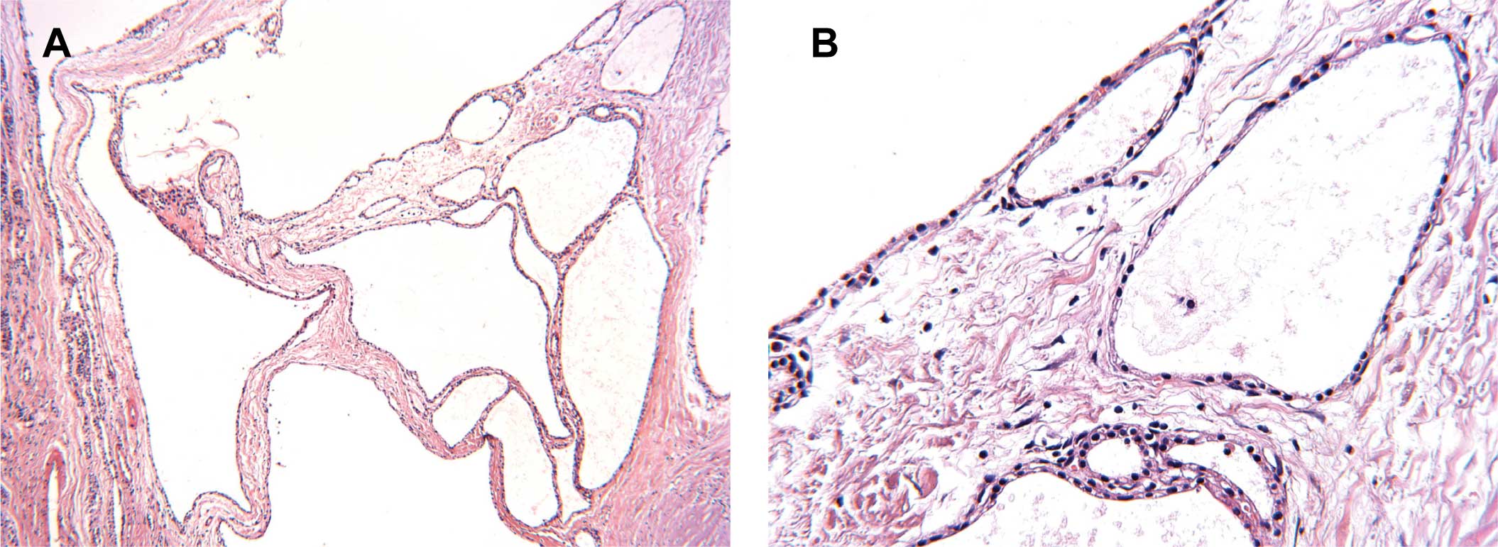
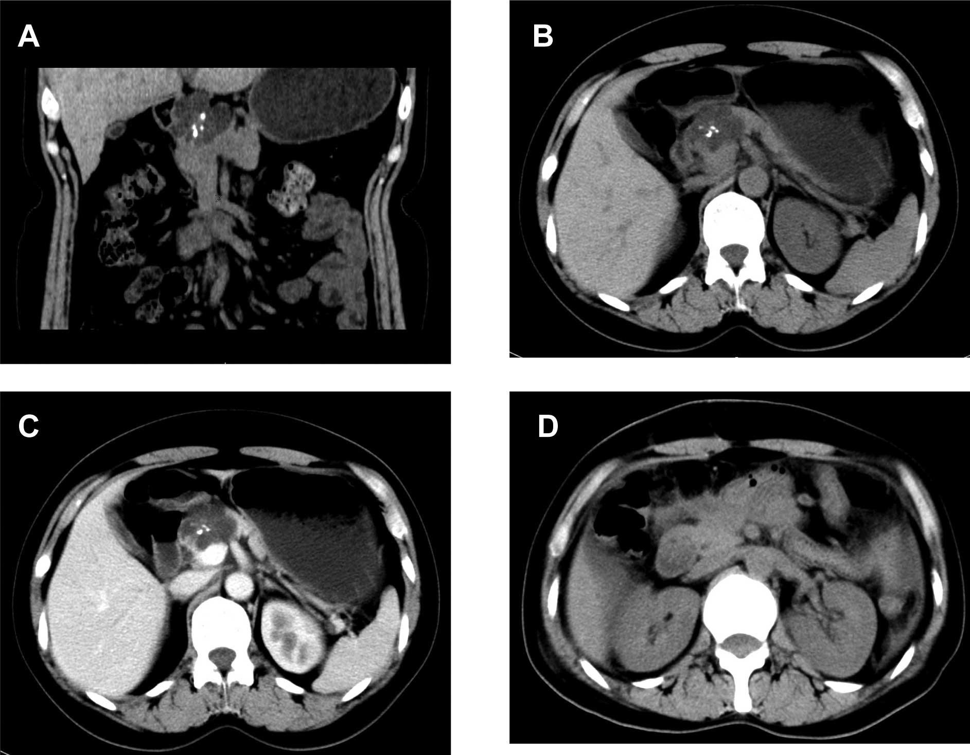
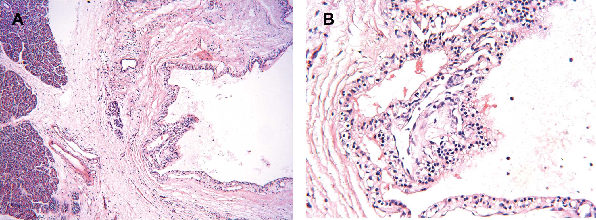
Serous microcystic adenomas (SMAs), also known as glycogen-rich cystadenomas, are uncommon exocrine tumors mainly involved in the body and tail of the pancreas. In this study, we report two SMA cases of the pancreatic head. In case 1, a 47-year-old woman was referred to our hospital for abdominal bloating and back pain; in case 2, a pancreatic space-occupying lesion was incidentally discovered in an asymptomatic 71-year-old female during an ultrasound. In both cases, a computer tomography scan showed well-demarcated and multilocular cysts in the head of the pancreas, and a central scar and calcifications were noted in case 1. During the exploratory laparotomies, the multiple cystic masses were found in the pancreatic head. A segmental resection of the pancreas and pancreatic jejunal anastomosis were performed. Histologically, the tumors were composed of tiny cysts that were filled with clear fluid and lined by a single layer of cuboidal cells with round, centrally located nuclei and clear cytoplasm, without cellular atypia. Immunohistochemical studies showed that the neoplastic cells were positive for cytokeratin 7 and vimentin but negative for synaptophysin and cytokeratin 20. A postoperative follow-up indicated no recurrence in the patients. We reviewed the literature regarding the etiology, clinical presentation, imaging characteristics, features and management of this lesion.
View References
1
|
Compagno J and Oertel JE: Mucinous cystic
neoplasms of the pancreas with overt and latent malignancy
(cystadenocarcinoma and cystadenoma). A clinicopathologic study of
41 cases. Am J Clin Pathol. 69:573–580. 1978.PubMed/NCBI
|
2
|
Morohoshi T, Held G and Kloppel G:
Exocrine pancreatic tumours and their histological classification.
A study based on 167 autopsy and 97 surgical cases. Histopathology.
7:645–661. 1983. View Article : Google Scholar : PubMed/NCBI
|
3
|
Subramanian S, Marappagounder S, Selvaraj
DR and Elangovan B: A rare association of serous cystadenoma of the
pancreas with mediastinal lipoma: a case report. Cases J.
2:71652009. View Article : Google Scholar : PubMed/NCBI
|
4
|
Sand J and Nordback I: The differentiation
between pancreatic neoplastic cysts and pancreatic pseudocyst.
Scand J Surg. 94:161–164. 2005.PubMed/NCBI
|
5
|
Tanaka M, Chari S, Adsay V, et al:
International consensus guidelines for management of intraductal
papillary mucinous neoplasms and mucinous cystic neoplasms of the
pancreas. Pancreatology. 6:17–32. 2006. View Article : Google Scholar : PubMed/NCBI
|
6
|
Goldsmith JD: Cystic neoplasms of the
pancreas. Am J Clin Pathol. 119(Suppl): 3–16. 2003.
|
7
|
Ng DZ, Goh BK, Tham EH, Young SM and Ooi
LL: Cystic neoplasms of the pancreas: current diagnostic modalities
and management. Ann Acad Med Singapore. 38:251–259. 2009.PubMed/NCBI
|
8
|
Kosmahl M, Wagner J, Peters K, Sipos B and
Kloppel G: Serous cystic neoplasms of the pancreas: an
immunohistochemical analysis revealing alpha-inhibin,
neuron-specific enolase, and muc6 as new markers. Am J Surg Pathol.
28:339–346. 2004. View Article : Google Scholar
|
9
|
Warshaw AL, Compton CC, Lewandrowski K,
Cardenosa G and Mueller PR: Cystic tumors of the pancreas. New
clinical, radiologic, and pathologic observations in 67 patients.
Ann Surg. 212:432–443; discussion 444–445. 1990.PubMed/NCBI
|
10
|
King JC, Ng TT, White SC, Cortina G, Reber
HA and Hines OJ: Pancreatic serous cystadenocarcinoma: a case
report and review of the literature. J Gastrointest Surg.
13:1864–1868. 2009. View Article : Google Scholar : PubMed/NCBI
|
11
|
Tseng JF, Warshaw AL, Sahani DV, Lauwers
GY, Rattner DW and Fernandez-del Castillo C: Serous cystadenoma of
the pancreas: tumor growth rates and recommendations for treatment.
Ann Surg. 242:413–419; discussion 419–421. 2005.PubMed/NCBI
|
12
|
Pyke CM, van Heerden JA, Colby TV, Sarr MG
and Weaver AL: The spectrum of serous cystadenoma of the pancreas.
Clinical, pathologic and surgical aspects. Ann Surg. 215:132–139.
1992. View Article : Google Scholar : PubMed/NCBI
|
13
|
Garcea G, Ong SL, Rajesh A, et al: Cystic
lesions of the pancreas. A diagnostic and management dilemma.
Pancreatology. 8:236–251. 2008. View Article : Google Scholar : PubMed/NCBI
|
14
|
Vernadakis S, Kaiser GM, Christodoulou E,
et al: Enormous serous microcystic adenoma of the pancreas. JOP.
10:332–334. 2009.PubMed/NCBI
|
15
|
Sahani DV, Saokar A, Hahn PF, Brugge WR
and Fernandez-Del Castillo C: Pancreatic cysts 3 cm or smaller: how
aggressive should treatment be? Radiology. 238:912–919. 2006.
View Article : Google Scholar : PubMed/NCBI
|