Usefulness of 18F-FDG positron emission tomography/computed tomography for the diagnosis of pyothorax-associated lymphoma: A report of three cases

  • Authors:
    • Yoshiyuki Abe
    • Katsumi Tamura
    • Ikuko Sakata
    • Jiro Ishida
    • Isao Fukuba
    • Rokuro Matsuoka
    • Seiichiro Shimizu
    • Hiroshi Murakami
    • Kikuo Machida
  • View Affiliations

  • Published online on: September 1, 2010     https://doi.org/10.3892/ol_00000146
  • Pages: 833-836
Metrics: Total Views: 0 (Spandidos Publications: | PMC Statistics: )
Total PDF Downloads: 0 (Spandidos Publications: | PMC Statistics: )


Abstract

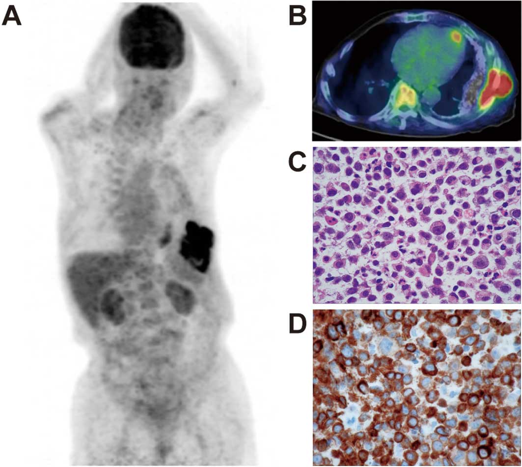
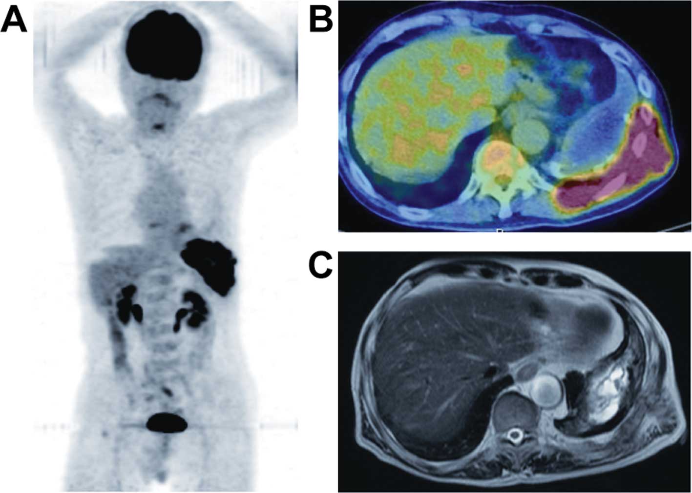
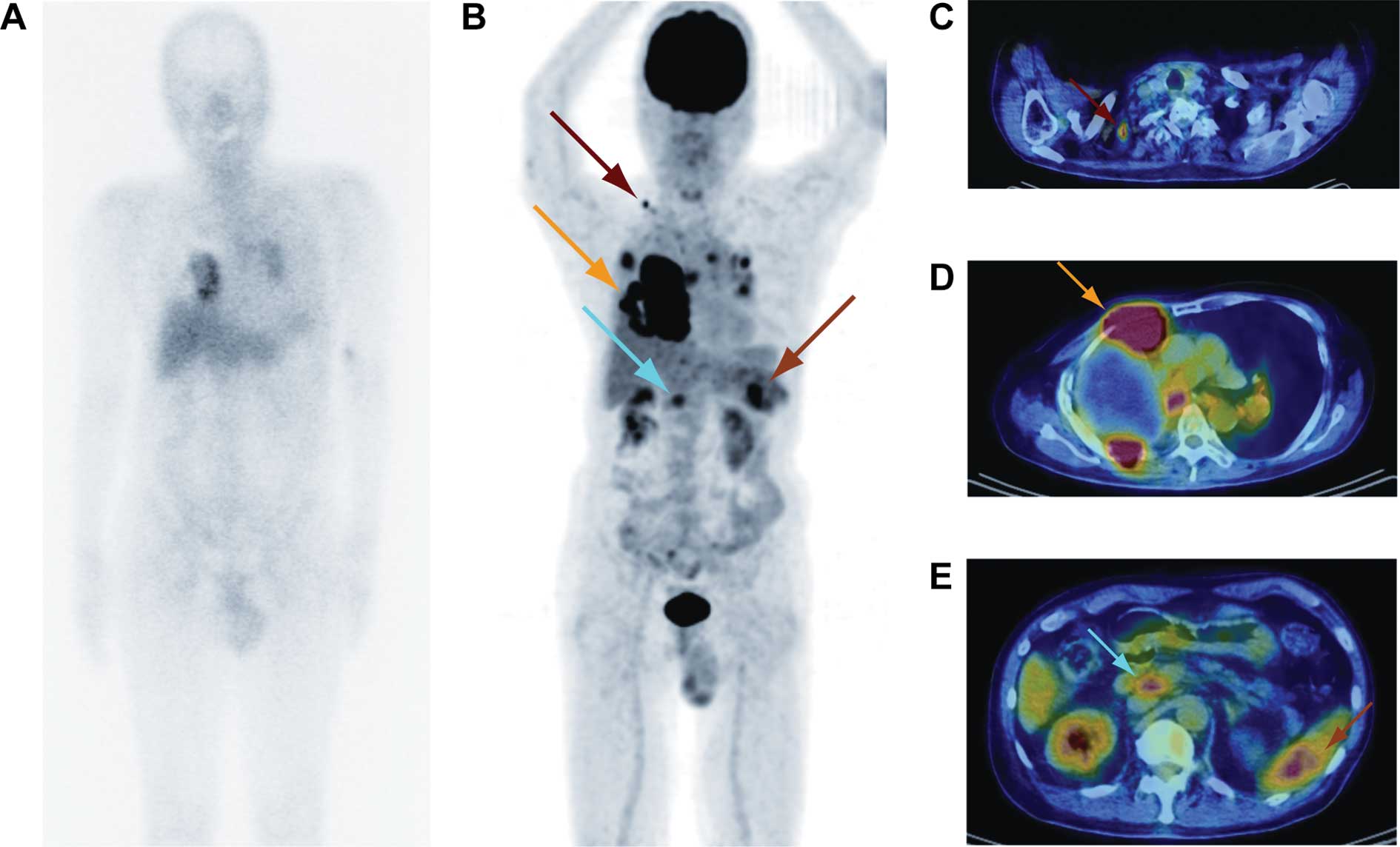
Pyothorax-associated lymphoma (PAL) is a unique and rare non-Hodgkin's lymphoma developing in the pleural cavity following a long-standing history of chronic pyothorax (CP). The development of F-18 2'-deoxy-2fluoro-D-glucose (FDG) positron emission tomography combined with computed tomography (PET/CT) has contributed to the evaluation of lymphoma staging. However, only a few studies describing FDG-PET/CT findings in PAL have been published. This study reported three cases of PAL; all 3 patients had previously undergone artificial collapse therapy for pulmonary tuberculosis. Both the first case (an 84-year-old male) and second case (an 83-year-old male) complained of abdominal pain. An ultrasound scan revealed a mass shadow in the left chest wall without abnormal findings in the abdomen, and the CT and magnetic resonance imaging scans suggested malignant lymphoma of the left chest. FDG-PET/CT imaging showed extremely intense FDG uptake only in the left pleura and chest wall. Diagnosis was CP in the two patients, showing a high maximum standardized uptake value (SUVmax: early, 14.8 and delayed, 19.4 in the first case; early, 20.8 and delayed, 27.3 in the second case, respectively). Histopathological analysis of the specimens obtained by biopsy of the PET/CT-positive pleural mass showed non-Hodgkin's, diffuse large B cell lymphoma in the two cases. The third case was a 79-year-old male with relapse after right pleuropneumonectomy for PAL (diffuse large B cell lymphoma) 4 years earlier. PET/CT showed intense FDG uptake (SUVmax: early, 19.9 and delayed, 35.7) in the right pleura and chest wall. Diagnosis was CP, suggesting the recurrence of PAL. Furthermore, abnormal intense FDG uptake was noted in the hilar, mediastinal and supraclavicular lymph nodes, as well as in the spleen. In conclusion, FDG-PET/CT imaging is useful in the evaluation of the area of invasion in PAL.
View Figures
View References

Related Articles

Journal Cover

September-October 2010
Volume 1 Issue 5

Print ISSN: 1792-1074
Online ISSN:1792-1082

Sign up for eToc alerts

Recommend to Library

Copy and paste a formatted citation
x
Spandidos Publications style
Abe Y, Tamura K, Sakata I, Ishida J, Fukuba I, Matsuoka R, Shimizu S, Murakami H and Machida K: Usefulness of 18F-FDG positron emission tomography/computed tomography for the diagnosis of pyothorax-associated lymphoma: A report of three cases . Oncol Lett 1: 833-836, 2010.
APA
Abe, Y., Tamura, K., Sakata, I., Ishida, J., Fukuba, I., Matsuoka, R. ... Machida, K. (2010). Usefulness of 18F-FDG positron emission tomography/computed tomography for the diagnosis of pyothorax-associated lymphoma: A report of three cases . Oncology Letters, 1, 833-836. https://doi.org/10.3892/ol_00000146
MLA
Abe, Y., Tamura, K., Sakata, I., Ishida, J., Fukuba, I., Matsuoka, R., Shimizu, S., Murakami, H., Machida, K."Usefulness of 18F-FDG positron emission tomography/computed tomography for the diagnosis of pyothorax-associated lymphoma: A report of three cases ". Oncology Letters 1.5 (2010): 833-836.
Chicago
Abe, Y., Tamura, K., Sakata, I., Ishida, J., Fukuba, I., Matsuoka, R., Shimizu, S., Murakami, H., Machida, K."Usefulness of 18F-FDG positron emission tomography/computed tomography for the diagnosis of pyothorax-associated lymphoma: A report of three cases ". Oncology Letters 1, no. 5 (2010): 833-836. https://doi.org/10.3892/ol_00000146