Introduction
The incidence and mortality of gastric cancer have
declined steadily over the past several decades. Nonetheless,
gastric cancer remains a major public health issue being the fourth
most common cancer and the second leading cause of cancer death
worldwide (1). Gastric neoplasia is
largely composed of adenocarcinomas, accounting for over 95% of
cases (2). Pathologically, gastric
adenorcarcinomas are most widely classified using Lauren and the
World Health Organisation (WHO) classification system. In the
Lauren classification, gastric cancers are stratified into
intestinal, diffuse, and mixed types, whereas the WHO
classification categorizes cancers according to features of
histopathological differentiation, namely papillary, tubular,
mucinous, and signet ring cell types (3). Genetically, gastric cancer is
generally considered to result from accumulation of genetic
alterations involving a variety of oncogenes and tumor suppressor
genes, however, concrete genetic sequences in the development of
gastric cancer still remain to be clarified (3,4).
Raf kinase inhibitor protein (RKIP; also known as
PEBP, for phosphatidylethanolamine-binding protein) is a widely
expressed and highly conserved protein (5–7), which
was firstly identified as a MAP kinase pathway inhibitor by
modulating the function of Raf-1 (8,9).
Currently, is known that RKIP also suppresses the activation of the
nuclear factor κB (NF-κB) (10) and
the regulator of G-protein coupled receptors (GRK-2) (11), and may be involved in regulation of
the cell cycle (12). Thus, RKIP
mediates important cellular mechanisms, including cell
differentiation, cell cycle, apoptosis and cell migration, and is
deregulated in several human disorders (13).
In cancer, RKIP is considered to be a signal
transduction modulator and a metastasis suppressor (14), and downregulated in several human
tumors, mainly in highly metastatic carcinomas (15–27).
Noteworthy, RKIP has been shown to be a prognostic marker in
prostate cancer, colorectal carcinomas, gliomas and GISTs (25,27–30).
Concerning RKIP expression in gastric carcinomas,
there are three, yet contradictory studies (31–33).
Thus, in the present work, using a large and clinically
well-characterized series of gastric carcinomas, we aimed first to
evaluate the frequency of RKIP expression not only in tumor but
also in normal and metastatic gastric tissues. Secondly, we aimed
to determine whether RKIP expression could be used to predict
clinical outcome of patients with gastric carcinomas.
Materials and methods
Patients and tissue samples
For the present study, the cases were
retrospectively selected from the University of São Paulo Gastric
Cancer Database: 152 patients with gastric adenocarcinoma operated
at the Stomach and Small Bowel Unit Hospital das Clinicas,
University of São Paulo, School of Medicine, São Paulo, Brazil,
between February 1993 and December 2002. In total 416 different
gastric tissues were analysed, including 163 normal mucosa, 59
metaplastic mucosa, 152 primary tumors and 42 lymph node metastasis
samples. The samples were retrieved from the files of the
Department of Pathology, and organized in tissue microarrays
(TMAs). To achieve representative sampling and minimize sample
loss, each case was included in duplicate in the TMAs.
Additionally, formalin-fixed and paraffin-embedded prostate
carcinoma and GISTs samples were used as positive and negative
controls for immunohistochemistry, as previously described
(27).
Relevant patient clinical data available included
patients age, gender, tumor size and location, WHO classification,
Lauren’s classification, TNM staging, depth of invasion, lymph node
metastases and lymphatic, vascular and neural invasions,
desmoplasia, inflammatory infiltrated and follow-up, as previously
described (34). The inclusion
criteria were: patients with primary gastric adenocarcinoma
submitted to subtotal or total gastrectomy with D2 lymphadenectomy,
submucosa layer invasion or deeper (T1b or higher), at least 25
lymph nodes retrieved per case, absence of distant metastasis (M0),
and available follow-up data. All patients were treated according
to a well-established surgical protocol following the Japanese
Gastric Cancer Association rules.
The histological sections, obtained from original
paraffin blocks and, after hematoxylin and eosin staining, were
submitted to histological review by senior pathologist. Primary
tumors were histologically classified according to World Health
Organization (35) in tubular,
intestinal, signet-cell and mucinous carcinomas. Lauren and Ming
(36,37) classifications were also applied to
primary tumors, stratifying lesions into diffuse or intestinal or
infiltrating or expansive types, respectively, the latter derived
from evaluation of the deepest tumor edge. Lymphatic, vascular or
perineural invasion was assessed as non-detected or present.
Pathological nodal status (pN) was determined histologically by
counting the affected lymph nodes and classified as pN0, pN1, pN2
or pN3 according to AJCC TNM Staging system (38). Information on the tumor size as well
its main location, classified as proximal or distal tumors, were
obtained from original surgical pathology reports.
Peri/intratumoral inflammatory infiltrate was semi-quantified as
absent, mild/moderate and intense whereas desmoplasmatic stromal
response as absent to mild or moderate to intense. Final
pathological TNM stage was also assessed.
Immunohistochemistry analysis for
RKIP
Tissue microarray (TMA) slides with 3 μm-thick
sections were subjected to immunohistochemical analysis according
to the streptavidin-biotin peroxidase complex system (UltraVision
Large Volume Detection System Anti-Polyvalent, HRP; LabVision
Corporation, CA, USA), as previously described (27,30,39).
Briefly, deparaffinised and rehydrated slides were submitted to
heat-induced antigen retrieval for 20 min at 98°C with 10 mM
citrate buffer (pH 6.0). After incubation with the primary antibody
raised against RKIP (dilution 1:800; incubation 2 h at RT; Upstate
Biotechnology, Lake Placid, NY, USA), the secondary biotinylated
goat anti-polyvalent antibody was applied for 10 min followed by
incubation with the streptavidin-peroxidase complex. The immune
reaction was visualized by 3,3′-diamonobenzidine (DAB) as a
chromogen. All sections were counterstained with Gill-2
haematoxylin. For negative controls, primary antibodies were
omitted and also replaced by a universal negative control antibody
(CEA, rabbit anti-human, Dako Corporation, Carpinteria, CA, USA).
Prostate carcinoma and GISTs cases, previously analysed (27,30,39),
were used as positive and negative controls. Sections were
independently scored by two of the authors (K.S. and A.L.F.),
following a semi-quantitative criterion: (−), 0% of immunoreactive
cells; (+), <5% of immunoreactive cells; (++), 5–50% of
immunoreactive cells; and (+++), >50% of immunoreactive cells.
Samples with scores (−) and (+) were considered negative, and those
with scores (++) and (+++) were considered positive.
Statistical analysis
Either the χ2 test or Fisher’s exact test
(when required) was done to determine the correlation between RKIP
expression status and clinicopathologic features. Survival curves
were estimated using the Kaplan-Meier product-limit method, and the
significance of the differences between survival curves was
determined using a log-rank test. Multivariate survival analyses
were done using the Cox proportional hazards model. Results were
considered to be statistically significant for p<0.05. All
statistical analyses were conducted using SPSS 16.0 statistical
software program.
Results
Patient data
We have available tissue in 152 patients, 114 with
advanced and 38 early gastric cancer. The mean age was 60.89
(range: 26–87 years). The majority of patients had tumors located
at the distal part of the stomach. The mean tumor size was 3.92
(range: 0.8–20 cm). There was a predominance of the Lauren
intestinal type. All patients were submitted to D2 lymphadenectomy
with a mean number of retrieved lymph nodes of 43.7 (range:
25–113). The mean follow-up was 62.3 months (range: 6–76 months),
and 13.4% was lost to follow-up.
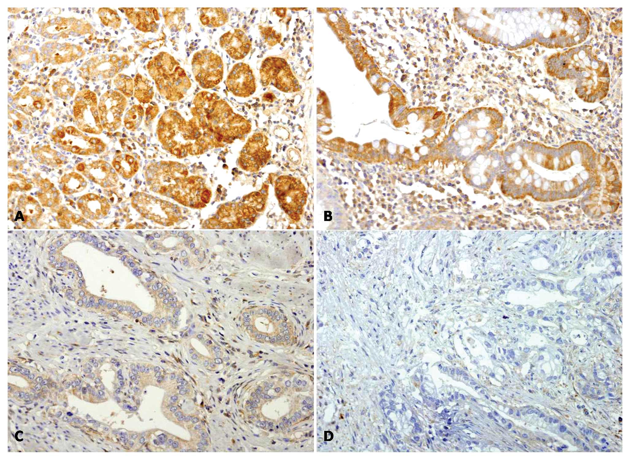
RKIP expression in gastric tissues
Four hundred and sixteen gastric tissue samples were
studied for RKIP expression by immunohistochemistry. RKIP staining
was always present in the cytoplasm of the cells and, according to
the immunohistochemistry score, we found RKIP-positive expression
in 84.7% (138/163) of normal gastric tissue, in 79.7% (47/59) of
gastric mucosa metaplasia, in 44.1% (67/152) of gastric primary
tumors, and in 9.5% (4/42) of gastric lymph node metastasis
(Table I and Fig. 1). There are no statistically
significant differences between RKIP expression in normal and
metaplasias (p=0.377). However, a statistically significant
(p<0.001) decrease of RKIP expression was found in primary
tumors, compared to normal and metaplasia tissues, and in
lymph-node metastasis, when compared to normal, metaplasia and
primary tumors (Table I).
Additionally, in 35 patients we had normal, tumor and metastatic
tissues available. We found that RKIP was positive in
non-neoplastic tissues in ~83% of the patients, but was absent ~69%
of the primary tumors and completely absent in metastatic tissues
(data not shown).
 | Table IFrequency of RKIP expression in
gastric tissues. |
Table I
Frequency of RKIP expression in
gastric tissues.
| | RKIP
Expression | | |
|---|
| |
| | |
|---|
| Gastric
tissues | N | Positive (%) | Negative (%) | p (vs. Normal) | p (vs. Primary
tumor) |
|---|
| Normal | 163 | 138 (84.7) | 25 (15.3) | - | <0.001 |
| Metaplasia | 59 | 47 (79.7) | 12 (20.3) | 0.377 | <0.001 |
| Primary tumor | 152 | 67 (44.1) | 85 (55.9) | <0.001 | - |
| Lymph node
metastasis | 42 | 4 (9.5) | 38 (90.5) | <0.001 | <0.001 |
RKIP expression and correlation with
clinical data
The correlations between RKIP expression and
clinicopathologic features are summarized in Table II. We found that RKIP is
differently expressed between the different WHO histological types
(p=0.03), being highly expressed in intestinal type, and lost in
tubular, mucinous and signet-ring cell carcinomas. At variance, no
statistically differences were observed among the Lauren subtypes.
RKIP is significantly lost in advanced gastric cancer when compared
with early tumors (p<0.001). Additionally, absence of RKIP
expression was statistically associated with tumors with higher
tumor size, with higher TNM stage, with the presence of vascular,
lymphatic and neuronal invasion and with the presence of lymph node
metastasis (Table II).
| Table IIAssociations between RKIP expression
and clinicopathologic features in gastric cancer patients. |
Table II
Associations between RKIP expression
and clinicopathologic features in gastric cancer patients.
| | RKIP
expression | |
|---|
| |
| |
|---|
| Parameter | N | Negative (%) | Positive (%) | p |
|---|
| Gender |
| Female | 48 | 27(56.2) | 21 (43.8) | 0.917 |
| Male | 103 | 57 (55.3) | 46 (44.7) | |
| Depth of
invasion |
| Early | 38 | 6 (15.8) | 32 (84.2) | <0.001 |
| Advanced | 114 | 79 (69.3) | 35 (30.7) | |
| Age (years) |
| <61 | 68 | 33 (48.5) | 35 (61.4) | 0.112 |
| ≥61 | 83 | 51 (51.5) | 32 (38.6) | |
| Tumor size
(cm) |
| <4 | 52 | 19 (36.5) | 33 (63.5) | <0.001 |
| ≥4 | 98 | 65 (66.3) | 33 (33.7) | |
| Tumor location |
| Proximal | 16 | 9 (56.2) | 7 (43.8) | 0.974 |
| Distal | 129 | 72 (55.8) | 57 (44.2) | |
| WHO
classification |
| Intestinal
adenocarcinoma | 14 | 1 (7.1) | 13 (92.9) | 0.030 |
| Tubular
adenocarcinoma | 66 | 46 (69.7) | 20 (30.3) | |
| Mucinous
adenocarcinoma | 14 | 9 (64.3) | 5 (35.7) | |
| Signet-ring cell
carcinoma | 36 | 23 (63.9) | 13 (36.1) | |
| pT |
| Muscular
propria/subserosa | 111 | 76 (68.5) | 35 (31.5) | <0.001 |
| Submucosa | 38 | 6 (15.8) | 32 (84.2) | |
| Lauren |
| Intestinal | 99 | 55 (55.6) | 44 (44.4) | 0.855 |
| Diffuse | 49 | 28 (57.1) | 21 (42.9) | |
| Lymphatic
Invasion |
| Absent | 84 | 39(46.4) | 45 (53.6) | 0.009 |
| Present | 68 | 46 (67.6) | 22 (32.4) | |
| Vascular
Invasion |
| Absent | 131 | 69 (52.7) | 62 (47.3) | 0.044 |
| Present | 21 | 16 (76.2) | 5 (23.8) | |
| Perineural
invasion |
| Absent | 86 | 40 (46.5) | 46 (53.5) | 0.008 |
| Present | 66 | 45 (67.2) | 21 (31.8) | |
| Inflammatory
infiltrated |
| Absent/mild | 118 | 62 (52.5) | 56 (47.5) | 0.233 |
|
Moderated/accentuated | 31 | 20 (64.5) | 11 (35.5) | |
| Desmoplasia |
|
Absent/discrete | 84 | 42(50.0) | 42 (50.0) | 0.081 |
|
Moderated/accentuated | 67 | 43 (64.2) | 24 (35.8) | |
| TNM |
| IA | 37 | 6 (16.2) | 31 (83.8) | <0.001 |
| IB | 34 | 24 (70.6) | 10 (29.4) | |
| II | 52 | 34 (65.4) | 18 (34.6) | |
| IIIA+IV | 27 | 20 (74.1) | 7 (25.9) | |
| Lymph node
metastasis |
| pN0 | 72 | 29 (40.3) | 43 (59.7) | 0.004 |
| pN1 | 55 | 37 (67.3) | 18 (32.7) | |
| pN2 | 23 | 17 (73.9) | 6 (26.1) | |
Correlations with patients survival
We found that additionally to the above mentioned
clinical factors, the absence of RKIP expression is also
significantly (p<0.001) associated with poor overall survival in
gastric carcinoma patients (Table
III and Fig. 2). To evaluate
whether RKIP expression is an independent prognostic factor, we
carried out a multivariate Cox regression analysis and found that
absence of RKIP expression is independently associated with
patients poor survival with a 4.53 hazard ratio (Table III). Additionally, male gender,
higher tumor size and presence of lymph node metastasis were also
found to be independent prognostic factors in our series of gastric
carcinomas (Table III).
| Table IIICorrelations between
clinicopathologic features and overall survival in gastric cancer
patients. |
Table III
Correlations between
clinicopathologic features and overall survival in gastric cancer
patients.
| | Univariate
analysis | Multivariate
analysis |
|---|
| |
|
|
|---|
| Parameter | N | (months ± SD) | pa | Hazard ratio (95%
CI) | pb |
|---|
| Gender |
| Female | 40 | 211.6±13.5 | 0.018 | 1.00 | |
| Male | 85 | 145.6±10.6 | | 2.95
(1.25–6.98) | 0.014 |
| Tumor size
(cm) |
| <4 | 74 | 203.1±10.9 | 0.002 | 1.00 | |
| ≥4 | 50 | 100.7±10.1 | | 2.35
(0.88–6.24) | 0.086 |
| WHO
classification |
| Intestinal
adenocarcinoma | 11 | 173.3±17.3 | 0.008 | | |
| Tubular
adenocarcinoma | 59 | 125.9±8.4 | | | |
| Mucinous
adenocarcinoma | 11 | 84.2±15.3 | | | |
| Signet-ring cell
carcinoma | 24 | 55.54±7.3 | | | |
| Lauren |
| Intestinal | 81 | 193.3±11.1 | 0.019 | 1.00 | |
| Diffuse | 39 | 139.5±17.2 | | 1.56
(0.74–3.28) | 0.244 |
| Lymph node
metastasis |
| pN0 | 59 | 231.4±8.3 | <0.001 | 1.00 | |
| pN1 | 47 | 106.2±10.3 | | 4.05
(1.45–11.27) | 0.007 |
| pN2 | 15 | 48.1±10.6 | | 6.63
(2.08–21.14) | 0.001 |
| Lymphatic
invasion |
| Absent | 70 | 206.9±10.9 | 0.002 | | |
| Present | 50 | 103.0±9.9 | | | |
| Vascular
invasion |
| Absent | 105 | 189.1±9.9 | 0.009 | 1.00 | |
| Present | 14 | 85.8±18.9 | | 1.01
(0.49–2.10) | 0.974 |
| Perineural
invasion |
| Absent | 68 | 222.7±9.3 | <0.001 | 1.00 | |
| Present | 52 | 82.8±7.9 | | 1.26
(0.52–3.06) | 0.605 |
| RKIP
expression |
| Positive | 58 | 230.2±8.8 | | 1.00 | |
| Negative | 68 | 84.5±7.1 | <0.001 | 4.53
(1.52–13.53) | 0.007 |
Discussion
Gastric carcinoma is still the fourth most common
cancer and the second leading cause of cancer-related death in the
world (1). Although excellent
long-term survival results for early-detected gastric cancer exist,
prognosis of advanced gastric cancer still remains poor (40). Prognosis of gastric carcinoma
patients depends on several pathological and genetic variables,
such as TNM grading and p53, MUC1, and E-cadherin (41–44).
However, patient outcome is difficult to predict using classic
histological and molecular classifications. Therefore, additional
markers are required to identify patients with risk to metastasize
and with poor prognosis.
Initial in vitro studies, showed that cell
lines derived from metastatic prostate cancer displayed decreased
levels of RKIP as compared with primary tumor cell lines, leading
to the suggestion of RKIP as a metastasis suppressor (18). Subsequent studies showed that
overexpression of RKIP in prostate and melanoma cell lines
suppresses metastasis by decreasing vascular invasion (18,20).
Previous studies have described low levels of RKIP in other
metastatic tumors, such as breast and colorectal carcinoma
(23,45), as well as in many other primary
tumors, including GISTs (27),
insulinoma (22), hepatocarcinoma
(24), ovarian carcinoma (26), merckel cell carcinoma (16) and thyroid carcinoma (15), cutaneous squamous cell carcinoma
(46) and nasopharyngeal carcinoma
(17). Furthermore, loss of
cytoplasmic RKIP has also been associated with poor prognosis in
prostate, colorectal, GISTs and glial tumors (25,27–30).
In gastric tumors, previous studies concerning RKIP
expression are contradictory. Chatterjee and collaborators reported
that in non-neoplastic gastric tissue RKIP cytoplasmic staining was
predominantly negative, and in tumor tissues only 29% (42/143) of
cases stained positive (31). In
contrast, Wang and collaborators described that RKIP is present in
~88% (35/40) of non-neoplastic tissues, in 52% (39/75) of the
primary tumors and only in 19% (5/26) of lymph node metastasis
(32). More recently, RKIP was
shown to be present in 38% (21/55) of gastric cancer tumor tissues
(33). In the present study, we
showed that RKIP is highly expressed in ~83% (185/222) of
non-neoplastic tissues (normal and metaplastic gastric mucosa), but
is significantly lost in 55% (93/168) of primary tumors and almost
absent in gastric lymph node metastasis with only ~10% (4/42) of
the sample staining positive, being in accordance to that described
by Wang et al(32) in a
smaller series. Our results suggested that RKIP could have an
important role in normal gastric mucosa and downregulation to
gastric cancer progression and metastatic mechanisms. To support
this hypothesis, we also found that absence of RKIP protein is
statistically associated with the presence of lymph node
metastasis, which fits well with described for gastric and other
epithelial tumors (18,23,33,45).
Additionally, the absence of RKIP expression is significantly
associated with clinical features that were associated with poor
prognosis in these patients (i.e., advanced tumors, higher tumor
size, WHO classification, muscular propria/subserosa invasion,
higher TNM stage, and lymphatic, neural and vascular invasion).
Significantly, we found that RKIP negativity is an independent
prognostic marker of worse prognosis in gastric cancer patients.
Despite the reported absence of RKIP expression in non-neoplastic
gastric tissue, Chatterjee et al also found that the absence
of RKIP expression was associated with poor prognosis, but only in
intestinal type of gastric cancer (31).
Despite the importance of RKIP as a metastasis and
prognostic marker in human cancer, the mechanisms of RKIP
downregulation remains to be unraveled (12). Some studies have investigated the
methylation status of RKIP promoter in colorectal cancer as a
possible mechanism, however, the results are discrepant (12,45,47).
In GISTs loss of RKIP expression was not associated with gene
promoter methylation (27). Of
note, in GISTs, the absence of RKIP was predominant (4 out of 6) in
tumors with a gastric location (27). Further studies are needed to
evaluate the possible mechanisms of RKIP downregulation in gastric
cancer.
Due to the pivot role of RKIP in tumor progression
and in metastasis, its re-activation can constitute an attractive
therapeutic strategy. In non-Hodgkin’s lymphoma cell lines it was
shown that treatment with Rituximab induced RKIP upregulation, with
further sensitization to chemotherapeutic induced apoptosis
(48). Other studies reported that
RKIP can be induced by nitric oxide or the proteasome inhibitor
NPI-0052, via NF-κB inhibition (49–51).
We herein reported the frequency of RKIP expression
in a large and clinically well-characterized series of different
gastric tissue samples. We showed that RKIP expression is lost
during gastric tumor progression, been practically absent in lymph
node metastasis. Importantly, we observed that the RKIP loss is
associated with other clinical characteristic of tumor
aggressiveness and constitutes an independent biomarker of poor
prognosis in gastric cancer patients.
Acknowledgements
Olga Martinho was recipient of a PhD fellowship
(SFRH/BD/36463/2007) from Fundação para a Ciência e Tecnologia
(FCT), Portugal.
References
|
1
|
Crew KD and Neugut AI: Epidemiology of
gastric cancer. World J Gastroenterol. 12:354–362. 2006.
|
|
2
|
Dicken BJ, Bigam DL, Cass C, Mackey JR,
Joy AA and Hamilton SM: Gastric adenocarcinoma - Review and
considerations for future directions. Ann Surg. 241:27–39.
2005.PubMed/NCBI
|
|
3
|
Vauhkonen M, Vauhkonen H and Sipponen P:
Pathology and molecular biology of gastric cancer. Best Pract Res
Clin Gastroenterol. 20:651–674. 2006. View Article : Google Scholar
|
|
4
|
Milne AN, Carneiro F, O’Morain C and
Offerhaus GJ: Nature meets nurture: molecular genetics of gastric
cancer. Hum Genet. 126:615–628. 2009. View Article : Google Scholar : PubMed/NCBI
|
|
5
|
Bernier I and Jolles P: Purification and
characterization of a basic 23 kDa cytosolic protein from bovine
brain. Biochim Biophys Acta. 790:174–181. 1984. View Article : Google Scholar : PubMed/NCBI
|
|
6
|
Hori N, Chae KS, Murakawa K, Matoba R,
Fukushima A, Okubo K, et al: A human cDNA sequence homologue of
bovine phosphatidylethanolamine-binding protein. Gene. 140:293–294.
1994. View Article : Google Scholar : PubMed/NCBI
|
|
7
|
Seddiqi N, Bollengier F, Alliel PM, Périn
JP, Bonnet F, Bucquoy S, et al: Amino acid sequence of the Homo
sapiens brain 21–23-kDa protein (neuropolypeptide h3), comparison
with its counterparts from Rattus norvegicus and Bos taurus
species, and expression of its mRNA in different tissues. J Mol
Evol. 39:655–660. 1994.
|
|
8
|
Yeung K, Seitz T, Li S, Janosch P,
McFerran B, Kaiser C, et al: Suppression of Raf-1 kinase activity
and MAP kinase signalling by RKIP. Nature. 401:173–177. 1999.
View Article : Google Scholar : PubMed/NCBI
|
|
9
|
Yeung K, Janosch P, McFerran B, Rose DW,
Mischak H, Sedivy JM, et al: Mechanism of suppression of the
Raf/MEK/extracellular signal-regulated kinase pathway by the raf
kinase inhibitor protein. Mol Cell Biol. 20:3079–3085. 2000.
View Article : Google Scholar : PubMed/NCBI
|
|
10
|
Yeung KC, Rose DW, Dhillon AS, Yaros D,
Gustafsson M, Chatterjee D, et al: Raf kinase inhibitor protein
interacts with NF-kappaB-inducing kinase and TAK1 and inhibits
NF-kappaB activation. Mol Cell Biol. 21:7207–7217. 2001. View Article : Google Scholar : PubMed/NCBI
|
|
11
|
Lorenz K, Lohse MJ and Quitterer U:
Protein kinase C switches the Raf kinase inhibitor from Raf-1 to
GRK-2. Nature. 426:574–579. 2003. View Article : Google Scholar : PubMed/NCBI
|
|
12
|
Al-Mulla F, Hagan S, Al-Ali W, Jacob SP,
Behbehani AI, Bitar MS, et al: Raf kinase inhibitor protein:
mechanism of loss of expression and association with genomic
instability. J Clin Pathol. 61:524–529. 2008. View Article : Google Scholar : PubMed/NCBI
|
|
13
|
Klysik J, Theroux SJ, Sedivy JM, Moffit JS
and Boekelheide K: Signaling crossroads: The function of Raf kinase
inhibitory protein in cancer, the central nervous system and
reproduction. Cell Signal. 20:1–9. 2008. View Article : Google Scholar : PubMed/NCBI
|
|
14
|
Granovsky AE and Rosner MR: Raf kinase
inhibitory protein: a signal transduction modulator and metastasis
suppressor. Cell Res. 18:452–457. 2008. View Article : Google Scholar : PubMed/NCBI
|
|
15
|
Akaishi J, Onda M, Asaka S, Okamoto J,
Miyamoto S, Nagahama M, et al: Growth-suppressive function of
phosphatidylethanolamine-binding protein in anaplastic thyroid
cancer. Anticancer Res. 26:4437–4442. 2006.PubMed/NCBI
|
|
16
|
Houben R, Michel B, Vetter-Kauczok CS,
Pföhler C, Laetsch B, Wolter MD, et al: Absence of classical MAP
kinase pathway signalling in Merkel cell carcinoma. J Invest
Dermatol. 126:1135–1142. 2006. View Article : Google Scholar : PubMed/NCBI
|
|
17
|
Chen Y, Ouyang GL, Yi H, Li MY, Zhang PF,
Li C, et al: Identification of RKIP as an Invasion Suppressor
Protein in nasopharyngeal carcinoma by proteomic analysis. J
Proteome Res. 7:5254–62. 2008. View Article : Google Scholar : PubMed/NCBI
|
|
18
|
Fu Z, Smith PC, Zhang L, Rubin MA, Dunn
RL, Yao Z, et al: Effects of raf kinase inhibitor protein
expression on suppression of prostate cancer metastasis. J Natl
Cancer Inst. 95:878–889. 2003. View Article : Google Scholar : PubMed/NCBI
|
|
19
|
Chatterjee D, Bai Y, Wang Z, Beach S, Mott
S, Roy R, et al: RKIP sensitizes prostate and breast cancer cells
to drug-induced apoptosis. J Biol Chem. 279:17515–17523. 2004.
View Article : Google Scholar : PubMed/NCBI
|
|
20
|
Schuierer MM, Bataille F, Hagan S, Kolch W
and Bosserhoff AK: Reduction in Raf kinase inhibitor protein
expression is associated with increased Ras-extracellular
signal-regulated kinase signaling in melanoma cell lines. Cancer
Res. 64:5186–5192. 2004. View Article : Google Scholar : PubMed/NCBI
|
|
21
|
Schuierer MM, Bataille F, Weiss TS,
Hellerbrand C and Bosserhoff AK: Raf kinase inhibitor protein is
downregulated in hepatocellular carcinoma. Oncol Rep. 16:451–456.
2006.PubMed/NCBI
|
|
22
|
Zhang L, Fu Z, Binkley C, Giordano T,
Burant CF, Logsdon CD, et al: Raf kinase inhibitory protein
inhibits beta-cell proliferation. Surgery. 136:708–715. 2004.
View Article : Google Scholar : PubMed/NCBI
|
|
23
|
Hagan S, Al-Mulla F, Mallon E, Oien K,
Ferrier R, Gusterson B, et al: Reduction of Raf-1 kinase inhibitor
protein expression correlates with breast cancer metastasis. Clin
Cancer Res. 11:7392–7397. 2005. View Article : Google Scholar : PubMed/NCBI
|
|
24
|
Lee HC, Tian B, Sedivy JM, Wands JR and
Kim M: Loss of Raf kinase inhibitor protein promotes cell
proliferation and migration of human hepatoma cells.
Gastroenterology. 131:1208–1217. 2006. View Article : Google Scholar : PubMed/NCBI
|
|
25
|
Al-Mulla F, Hagan S, Behbehani AI, Bitar
MS, George SS, Going JJ, et al: Raf kinase inhibitor protein
expression in a survival analysis of colorectal cancer patients. J
Clin Oncol. 24:5672–5679. 2006. View Article : Google Scholar : PubMed/NCBI
|
|
26
|
Li HZ, Wang Y, Gao Y, Shao J, Zhao XL,
Deng WM, et al: Effects of raf kinase inhibitor protein expression
on metastasis and progression of human epithelial ovarian cancer.
Mol Cancer Res. 6:917–928. 2008. View Article : Google Scholar : PubMed/NCBI
|
|
27
|
Martinho O, Gouveia A, Silva P, Pimenta A,
Reis RM and Lopes JM: Loss of RKIP expression is associated with
poor survival in GISTs. Virchows Arch. 455:277–284. 2009.
View Article : Google Scholar : PubMed/NCBI
|
|
28
|
Fu Z, Kitagawa Y, Shen R, Shah R, Mehra R,
Rhodes D, et al: Metastasis suppressor gene Raf kinase inhibitor
protein (RKIP) is a novel prognostic marker in prostate cancer.
Prostate. 66:248–256. 2006. View Article : Google Scholar : PubMed/NCBI
|
|
29
|
Zlobec I, Baker K, Minoo P, Jass JR,
Terracciano L and Lugli A: Node-negative colorectal cancer at high
risk of distant metastasis identified by combined analysis of lymph
node status, vascular invasion, and Raf-1 kinase inhibitor protein
expression. Clin Cancer Res. 14:143–148. 2008. View Article : Google Scholar
|
|
30
|
Martinho O, Granja S, Jaraquemada T,
Caeiro C, Miranda-Gonçalves V, Honavar M, et al: Downregulation of
RKIP is associated with poor outcome and malignant progression in
gliomas. PLoS One. 7:e307692012. View Article : Google Scholar : PubMed/NCBI
|
|
31
|
Chatterjee D, Sabo E, Tavares R and
Resnick MB: Inverse association between Raf Kinase Inhibitory
Protein and signal transducers and activators of transcription 3
expression in gastric adenocarcinoma patients: implications for
clinical outcome. Clin Cancer Res. 14:2994–3001. 2008. View Article : Google Scholar
|
|
32
|
Wang J, Yang YH, Wang AQ, Yao B, Xie G,
Feng G, et al: Immunohistochemical detection of the Raf kinase
inhibitor protein in nonneoplastic gastric tissue and gastric
cancer tissue. Med Oncol. 27:219–23. 2010. View Article : Google Scholar : PubMed/NCBI
|
|
33
|
Jia B, Liu H, Kong Q and Li B: RKIP
expression associated with gastric cancer cell invasion and
metastasis. Tumour Biol. 33:919–925. 2012. View Article : Google Scholar : PubMed/NCBI
|
|
34
|
Pinheiro C, Longatto-Filho A, Simões K,
Jacob CE, Bresciani CJ, Zilberstein B, et al: The prognostic value
of CD147/EMMPRIN is associated with monocarboxylate transporter 1
co-expression in gastric cancer. Eur J Cancer. 45:2418–2424. 2009.
View Article : Google Scholar : PubMed/NCBI
|
|
35
|
Fenoglio-Preiser C, Carneiro F, Powell SM,
et al: Tumors of the stomach. Pathology and Genetics of Tumors of
the Digestive System. Hamilton LAA: Lyon: IARC Press; pp. 37–66.
2000
|
|
36
|
Lauren T: The two histologic main types of
gastric carcinoma. Acta Pathol Microbiol Scand. 64:341962.
|
|
37
|
Ming SC: Gastric carcinoma. A
pathobiological classification. Cancer. 39:2475–85. 1977.
View Article : Google Scholar : PubMed/NCBI
|
|
38
|
Greene FL, Page DL, Fleming ID, et al:
AJCC Cancer Staging Manual. 6th edition. Springer; New York: 2002,
View Article : Google Scholar
|
|
39
|
Martinho O, Faloppa CC, Scapulatempo Neto
C, Longatto-Filho A, Baiocchi G, Werneck da Cunha I, et al: Loss of
RKIP expression during the carcinogenic evolution of endometrial
cancer. J Clin Pathol. 65:122–8. 2012. View Article : Google Scholar : PubMed/NCBI
|
|
40
|
Desai AM, Pareek M, Nightingale PG and
Fielding JW: Improving outcomes in gastric cancer over 20 years.
Gastric Cancer. 7:196–203. 2004.PubMed/NCBI
|
|
41
|
Gabbert HE, Muller W, Schneiders A, Meier
S and Hommel G: The relationship of p53 expression to the prognosis
of 418 patients with gastric carcinoma. Cancer. 76:720–726. 1995.
View Article : Google Scholar : PubMed/NCBI
|
|
42
|
Utsunomiya T, Yonezawa S, Sakamoto H,
Kitamura H, Hokita S, Aiko T, et al: Expression of MUC1 and MUC2
mucins in gastric carcinomas: its relationship with the prognosis
of the patients. Clin Cancer Res. 4:2605–2614. 1998.PubMed/NCBI
|
|
43
|
Yonemura Y, Nojima N, Kaji M, Fujimura T,
Itoh H, Ninomiya I, et al: E-cadherin and urokinase-type
plasminogen activator tissue status in gastric carcinoma. Cancer.
76:941–953. 1995. View Article : Google Scholar : PubMed/NCBI
|
|
44
|
Washington K, Gottfried MR and Telen MJ:
Expression of the cell adhesion molecule CD44 in gastric
adenocarcinomas. Hum Pathol. 25:1043–1049. 1994. View Article : Google Scholar : PubMed/NCBI
|
|
45
|
Minoo P, Zlobec I, Baker K, Tornillo L,
Terracciano L, Jass JR, et al: Loss of Raf-1 kinase inhibitor
protein expression is associated with tumor progression and
metastasis in colorectal cancer. Am J Clin Pathol. 127:820–827.
2007. View Article : Google Scholar : PubMed/NCBI
|
|
46
|
Zaravinos A, Kanellou P, Baritaki S,
Bonavida B and Spandidos DA: BRAF and RKIP are significantly
decreased in cutaneous squamous cell carcinoma. Cell Cycle.
8:1402–1408. 2009. View Article : Google Scholar : PubMed/NCBI
|
|
47
|
Minoo P, Baker K, Goswami R, Chong G,
Foulkes WD, Ruszkiewicz AR, et al: Extensive DNA methylation in
normal colorectal mucosa in hyperplastic polyposis. Gut.
55:1467–74. 2006. View Article : Google Scholar : PubMed/NCBI
|
|
48
|
Jazirehi AR, Vega MI, Chatterjee D,
Goodglick L and Bonavida B: Inhibition of the Raf-MEK1/2-ERK1/2
signaling pathway, Bcl-xL down-regulation, and chemosensitization
of non-Hodgkin’s lymphoma B cells by Rituximab. Cancer Res.
64:7117–7126. 2004.PubMed/NCBI
|
|
49
|
Bonavida B, Baritaki S, Huerta-Yepez S,
Vega MI, Chatterjee D and Yeung K: Novel therapeutic applications
of nitric oxide donors in cancer: roles in chemo- and
immunosensitization to apoptosis and inhibition of metastases.
Nitric Oxide. 19:152–157. 2008. View Article : Google Scholar : PubMed/NCBI
|
|
50
|
Baritaki S, Yeung K, Palladino M, Berenson
J and Bonavida B: Pivotal roles of snail inhibition and RKIP
induction by the proteasome inhibitor NPI-0052 in tumor cell
chemoimmunosensitization. Cancer Res. 69:8376–8385. 2009.
View Article : Google Scholar : PubMed/NCBI
|
|
51
|
Baritaki S, Chapman A, Yeung K, Spandidos
DA, Palladino M and Bonavida B: Inhibition of epithelial to
mesenchymal transition in metastatic prostate cancer cells by the
novel proteasome inhibitor, NPI-0052: pivotal roles of Snail
repression and RKIP induction. Oncogene. 28:3573–3585. 2009.
View Article : Google Scholar
|