Human chorionic gonadotropin β regulates epithelial-mesenchymal transition and metastasis in human ovarian cancer
- Authors:
- Published online on: July 14, 2017 https://doi.org/10.3892/or.2017.5818
- Pages: 1464-1472
-
Copyright: © Liu et al. This is an open access article distributed under the terms of Creative Commons Attribution License.
Metrics: Total
Views: 0 (Spandidos Publications: | PMC Statistics: )
Total PDF Downloads: 0 (Spandidos Publications: | PMC Statistics: )
Abstract
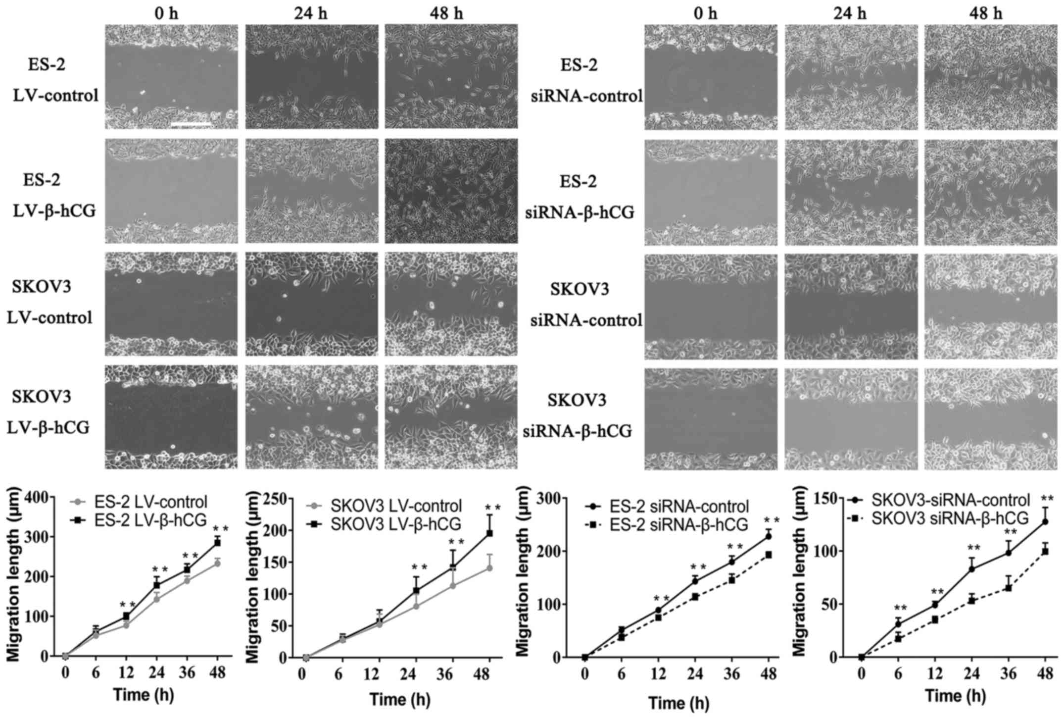
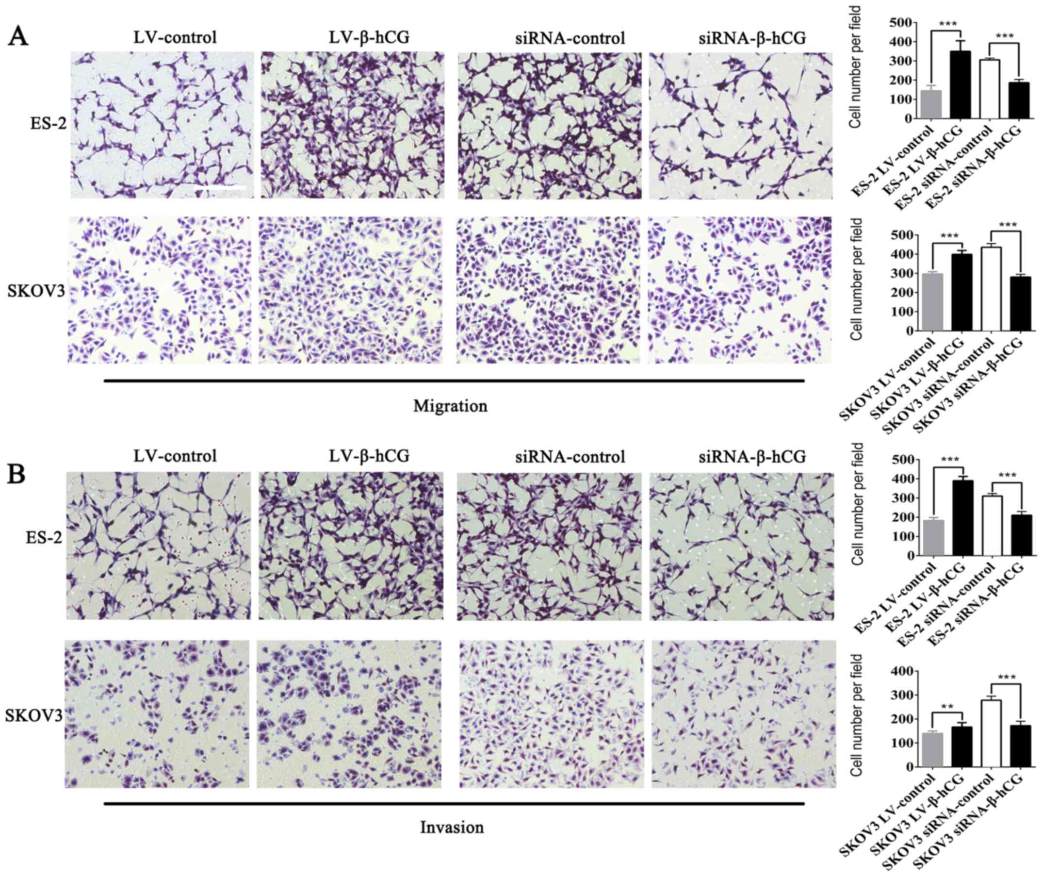
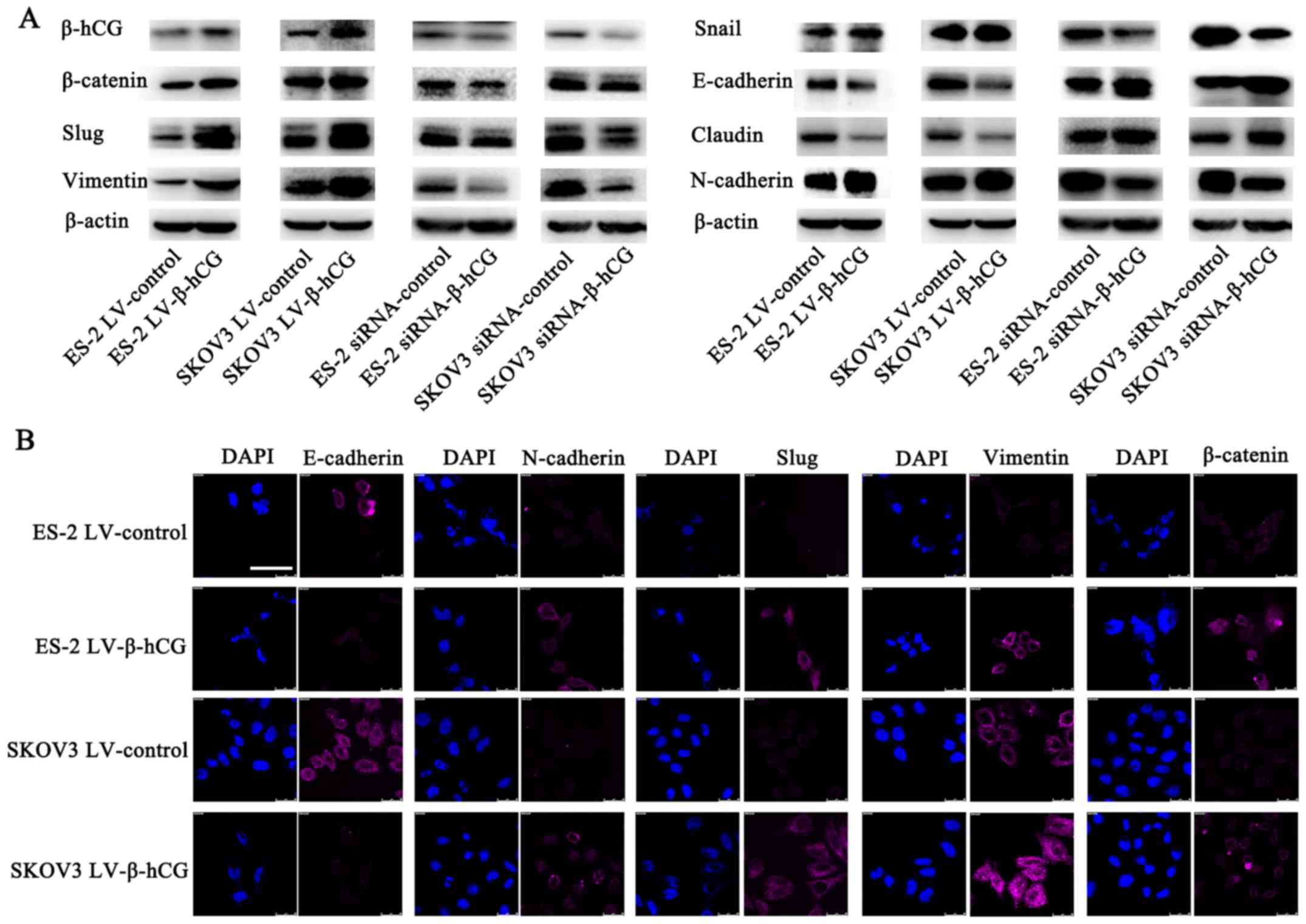
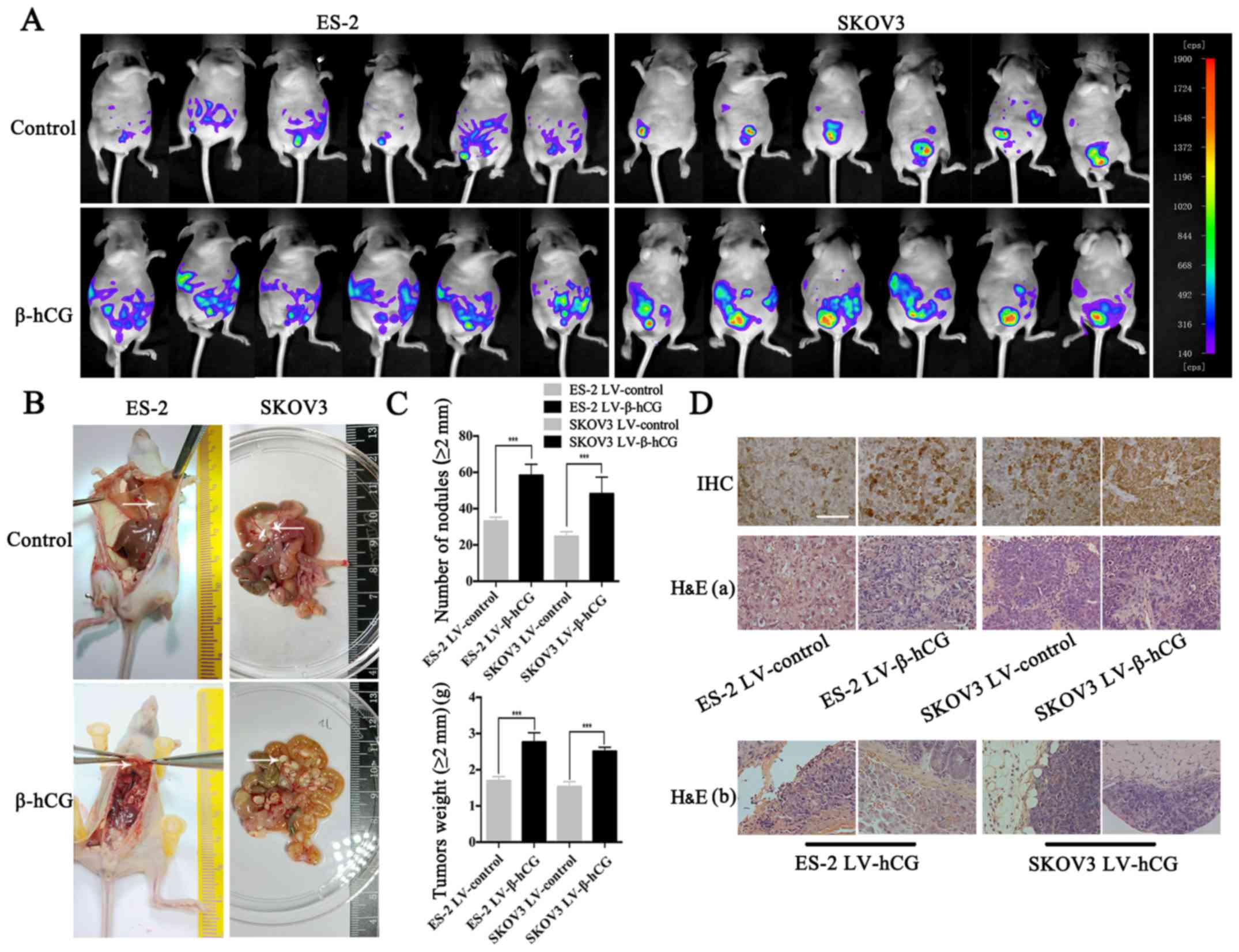
Human chorionic gonadotropin β (β-hCG) is a well-known and accurate marker for the diagnosis and monitoring of pregnancy, trophoblastic tumors and ovarian germ cell tumors. Recently, β-hCG has been found to be closely related to poor prognosis and metastasis in various other malignant tumors, while its role and mechanism in ovarian cancer is still unclear. In the present study, lentiviral‑mediated transfection and small interfering RNA (siRNA) were used to alter β-hCG expression in the ovarian cancer cell lines ES-2 and SKOV3, respectively. Then, migration and invasion activity regulated by β-hCG were evaluated by wound-healing and Transwell assays in vitro and in a peritoneal xenograft nude mouse model in vivo. EDTA and trypsin were utilized to investigate the attachment ability of these cells. Moreover, the expression of epithelial mesenchymal transition (EMT) markers (β-catenin, Slug, vimentin, Snail, claudin, E-cadherin and N-cadherin) was assessed by western blotting and immunofluorescence in ES-2 and SKOV3 cells. Furthermore, β-hCG and EMT markers were evaluated in human ovarian cancer specimens by IHC. The results showed that overexpression of β-hCG clearly promoted migration and invasion in ES-2 and SKOV3 cells (P<0.05) and facilitated metastasis in peritoneal xenografts, while silencing of β-hCG led to the opposite effect. Moreover, β-hCG was closely associated with cell morphology, attachment ability and EMT marker expression in ES-2 and SKOV3 cells and human ovarian cancer specimens. Upregulation of β-hCG promoted cells from an epithelial-like morphology to a mesenchymal-like phenotype, decreased the adhesion ability (P<0.05), and reduced the expression of epithelial markers (E-cadherin) while inducing the expression of mesenchymal markers (vimentin, N-cadherin, β-catenin and Slug). Furthermore, the converse effects were confirmed by knockdown of β-hCG. These findings strongly suggest that β-hCG may regulate metastasis of ovarian cancer through EMT, and it may become a new target for therapeutic intervention.