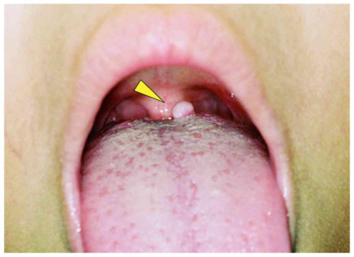
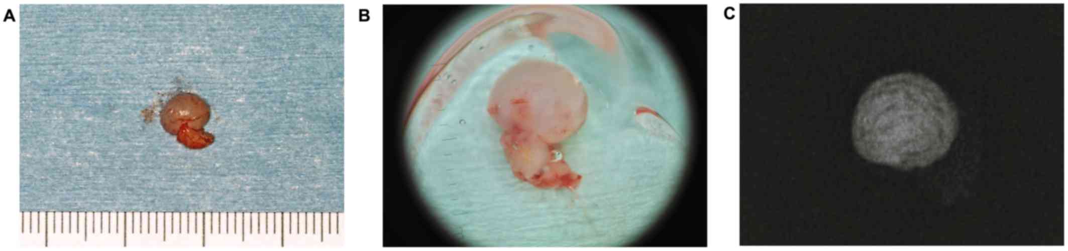
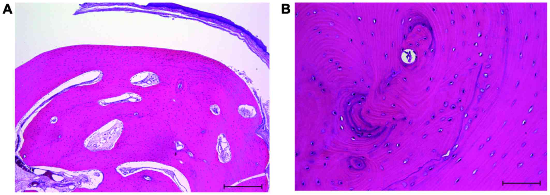
|
1
|
Neville BW, Damm DD, Allen CM and Bouquot
JE: Oral and Maxillofacial Pathology. 3rd. Saunders Elsevier; St.
Louis: pp. 5522009
|
|
2
|
Chou LS, Hansen LS and Daniels TE:
Choristomas of the oral cavity: A review. Oral Surg Oral Med Oral
Pathol. 72:584–593. 1991. View Article : Google Scholar : PubMed/NCBI
|
|
3
|
Krolls SO, Jacoway JR and Alexander WN:
Osseous choristomas (osteomas) of intraoral soft tissues. Oral Surg
Oral Med Oral Pathol. 32:588–595. 1971. View Article : Google Scholar : PubMed/NCBI
|
|
4
|
Tohill MJ, Green JG and Cohen DM:
Intraoral osseous and cartilaginous choristomas: Report of three
cases and review of the literature. Oral Surg Oral Med Oral Pathol.
63:506–510. 1987. View Article : Google Scholar : PubMed/NCBI
|
|
5
|
Psimopoulou M and Antoniades K: Submental
osseous choristoma: A case report. J Oral Maxillofac Surg.
56:666–667. 1998. View Article : Google Scholar : PubMed/NCBI
|
|
6
|
Dalkiz M, Hakan Yurdakul R, Pakdemirli E
and Beydemir B: Recurrent osseous choristoma of the masseter
muscle: Case report. J Oral Maxillofac Surg. 59:836–839. 2001.
View Article : Google Scholar : PubMed/NCBI
|
|
7
|
Yamamoto N, Ishikawa A, Yamauchi K,
Miyamoto I, Tanaka T, Kito S, Matsuo K, Yamashita Y, Morimoto Y and
Takahashi T: Osteolipoma of the lower lip: A case report. Asian J
Oral Maxillofac Surg. 23:143–145. 2011. View Article : Google Scholar
|
|
8
|
Russo T, Piccolo V, Lallas A and
Argenziano G: Recent advances in dermoscopy. F1000Res.
5:1842016.
|
|
9
|
Lallas A, Zalaudek I, Argenziano G, Longo
C, Moscarella E, Di Lernia V, Al Jalbout S and Apalla Z: Dermoscopy
in general dermatology. Dermatol Clin. 31:679–694. 2013. View Article : Google Scholar : PubMed/NCBI
|
|
10
|
De Giorgi V, Massi D and Carli P:
Dermoscopy in the management of pigmented lesions of the oral
mucosa. Oral Oncol. 39:534–535. 2003. View Article : Google Scholar : PubMed/NCBI
|
|
11
|
Olszewska M, Banka A, Gorska R and
Warszawik O: Dermoscopy of pigmented oral lesions. J Dermatol Case
Rep. 2:43–48. 2008. View Article : Google Scholar : PubMed/NCBI
|
|
12
|
Strumia R: Videodermatoscopy: A useful
tool for diagnosing cutaneous dystrophic calcifications. Dermatol
Online J. 11:282005.PubMed/NCBI
|
|
13
|
Lallas A, Moscarella E, Argenziano G,
Longo C, Apalla Z, Ferrara G, Piana S, Rosato S and Zalaudek I:
Dermoscopy of uncommon skin tumours. Australas J Dermatol.
55:53–62. 2014. View Article : Google Scholar : PubMed/NCBI
|
|
14
|
Zaballos P, Llambrich A, Puig S and
Malvehy J: Dermoscopic findings of pilomatricomas. Dermatology.
217:225–230. 2008. View Article : Google Scholar : PubMed/NCBI
|
|
15
|
Okamoto T, Sasaki R, Kataoka T, Kumasaka
A, Kaibuchi N, Naganawa T, Fukada K and Ando T: Dermoscopy imaging
findings in the normal Oral Mucosa. Oral Oncol. 51:e69–e70. 2015.
View Article : Google Scholar : PubMed/NCBI
|
|
16
|
Warszawik-Hendzel O, Słowińska M,
Olszewska M and Rudnicka L: Melanoma of the oral cavity:
Pathogenesis, dermoscopy, clinical features, staging and
management. J Dermatol Case Rep. 8:60–66. 2014. View Article : Google Scholar : PubMed/NCBI
|
|
17
|
Güleç AT: Dermoscopic features of squamous
cell carcinoma of the tongue: It looks similar to cutaneous
squamous cell carcinoma. J Am Acad Dermatol. 75:e53–e54. 2016.
View Article : Google Scholar : PubMed/NCBI
|
|
18
|
Drogoszewska B, Chomik P, Polcyn A and
Michcik A: Clinical diagnosis of oral erosive lichen planus by
direct oral microscopy. Postepy Dermatol Alergol. 31:222–228. 2014.
View Article : Google Scholar : PubMed/NCBI
|
|
19
|
Lee DL, Wong KT, Mak SM, Soo G and Tong
MC: Lingual osteoma: Case report and literature review. Arch
Otolaryngol Head Neck Surg. 135:308–310. 2009. View Article : Google Scholar : PubMed/NCBI
|
|
20
|
Yoshimura H, Ohba S, Matsuda S, Kobayashi
J, Ishimaru K, Imamura Y and Sano K: Osseous choristoma of the
buccal mucosa: A case report with immunohistochemical study of bone
morphogenetic protein-2 and −4 and a review of the literature. J
Oral Maxillofac Surg Med Pathol. 26:351–355. 2014. View Article : Google Scholar
|
|
21
|
Yamamoto M, Migita M, Ogane S, Narita M,
Yamamoto N, Takaki T, Matsuzaka K and Shibahara T: Osseous
choristoma in child with strong vomiting reflex. Bull Tokyo Dent
Coll. 55:207–215. 2014. View Article : Google Scholar : PubMed/NCBI
|
|
22
|
Monserrat M: Ostéome de la langue. Bull
Soc Anat. 88:282–283. 1913.
|
|
23
|
Church LE: Osteoma of the tongue. Report
of a case. Oral Surg Oral Med Oral Pathol. 17:768–770. 1964.
View Article : Google Scholar : PubMed/NCBI
|
|
24
|
Roy JJ, Klein HZ and Tipton DL:
Osteochondroma of the tongue. Arch Pathol. 89:565–568.
1970.PubMed/NCBI
|
|
25
|
Xiao YT, Xiang LX and Shao JZ: Bone
morphogenetic protein. Biochem Biophys Res Commun. 362:550–553.
2007. View Article : Google Scholar : PubMed/NCBI
|
|
26
|
Bragdon B, Moseychuk O, Saldanha S, King
D, Julian J and Nohe A: Bone morphogenetic proteins: A critical
review. Cell Signal. 23:609–620. 2011. View Article : Google Scholar : PubMed/NCBI
|
|
27
|
Kusumoto K, Bessho K, Fujimura K, Akioka
J, Ogawa Y and Iizuka T: Comparison of ectopic osteoinduction in
vivo by recombinant human BMP-2 and recombinant Xenopus BMP-4/7
heterodimer. Biochem Biophys Res Commun. 239:575–579. 1997.
View Article : Google Scholar : PubMed/NCBI
|
|
28
|
Kim SY, Choi HY, Myung KB and Choi YW: The
expression of molecular mediators in the idiopathic cutaneous
calcification and ossification. J Cutan Pathol. 35:826–831. 2008.
View Article : Google Scholar : PubMed/NCBI
|