Introduction
Intervertebral disc degeneration (IVDD) has been
associated with a number of factors, including mechanical loading,
aging, genetic factors and nutrition. IVDD is considered to be an
important factor for neck and back pain (1). The age of occurrence of IVDD is
becoming younger as current changes in human lifestyles ultimately
affect daily life and work (2).
Although medication or surgical treatment may be able to eliminate
some of the symptoms, it is still difficult to radically prevent or
stop the progression of IVDD (2).
IVDD manifests extensive histomorphological alterations, including
fibrosis of the nucleus pulposus, disorganization of the annulus
fibrosus lamellae and thinning and calcification of the cartilage
end plate (1). However, the
underlying mechanism of pathophysiology of the disc degenerative
process remains to be elucidated (3). Therefore, it is important to
determine the pathophysiological changes of IVDD progression.
Intervertebral discs acquire nutrition mainly through the endplate
pathway, and the protection of endplate cartilage may effectively
delay the occurrence of IVDD (2).
An intervertebral disc is located between two neighboring vertebral
bodies and functions to buffer the tension induced by body weight,
tensile force and shear force generated by spinal activity
(3). Therefore, external
mechanical stimulation is regarded as one of the most important
factors leading to IVDD (4). In
our previous study, the synthesis, deposition and degradation of
the extracellular matrix (ECM) components of endplate chondrocytes
were demonstrated to change following applied intermittent cyclic
mechanical tension in vitro, which subsequently led to the
degeneration of endplate chondrocytes (5). SRY box 9 (SOX9) is a transcription
factor regulating the synthesis of extracellular matrix (5). Collagen type II α1 (COL2A1) and
aggrecan (ACAN) are components in the extracellular matrix, and
matrix metalloproteinase 13 (MMP13) is a marker of cartilage
degeneration (5). SOX9, COL2A1 and
ACAN alleviates IVDD, while MMP13 promotes the progression of IVDD
(5).
The present study used an FX-5000T tension system to
effectively simulate the stress condition of cells in vitro.
This system has been widely used in research on various tissues,
including bone healing, cartilage cells, periodontal ligament
cells, lung epithelial cells, myocardial cells, skin cells and
tendon cells (6–10). The present study aimed to clarify
the relationship between chondrocyte morphological changes and the
degree of degeneration, which may provide insight for the
examination of related diseases that are induced by mechanical
stimulation.
Materials and methods
Primary culture of rat endplate
chondrocytes
A total of 12 (2-month-old, male, weight, 150–180 g)
Sprague-Dawley rats were purchased from Shanghai SLAC Laboratory
Animal Co., Ltd. (Shanghai, China) and were individually caged
under standard laboratory conditions (12 h light/dark cycle, 21°C
controlled temperature, relative humidity 40–70%, free access to
food and water, 0.03% CO2). All experimental procedures
were approved by the Committees of the Yijishan Hospital, The First
Affiliated Hospital of Wannan Medical College. Endplate cartilage
was obtained from Sprague-Dawley rats that were anesthetized by
intraperitoneal injection with 4% chloral hydrate (500 mg/kg) and
sacrificed by cervical dislocation. The whole lumbar spine was
removed and washed twice with 1X PBS, containing 1×10⁸
U/l penicillin and 1 g/l streptomycin. The annulus fibrous and
nucleus pulposus were incised to expose the shallow disc-like
translucent endplate cartilage, which was subsequently cut into a
small piece (1 mm3) with ophthalmic scissors.
Chondrocytes were obtained by digestion with trypsin and
collagenase II (Gibco; Thermo Fisher Scientific, Inc., Waltham, MA,
USA), as previously described (11). The cells were washed twice with 1X
PBS and cultured in a 10 cm Petri dish with Dulbecco's modified
Eagle's medium (DMEM)/F-12 medium (HyClone; GE Healthcare Life
Sciences, Logan, UT, USA) supplemented with 10% fetal bovine serum
(FBS: Gibco; Thermo Fisher Scientific, Inc.) in a humidified
incubator containing 5% CO2 at 37°C. Culture medium was
replaced every three days and endplate chondrocytes were collected
at the third passage (P3). This study was approved by the Research
Ethics Committee of Yijishan Hospital affiliated to Wannan Medical
College (Wuhu, China).
Application of intermittent cyclic
mechanical stretch
P3 rat endplate chondrocytes were seeded
(2×05 cells/well) in 2 ml DMEM/F-12 medium with 10% FBS
on 6-well flexible silicone rubber BioFlex plates that were covered
with collagen type I (Flexcell International Corporation,
Burlington, NC, USA), and incubated at 37°C in a humidified
incubator containing 5% CO2. At 70–80% confluence, cells
in the intermittent cyclic mechanical tension (ICMT) group received
mechanical loading (0.5 Hz sinusoidal curve; 10% elongation; 8
h/day) that was applied using the FX-5000T cell stretch tensile
stress loading system (Flexcell International Corporation); cells
in the negative control (NC) group did not receive mechanical
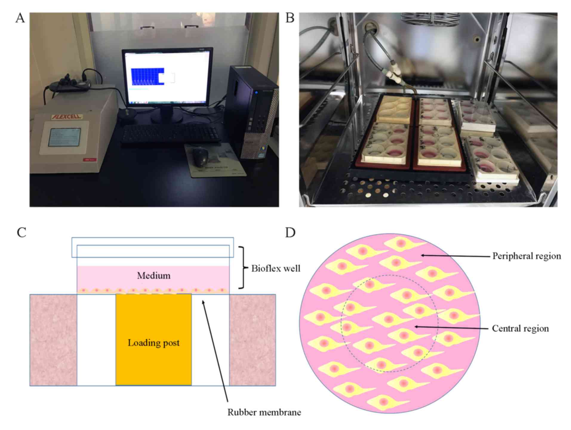
loading. The FX-5000T cell stretch tensile stress loading system
was manufactured by Flexcell International Corporation (Fig. 1). The basic principle of the system
(negative pressure exchange mode) is to form an effective closed
cavity in the cell culture plate between the basement membrane and
the substrate through a rubber gasket. The inlet and outlet of the
sealed chamber are inserted into the CO2 incubator and
the inlet and outlet pipes are connected to the sealed chamber in
the incubator. The vacuum pump is used to extract the negative
pressure generated by the enclosed chamber to pull the
three-dimensional top column to cause the deformation of the
elastic base film. The deformation of the basement membrane is
altered by the computer control system to monitor the pressure of
the regulating gas and the adherent growing cells are subjected to
stretch loading mechanic stimulus. Owing to the loading system gas
pressure adjustment of the top pillar and the BioFlex six-hole
multi-directional plate not being in full contact as presented in
Fig. 1C, with the extension of
mechanical stimulation time, the morphologies of cells in the
central and periphery regions were different.
Flow cytometry
P3 endplate chondrocytes were seeded
(2×10⁵ cells/well) on 6-well flexible silicone rubber
BioFlex plates coated with collagen I. ICMT (0.5 Hz sinusoidal
curve; 10% elongation; 8 h/day) was applied when the cells reached
70–80% confluence for 1 day and 2 days. The membrane of the
six-well flexible silicone rubber BioFlex plates was cut along the
dotted line (Fig. 1) following the
applied mechanical loading. Cells in the central and peripheral
region of each well were separately scraped with a cell scraper and
collected by centrifugation following two washes with cold PBS. The
supernatant was removed and 1X Annexin-binding buffer (Thermo
Fisher Scientific, Inc.) was added to resuspend the cells at a
density of 1×10⁶ cells/ml. Annexin V-Alexa Fluor 488 (5
µl; Thermo Fisher Scientific, Inc.) and propidium iodide (PI; 1 µl;
100 µg/ml) working solution (Thermo Fisher Scientific, Inc.) were
added to each 100 µl of the cells suspension, and 200 µl of 1X
Annexin-binding buffer was added following incubation at room
temperature for 15 min. Unless indicated, all of the above
procedures were maintained on ice. Stained cells were immediately
analyzed by flow cytometry using a FACSCalibur flow cytometer (BD
Biosciences, San Jose, CA, USA) with FACSDiva software (BD
Biosciences; v 8.0.1). PI and annexin V-Alexa Fluor 388
double-positive cells were defined as apoptotic.
Toluidine blue staining
ICMT cells received mechanical loading for 3 days.
The cells were collected and washed twice with PBS, fixed with 4%
paraformaldehyde for 15 min at 25°C followed by 2 washes with PBS.
Subsequently, the cells were stained with 0.5% (W/W) toluidine blue
for 30 min at 25°C. An light inverted microscope was used to
capture images (5 fields/slide) following removal of the staining
dye.
Phalloidin cytoskeleton staining
P3 endplate chondrocytes were seeded
(1×10³ cells/well; 6-well plates) on confocal glass
culture dishes and grown until 40–50% confluent at 37°C. The
culture medium was removed and the cells were washed twice with PBS
and fixed in 4% paraformaldehyde solution at room temperature for
15 min. Cells were washed twice with PBS and permeabilized in 5%
Triton X-100 solution (Yeasen, Shanghai, China) for 10 min at 25°C.
Following three washes with PBS, fluorescein isothiocyanate
(FITC)-labeled phalloidin solution (200 µl; Yeasen) was added to
each well and cells were incubated in the dark at room temperature
for 30 min. Nuclei were stained with DAPI solution (200 µl; Yeasen)
for 30 sec at 25°C. Images were captured using a laser confocal
microscope with FITC excitation (Ex) and emission (Em) filters at
496 and 516 nm, respectively, and DAPI Ex and Em at 364 and 454 nm,
respectively.
RNA extraction and reverse
transcription-quantitative polymerase chain reaction (RT-qPCR)
Cells in the central and peripheral regions of the
BioFlex plates were collected separately, as aforementioned. Total
RNA was isolated using TRIzol reagent (Invitrogen; Thermo Fisher
Scientific, Inc.), according to the manufacturer's instructions.
First-strand cDNA was synthesized from 1 µg of total RNA by
incubating for 1 h at 42°C with Superscript III reverse
transcriptase (Invitrogen; Thermo Fisher Scientific, Inc.)
following oligo (dT) priming. Following reverse transcription, qPCR
was performed using a LightCycler 480 Real-Time PCR System (Roche
Diagnostics, Basel, Switzerland) using SYBR1 Premix Ex Taq (Takara
Biotechnology Co., Ltd., Dalian, China), according to the
manufacturer's protocol. Primer sequences are provided in Table I; GAPDH was used as internal
control. qPCR thermocycling conditions were as follows: Initial
denaturation at 95°C for 10 sec, 50 cycles at 95°C for 10 sec and
60°C for 30 sec; and final extension at 95°C for 15 sec and 60°C
for 30 sec. The data were analyzed using the comparison
2−ΔΔCq method and expressed as the fold change relative
to the respective control (12).
Each sample was analyzed in triplicate.
 | Table I.Primer sequences for reverse
transcription-quantitative polymerase chain reaction. |
Table I.
Primer sequences for reverse
transcription-quantitative polymerase chain reaction.
| Gene | Primer sequence
(5′→3′) | Length (bp) |
|---|
| SOX9 | F:
CGTCAACGGCTCCAGCA | 69 |
|
| R:
TGCGCCCACACCATGA |
|
| COL2A1 | F:
CCTGAAACTCTGCCACCCAG | 151 |
|
| R:
GTTCTTCCGAGGCACAGTCG |
|
| ACAN | F:
ACACCCCTACCCTTGCTTCT | 124 |
|
| R:
AAAGTGTCCAAGGCATCCAC |
|
| MMP13 | F:
CCCTGGAGCCCTGATGTTT | 109 |
|
| R:
CTCTGGTGTTTTGGGGTGCT |
|
| GAPDH | F:
CTCAACTACATGGTCTACATGTTCCA | 81 |
|
| R:
TGCGCCCACACCATGA |
|
Western blotting
Endplate chondrocytes received mechanical
stimulation for 7 days, and were collected as aforementioned.
Protein was extracted from the cells (1×10⁴ cells/well;
6-well plates) following cell lysis with 100 µl
radioimmunoprecipitation buffer containing 1%
phenylmethanesulfonyl, then the cell lysates were placed on ice for
30 min followed by a high-speed centrifuge at 13,500 × g for 15 min
at 4°C. The supernatant was collected, and protein concentration
was determined using a BCA Protein Assay kit. The protein was
denatured with loading buffer and incubated at 95°C for 15 min. A
total of 20 µg protein was loaded per lane using 10% SDS-PAGE and
electrotransferred onto nitrocellulose membranes (Whatman; GE
Healthcare Life Sciences). Membranes were blocked with 5% bovine
serum albumin (BIOSHARP Technology, Inc., Shanghai, China) for 1 h
at 25°C and incubated overnight at 4°C with the following primary
antibodies (all at 1:800): Monoclonal rabbit anti-GAPDH (catalog
no. 14C10; Cell Signaling Technology, Inc., Danvers, MA, USA),
polyclonal rabbit anti-SRY box 9 (SOX9; catalog no. CY5400;
Shanghai Abways Biotechnology Co., Ltd., Shanghai, China),
polyclonal rabbit anti-matrix metalloproteinase 13 (MMP13; catalog
no. AY3010; Shanghai Abways Biotechnology Co., Ltd.) and monoclonal
rabbit anti-collagen type II α1 (COL2A1; catalog no. P02458;
Bioworld Technology, Inc., St. Louis Park, MN, USA). The membranes
were washed three times with TBS with 0.5% Tween-20 (TBST) and
incubated with horseradish peroxidase-conjugated goat anti-rabbit
immunoglobulin G secondary antibodies (catalog no. 7074; Cell
Signaling Technology, Inc., Danvers, MA, USA) were used at a
1:5,000 dilution at room temperature for 2 h. The antigen-antibody
complexes were visualized with the enhanced chemiluminescence
detection system (EMD Millipore, Billerica, MA, USA).
Immunoreactive bands were quantified on autoradiography films in
triplicate with using Image J software (version 4.8; National
Institutes of Health, Bethesda, MD, USA) by normalizing the band
intensities to GAPDH.
Statistical analysis
All data are expressed as the mean ± standard error
of the mean and analyzed by one-way analysis of variance followed
by Bonferroni post hoc test as appropriate. All tests were
two-tailed; P<0.05 was considered to indicate a statistically
significant difference. All statistical analyses were conducted
with SPSS 18.0 (SPSS Inc., Chicago, IL, USA).
Results
Degeneration of rat endplate
chondrocytes induced in vitro
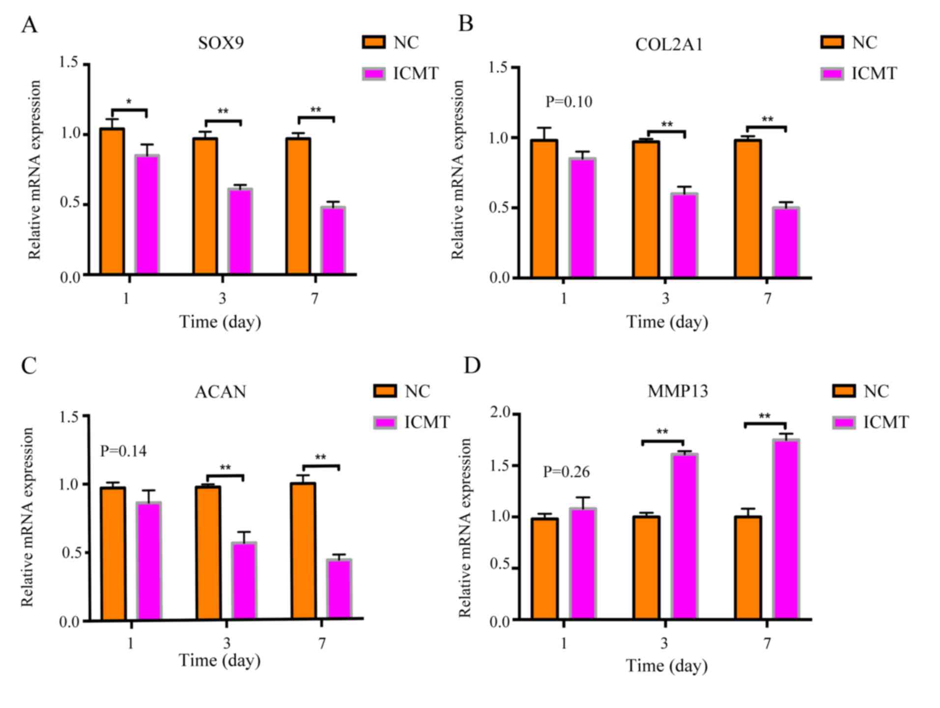
RT-qPCR results revealed that the mRNA expression
levels of cartilage-related anabolic genes SOX9, COL2Al and ACAN in
the ICMT-treatment group gradually decreased on days 1, 3 and 7
compared with NC (Fig. 2A-C),
whereas the expression levels of MMP13 gradually increased compared
with NC at the same time points (Fig.
2D). Rat endplate chondrocytes exhibited degenerative
alterations following ICMT including decreased expression of SOX9,
COL2A1 and ACAN and elevated expression of MMP13.
Cell morphological changes in the
central and peripheral regions
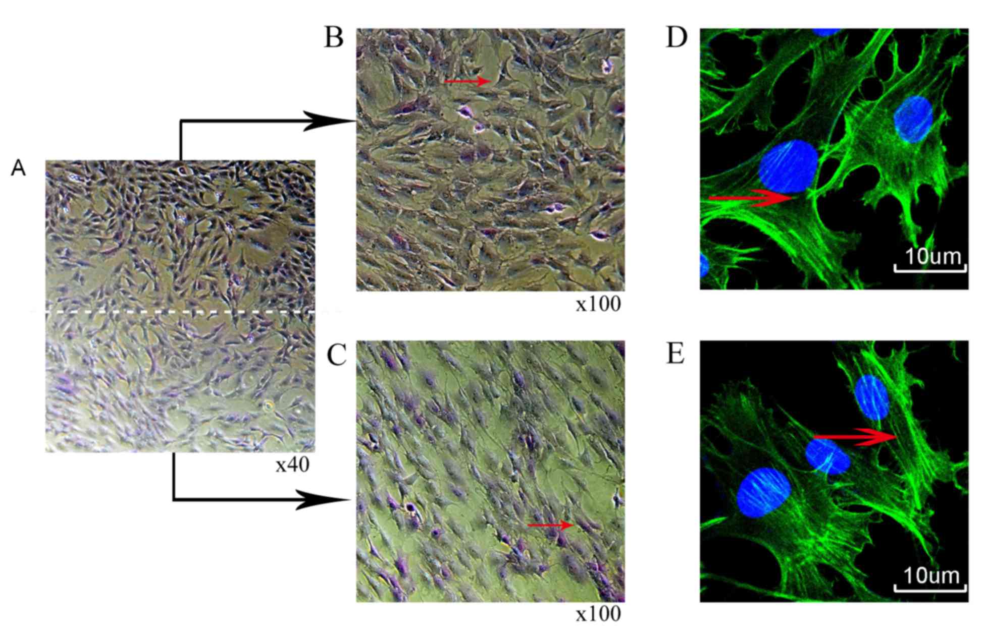
Rat chondrocytes were treated with mechanical
stimulation (10% ICMT, 0.5 Hz and 8 h/day) for 3 days. Toluidine
blue staining demonstrated a long shuttle change of cells in the
peripheral region of the rubber membrane compared with cells in the
central region (Fig. 3A-C).
Phalloidin cytoskeleton staining also revealed that the filamentous
(F)-actin in the peripheral cells were more notably stretched and
elongated compared with the F-actin in cells in the central region
as indicated by the red arrow (Fig. 3D
and E).
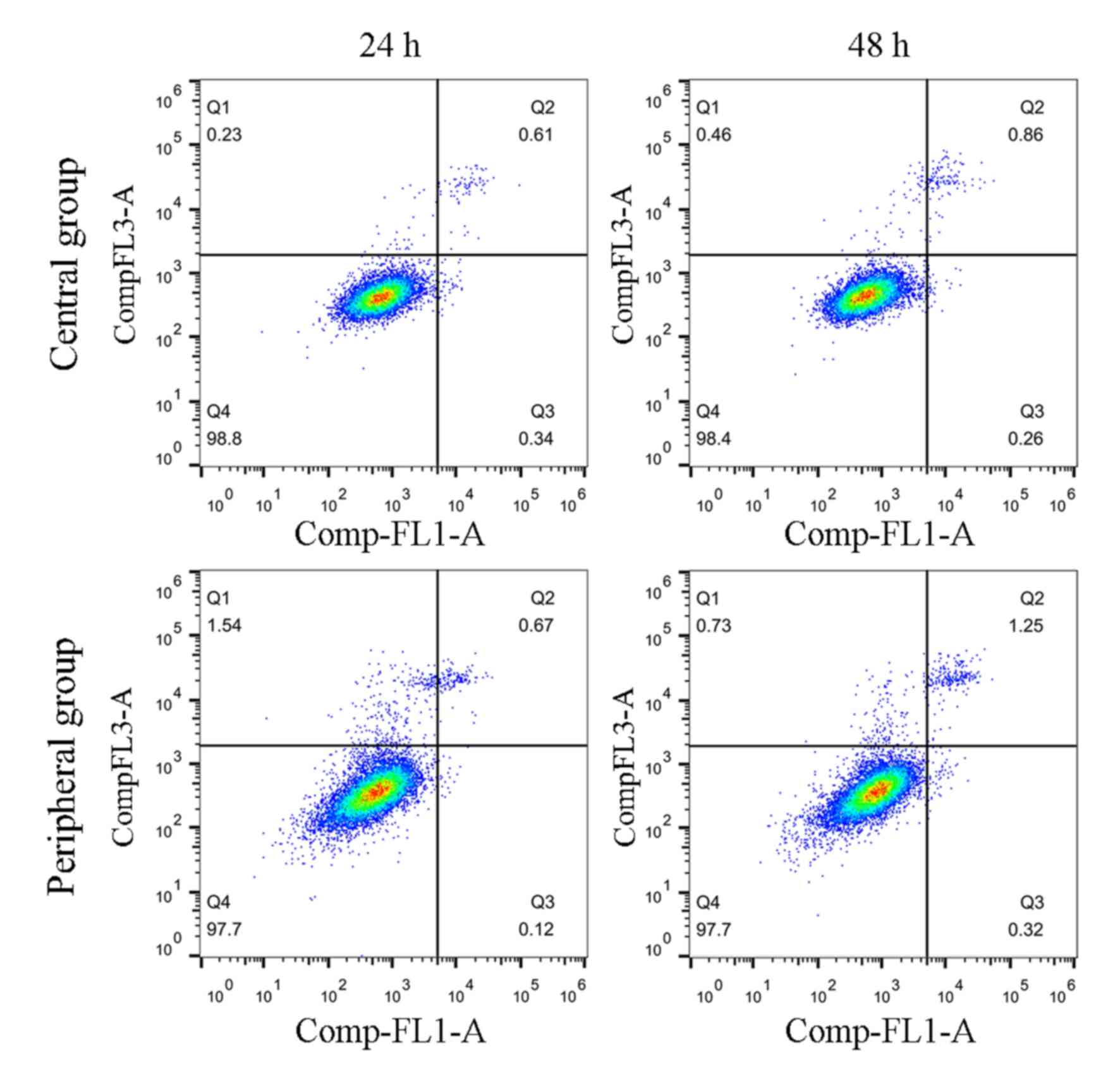
Effects of ICMT on apoptosis in the
central and peripheral regions
The levels of apoptosis were also examined to
further determine the effects of ICMT induction on endplate
chondrocytes; cells received mechanical stimulation for 24 and 48
h. No differences were identified for the levels of apoptosis
between the ICMT and NC groups at either 24 or 48 h (Fig. 4).
Degree of cell degeneration between
the central and peripheral regions
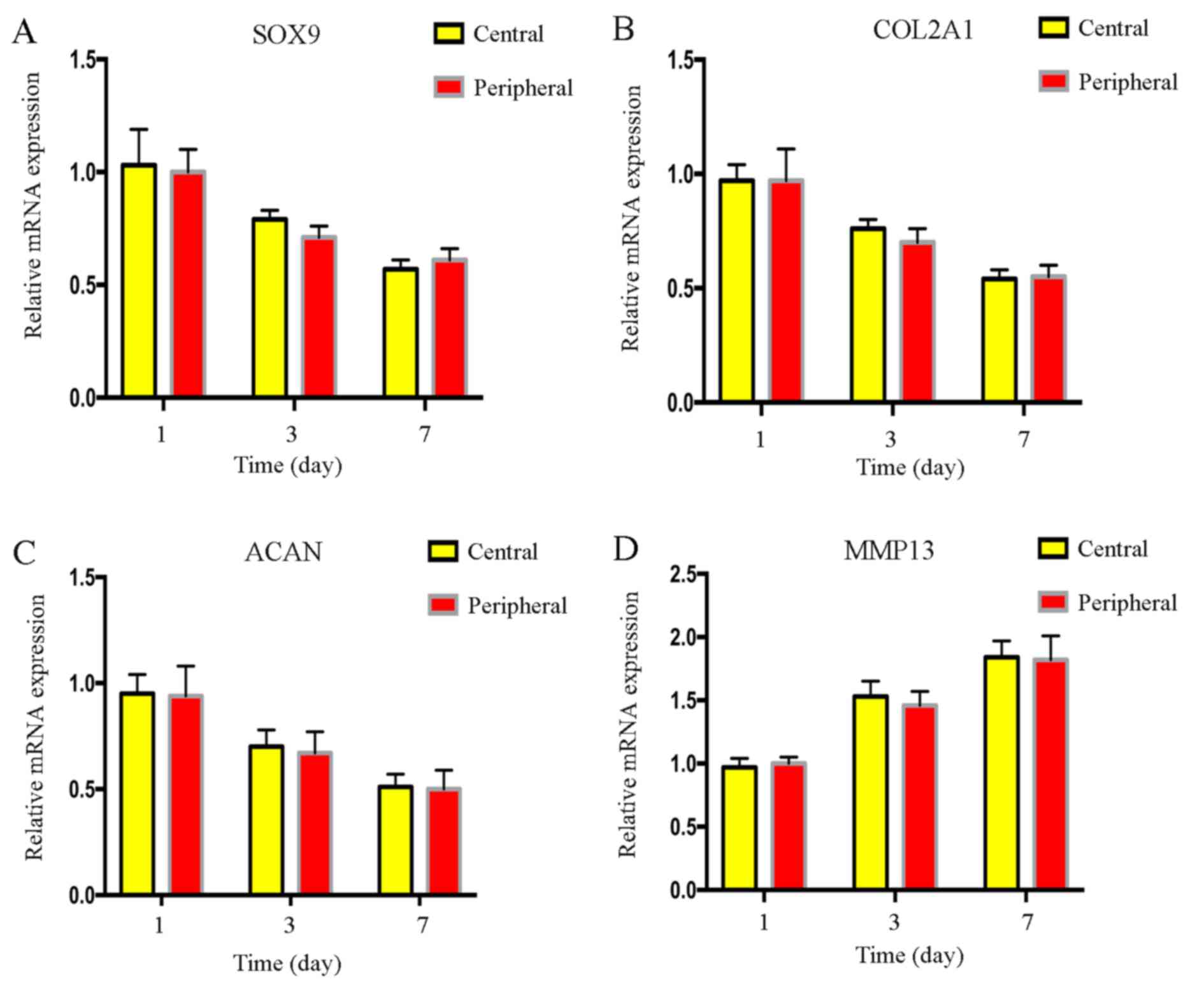
Results from RT-qPCR indicated that there were no
significant differences in the mRNA expression levels of the
cartilage-related genes, SOX9, COL2A1, ACAN and MMP13, between
cells in the central and peripheral regions following mechanical
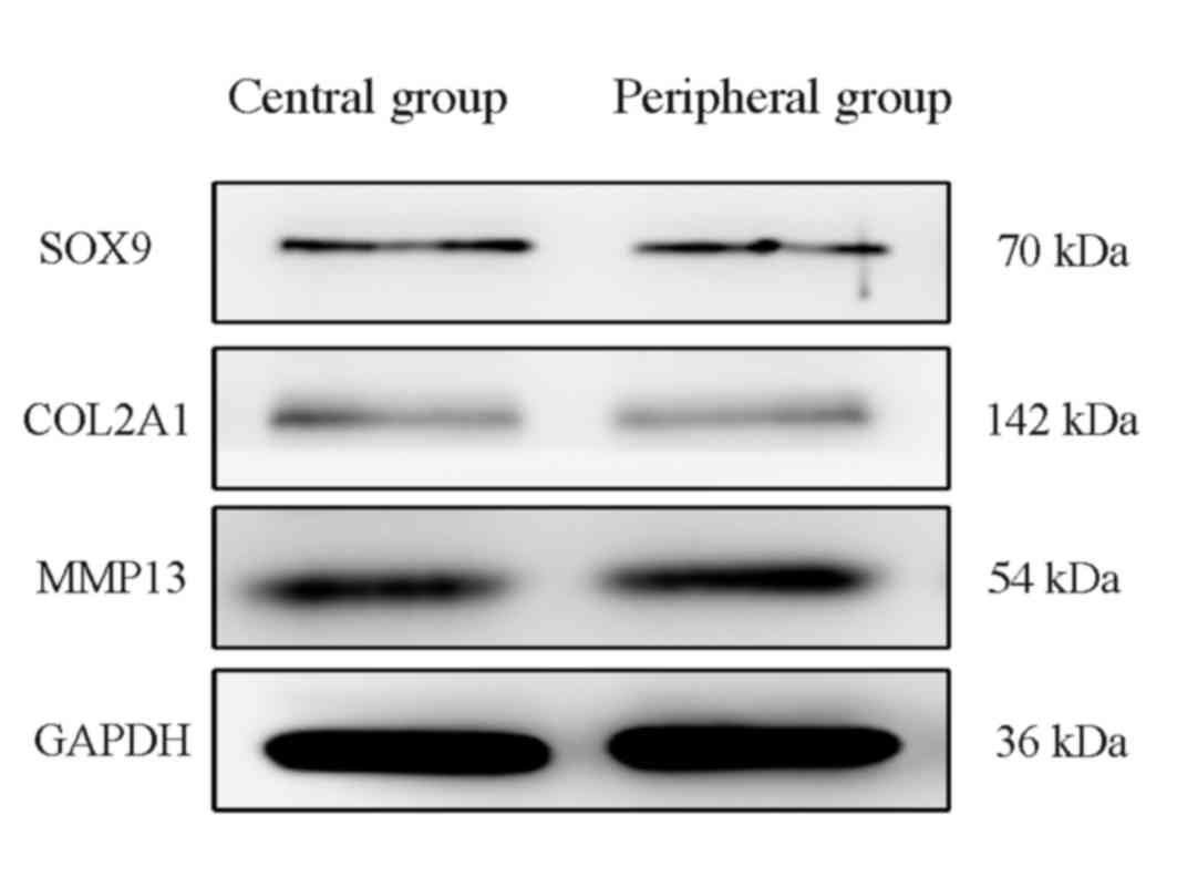
stimulation (Fig. 5). Similarly,
western blotting results indicated no notable differences in SOX9,
COL2A1 and MMP13 protein expression levels between cells in the two
regions, however, due to the lack of a suitable ACAN antibody,
alterations in protein expression of ACAN were not detected
(Fig. 6). There was no difference
in the degree of the endplate chondrocyte degeneration between the
two groups.
Discussion
Endplate cartilage is a thin layer of hyaline
cartilage that lies between the vertebrae and the intervertebral
disc, and mainly comprises chondrocytes and ECM. Endplate cartilage
effectively regulates the mechanical stimulation in the course of
physiological activities (13).
Excessive mechanical stimulation may cause metabolic imbalances of
the cartilage ECM through the relevant signaling pathways,
including Bone morphogenetic proteins (BMPs) and transforming
growth factor-β (TGF-β), and may result in lower back pain and
neurological dysfunction (14,15).
The ECM of endplate cartilage is mainly composed of collagen fibers
and cartilage proteoglycans; the main collagen fiber is type II
collagen (16). It was previously
reported that the ECM components of endplate chondrocytes were
altered following mechanical stimulation, and the cytoskeleton
became more slender (7,17). The cytoskeleton consists of
microfilaments, microtubules and intermediate filaments, comprising
actin, tubulin and vimentin. In chondrocytes, actin filaments
function to maintain the chondrocyte phenotype by modulating cell
shape and to provide mechanical integrity to withstand compression
(18,19). A previous study demonstrated that
mechanical stimulation may lead to alterations in the structure and
morphology of intracellular actin (20); it was confirmed that cell
degeneration is closely related to cytoskeletal actin, but the
specific mechanism remains unknown.
Results from the presents study demonstrated a
change in cell morphology in different regions (central and
peripheral) following mechanical stimulation. Immunocytochemical
staining demonstrated that the cells in the peripheral region were
particularly elongated following ICMT, and that the F-actin
cytoskeleton was stretched and elongated, which indicated that
mechanical stimulation caused notable morphological changes of the
cytoskeleton protein actin. RT-qPCR and western blotting results
demonstrated that the mRNA and protein expression levels,
respectively, of the cartilage-related genes SOX9, COL2A1, ACAN and
MMP13 were not different between cells in the central and
peripheral regions following mechanical stimulation, which
indicated that the degree of cell degeneration induced by
mechanical stimulation between the two groups was consistent. In
addition, although the morphological changes between the two
regions were different, no differences in apoptosis between the two
groups were observed following stimulation for 24 and 48 h.
A previous study demonstrated that actin in the
cytoskeleton regulates the metabolism of the ECM through relevant
mechanical signaling pathway, including BMPs and TGF-β, and
ultimately induces changes in chondrocyte degeneration (21). In the present study, the
morphological changes of cells did not necessarily coincide with
the degree of cell degeneration. Although these results differed
from previous studies, it was demonstrated that cell morphological
changes may occur 3 days following mechanical stimulation. The
present study was not able to validate the relationship between
cell morphological changes and degeneration by extending the
loading time, which requires further investigation. It is proposed
to induce the degeneration of chondrocytes in a short time by
changing the intensity of mechanical stimulation. Further study on
the intrinsic relationship between cell morphology and cell
degeneration may improve our understanding of the pathogenesis of
intervertebral disc degeneration disease and may provide a more
comprehensive theoretical and experimental basis for the study of
intervertebral disc degeneration caused by mechanical factors.
Acknowledgements
The present study was supported by a grant from The
National Natural Science Foundation of China (grant no. 81572185),
The Natural Science Foundation of Anhui Province of China (grant
no. 1708085MH185) and The Scientific and Technological Achievements
Transformation Project of Wuhu City (grant no. 2017CG27).
References
|
1
|
Manek NJ and MacGregor AJ: Epidemiology of
back disorders: Prevalence, risk factors, and prognosis. Curr Opin
Rheumatol. 17:134–140. 2005.PubMed/NCBI
|
|
2
|
Rodriguez AG, Rodriguez-Soto AE, Burghardt
AJ, Berven S, Majumdar S and Lotz JC: Morphology of the human
vertebral endplate. J Orthop Res. 30:280–287. 2012. View Article : Google Scholar : PubMed/NCBI
|
|
3
|
Walter BA, Korecki CL, Purmessur D,
Roughley PJ, Michalek AJ and Iatridis JC: Complex loading affects
intervertebral disc mechanics and biology. Osteoarthritis
Cartilage. 19:1011–1018. 2011. View Article : Google Scholar : PubMed/NCBI
|
|
4
|
Vergroesen PP, Kingma I, Emanuel KS,
Hoogendoorn RJ, Welting TJ, van Royen BJ, van Dieën JH and Smit TH:
Mechanics and biology in intervertebral disc degeneration: A
vicious circle. Osteoarthritis Cartilage. 23:1057–1070. 2015.
View Article : Google Scholar : PubMed/NCBI
|
|
5
|
Xu HG, Yu YF, Zheng Q, Zhang W, Wang CD,
Zhao XY, Tong WX, Wang H, Liu P and Zhang XL: Autophagy protects
end plate chondrocytes from intermittent cyclic mechanical tension
induced calcification. Bone. 66:232–239. 2014. View Article : Google Scholar : PubMed/NCBI
|
|
6
|
Pan J, Wang T, Wang L, Chen W and Song M:
Cyclic strain-induced cytoskeletal rearrangement of human
periodontal ligament cells via the Rho signaling pathway. PLoS One.
9:e915802014. View Article : Google Scholar : PubMed/NCBI
|
|
7
|
Xu HG, Ma MM, Zheng Q, Shen X, Wang H,
Zhang SF, Xu JJ, Wang CD and Zhang XL: P120-catenin protects
endplate chondrocytes from intermittent cyclic mechanical tension
induced degeneration by inhibiting the expression of RhoA/ROCK-1
signaling pathway. Spine (Phila Pa 1976). 41:1261–1271. 2016.
View Article : Google Scholar : PubMed/NCBI
|
|
8
|
Wang J, Wang CD, Zhang N, Tong WX, Zhang
YF, Shan SZ, Zhang XL and Li QF: Mechanical stimulation
orchestrates the osteogenic differentiation of human bone marrow
stromal cells by regulating HDAC1. Cell Death Dis. 7:e22212016.
View Article : Google Scholar : PubMed/NCBI
|
|
9
|
Zhang R, Mao P, Fu W, Pang XQ, Wang YY,
Yang C, He WQ, Liu XQ and Li YM: Effects of cyclic stretch on the
induction of the transdifferentiation in human lung epithelial
cells. Zhonghua Wei Zhong Bing Ji Jiu Yi Xue. 25:455–459. 2013.(In
Chinese). PubMed/NCBI
|
|
10
|
Witt F, Duda GN, Bergmann C and Petersen
A: Cyclic mechanical loading enables solute transport and oxygen
supply in bone healing: An in vitro investigation. Tissue Eng Part
A. 20:486–493. 2014.PubMed/NCBI
|
|
11
|
Xu HG, Hu CJ, Wang H, Liu P, Yang XM,
Zhang Y and Wang LT: Effects of mechanical strain on ANK, ENPP1 and
TGF-β1 expression in rat endplate chondrocytes in vitro. Mol Med
Rep. 4:831–835. 2011.PubMed/NCBI
|
|
12
|
Livak KJ and Schmittgen TD: Analysis of
relative gene expression data using real-time quantitative PCR and
the 2(-Delta Delta C(T)) method. Methods. 25:402–408. 2001.
View Article : Google Scholar : PubMed/NCBI
|
|
13
|
Ferguson SJ and Steffen T: Biomechanics of
the aging spine. Eur Spine J. 12 Suppl 2:S97–S103. 2003. View Article : Google Scholar : PubMed/NCBI
|
|
14
|
Alkhatib B, Rosenzweig DH, Krock E,
Roughley PJ, Beckman L, Steffen T, Weber MH, Ouellet JA and Haglund
L: Acute mechanical injury of the human intervertebral disc: Link
to degeneration and pain. Eur Cells Mater. 28:98–111. 2014.
View Article : Google Scholar
|
|
15
|
Sowa GA, Coelho JP, Vo NV, Pacek C,
Westrick E and Kang JD: Cells from degenerative intervertebral
discs demonstrate unfavorable responses to mechanical and
inflammatory stimuli: A pilot study. Am J Phys Med Rehabil.
91:846–855. 2012. View Article : Google Scholar : PubMed/NCBI
|
|
16
|
Bruckner P and van der Rest M: Structure
and function of cartilage collagens. Microsc Res Tech. 28:378–384.
1994. View Article : Google Scholar : PubMed/NCBI
|
|
17
|
Xu HG, Zheng Q, Song JX, Li J, Wang H, Liu
P, Wang J, Wang CD and Zhang XL: Intermittent cyclic mechanical
tension promotes endplate cartilage degeneration via canonical Wnt
signaling pathway and E-cadherin/β-catenin complex cross-talk.
Osteoarthritis Cartilage. 24:158–168. 2016. View Article : Google Scholar : PubMed/NCBI
|
|
18
|
Benya PD, Brown PD and Padilla SR:
Microfilament modification by dihydrocytochalasin B causes retinoic
acid-modulated chondrocytes to reexpress the differentiated
collagen phenotype without a change in shape. J Cell Biol.
106:161–170. 1988. View Article : Google Scholar : PubMed/NCBI
|
|
19
|
Guilak F: Compression-induced changes in
the shape and volume of the chondrocyte nucleus. J Biomech.
28:1529–1541. 1995. View Article : Google Scholar : PubMed/NCBI
|
|
20
|
Li S, Jia X, Duance VC and Blain EJ: The
effects of cyclic tensile strain on the organisation and expression
of cytoskeletal elements in bovine intervertebral disc cells: An in
vitro study. Eur Cell Mater. 21:508–522. 2011. View Article : Google Scholar : PubMed/NCBI
|
|
21
|
Nofal GA and Knudson CB: Latrunculin and
cytochalasin decrease chondrocyte matrix retention. J Histochem
Cytochem. 50:1313–1324. 2002. View Article : Google Scholar : PubMed/NCBI
|