Introduction
Oral squamous cell carcinoma cells (OSCC) is the
most common oral cancer, and accounts for ~90% total oral cancers
(1). Despite considerable
improvements in therapeutic strategies for OSCC over the past two
decades, the 5-year survival rate of OSCC has not improved
significantly and its incidence continues to increase among young
people and women (2,3). Therefore, exploring novel molecular
mechanisms involved in development and progression of OSCC is
urgently needed for identifying novel and effective therapeutic
targets.
MicroRNAs (miRNAs) are a class of highly conserved
endogenous, single-stranded, small non-protein-coding RNAs
comprising ~18–25 nucleotides that inhibit gene expression at the
post-transcriptional level by directly binding to the
3′-untranslated region (3′UTR) of messenger RNAs (mRNAs) (4). Through repressing the target gene,
miRNAs are able to regulate diverse biological processes, such as
cell proliferation, cell migration, apoptosis, differentiation,
development, immunity, and metabolism (5,6).
Importantly, miRNAs are dysregulated in various types of cancer as
oncogenes or tumor suppressors (7,8). For
OSCC, number of miRNAs have been identified to be involved in
progression and development of OSCC (9,10), and
to be used as diagnosis marker and therapy target.
MicroRNA-152 (miR-152) has been shown to be
downregulated, and function as a tumor suppressor in many types of
cancers, including gastric cancer (11), colorectal cancer (12), breast cancer (13), non-small lung cancer (14), cervical cancer (15), and prostate cancer (16). However, its potential role and
mechanism in regulating OSCC carcinogenesis and progression remains
unclear. This study aimed to reveal the exact role as well as the
relevant mechanism of miR-152 in the regulation of OSCC cell
proliferation and invasion.
Materials and methods
Tissue samples
Total of 40 paired fresh surgically resected OSCC
tumors and matched adjacent normal tissues were obtained from OSCC
patients who were diagnosed, treated, and followed-up at the
Department of Oral and Maxillofacial Surgery, School and Hospital
of Stomatology, Jilin University (Changchun, China). Fresh frozen
tissues were immediately frozen in liquid nitrogen and stored in
liquid nitrogen until RNA extraction. All samples were confirmed by
pathological examinations. Written informed consent was obtained
from all participants. This study was approved by the Ethics
Committee of School and Hospital of Stomatology, Jilin University
(Changchun, China). Patients who had received chemotherapy or
radiotherapy prior to surgery were excluded. Clinical parameters,
including pathological features and clinical stage, were
retrospectively harvested and are presented in Table I.
 | Table I.Association of miR-152 expression with
clinicopathological factors of 40 OSCC patients. |
Table I.
Association of miR-152 expression with
clinicopathological factors of 40 OSCC patients.
|
|
| miR-152
expression |
|
|---|
|
|
|
|
|
|---|
| Variables | No. of cases | Low (n %) | High (n %) | P-value |
|---|
| Age (years) |
|
|
| >0.05 |
|
<60 | 17 | 10 (58.8) | 7
(41.2) |
|
|
≥60 | 23 | 14 (60.9) | 9
(39.1) |
|
| Sex |
|
|
| >0.05 |
|
Male | 22 | 13 (59.1) | 9
(40.9) |
|
|
Female | 18 | 11 (61.1) | 7
(38.9) |
|
| Clinical stage |
|
|
| >0.05 |
|
I–II | 29 | 14 (48.2) | 15 (51.8) |
|
|
III–IV | 11 | 10 (90.9) | 1
(9.1) |
|
| Differentiated |
|
|
| >0.05 |
|
Well/moderate | 25 | 14 (56.0) | 11 (44.0) |
|
|
Poor | 15 | 10 (66.7) | 5
(33.3) |
|
| Lymph node
metastasis |
|
|
| <0.05 |
| No | 30 | 14 (46.7) | 16 (53.3) |
|
|
Yes | 10 | 10 (100) | 0
(0) |
|
Cell culture and transfection
The human normal oral keratinocytes (hNOKs) and four
human OSCC cell lines, including Tca8113, OSCC-15, SCC-9, and
SCC-25, were obtained from American Type Culture Collection
(Manassas, VA, USA), and cultured in DMEM/Nutrient Mixture F12
(Hyclone, Logan, UT, USA) supplemented with 10% fetal bovine serum
(FBS, Gibco, USA), 100 U/ml penicillin (Sigma-Aldrich, St. Louis,
MO, USA), or 100 µg/ml streptomycin (Sigma-Aldrich) in a humidified
incubator containing 5% CO2 at 37°C. The cells from
passage 4 were used for all the experiments.
miR-152 mimic or corresponding negative control
(miR-NC) was brought from GenePharma (Shanghai, China). The c-MET
overexpression vector (pCDNA3.1-c-MET, lack of 3′UTR) was granted
from Yue Zhao (Jilin University). Transfection was performed using
the Lipofectamine™ 2000 reagent (Invitrogen, Carlsbad, CA, USA)
following the manufacturer's protocol.
Quantitative real-time PCR
Total RNA was extracted from the cultured cells or
tissues using TRIzol (Invitrogen) according to the manufacturer's
instructions. A dual-beam ultraviolet spectrophotometer (Eppendorf,
Hamburg, Germany) were used to determine purity and concentration
of total RNA. Then, total RNA was reverse transcribed to cDNA using
a PrimeScript® RT reagent kit (Takara Biotechnology Co.,
Ltd., Dalin, China) or the miScript Reverse Transcription kit
(Qiagen, Hilden, Germany). The PCR were performed using SYBR premix
real-time PCR reagent (Takara) under an ABI7900 real-time PCR
system (Applied Biosystems, Foster City, CA, USA). The primers for
miR-152 and U6 were brought from Applied Biosystems. The primers
for c-MET and GAPDH were used in this study as described previously
(17). U6 and GAPDH were applied as
internal controls for miR-152 and c-MET mRNA, respectively.
Relative quantification was analyzed using the comparative
2−ΔΔCt method.
Cell proliferation assay
3-(4,5-dimethylthiazol-2-yl)-2,5-diphenyl
tetrazolium bromide (MTT) assay kit (Sigma-Aldrich) were used to
measure cell proliferation. In brief, the transfected cells were
seeded in a 96-well plate (1×10³ cells/well) and
cultured for 1–3 days. After incubated for 24, 48 and 72 h
respectively, 25 µl of MTT (10 mg/ml) was added to each well and
cultured for 4 h. Then, the supernatant was removed, and 150 µl of
dimethyl sulfoxide (DMSO, Sigma-Aldrich, St. Louis, MO, USA) was
added to each well for 10 min at room temperature. The absorbance
at 490 nm was detected with a microplate reader (Bio-Rad, Hercules,
CA, USA).
Colony formation assay
Transfected cells were seeded in 6-well plates (500
cells per well) and allowed to grow for 10 days to determine their
colony-forming ability. After 70% ethanol fixation, the plate was
stained with 0.1% crystal violet for 5 min (Sigma). Cell colonies
were counted under a light microscope (Zeiss, Jena, Germany).
Migration and invasion assays
To determine cell migration, wound healing assay was
performed when transfected cells grew to 100% confluence in 6-well
plates. Wounds were created with a 10-µl pipette tip to scratch the
culture. After cells were rinsed with PBS, the medium was then
added, and cultured for 24 h at 37°C. At 0 and 24 h after the
scratch, the cultures were examined and photographed under a light
microscope (Zeiss).
For invasion assay, 1×10⁵ transfected
cells were seeded into the upper chambers with Matrigel™ Basement
Membrane Matrix (BD Biosciences) in free serum. Medium with 20% FBS
was added to the lower chambers to stimulate cell invasion.
Twenty-four hours later, cells in the upper chambers were removed,
while cells that migrated to the lower surface were fixed with 70%
ethanol for 10 min and stained with 0.1% crystal violet
(MedChemExpress, Princeton, NJ, USA) for 15 min. The numbers of
invaded cells were observed using a light microscope
(magnification, ×200, Zeiss) and counted in five randomly selected
fields.
Luciferase reporter assay
miR-152 target gene was predicted by three publicly
available algorithms: TargetScan, PicTar, and miRBase. c-MET was
chosen as a target of miR-152. c-MET 3′UTR regions
containing predicted miR-152 binding sites were amplified by PCR,
and the production was subcloned into the pGL3 vector (Ambion,
Austin, TX, USA), named as WT-c-MET-3′UTR. In addition, the binding
sequence of c-MET 3′UTR was mutated using a Quik-Change™
Site-Directed Mutagenesis kit (Stratagene; Agilent Technologies,
Inc., Santa Clara, CA, USA), and also cloned into pGL3 vector,
named as: MUT-c-MET-3′UTR. For the luciferase reporter assay, SCC-9
cells were cotransfected with miR-152 mimic or miR-NC and
WT-c-MET-3′UTR or MUT-c-MET-3′UTR reporter plasmid by Lipofectamine
2000. After 48 h cell transfection, luciferase activities were
detected using a Dual-Luciferase® Reporter assay kit
(Promega, Madison, WI, USA) according to the manufacturer's
protocols. The results were presented as the ratio of firefly
luciferase activity to renilla luciferase activity.
Western blotting
The total cell lysates were harvested using a
detergent lysis buffer (Sigma-Aldrich). The protein concentration
of each sample was measured using a BCA protein assay kit
(Bio-Rad). The protein was separated on 10% SDS polyacrylamide
gels, subsequently transferred to polyvinylidene difluoride (PVDF)
membranes (Merck, Millipore, Germany). Then, the membrane were
blocked with a 5% skim milk solution and incubated with the
following antibodies overnight at 4°C: anti-c-MET (1:1,000, Santa
Cruz Biotechnology, Inc., Santa Cruz, CA, USA), anti-AKT (1:2,000,
Santa Cruz Biotechnology, Inc.) and anti-p-AKT (1:1,500, Santa Cruz
Biotechnology, Inc.), anti-PI3K (1:2,000, Santa Cruz Biotechnology,
Inc.) and anti-p-PI3K (1:1,000, Santa Cruz Biotechnology, Inc.),
anti-GAPDH (1:5,000, Santa Cruz Biotechnology, Inc.). The membranes
were then incubated with the corresponding secondary HRP-conjugated
antibodies (1:5,000; Cell Signaling Technology, Inc., Danvers, MA,
USA) for 2 h at room temperature. GAPDH was used as a loading
control. Protein bands were observed by the enhanced
chemiluminescence system (ECL kit, Millipore, Billerica, MA, USA)
and were analyzed with the Bio-Rad ChemiDoc XRS system
(Bio-Rad).
Mouse xenograft models
All experimental procedures were approved by the
experimental animal ethics committee of Jilin University
(Changchun, China). Twenty male BALB/c nude mice (6-week-old) were
housed and maintained in a pathogen-free (SPF) environment. SCC-9
cells transfected with miR-152 mimic or miR-NC (2×10⁶)
were mixed with Matrigel (BD Lifesciences) and injected
subcutaneously into the right flank of each animal (n=10 each
group), respectively. The tumor volume was measured once every five
days until the mice were sacrificed using the following formula:
tumor volume (V)= (length × width2)/2. The mice were
sacrificed on day 30 after inoculation, then tumor tissues were
stripped and weighted. The tumor xenografts were fixed and prepared
for immunohistochemistry (IHC). IHC was performed as described
previously (18).
Statistical analysis
Results are presented as mean ± standard deviation
(SD) from at least three independent experiments, and was analyzed
using version 19 SPSS statistical software (SPSS, Chicago, IL,
USA). The data were analyzed between two groups using Student's
t-test or more than two groups using one-way analysis of variance
(ANOVA). Spearman's correlation coefficient was used to assess
correlations between miR-152 and c-MET. The survival ratio
was analyzed by Kaplan-Meier plots. For all statistical analyses,
P-values of <0.05 were considered to be statistically
significant.
Results
miR-152 is downregulated in both OSCC
cells and clinical specimens
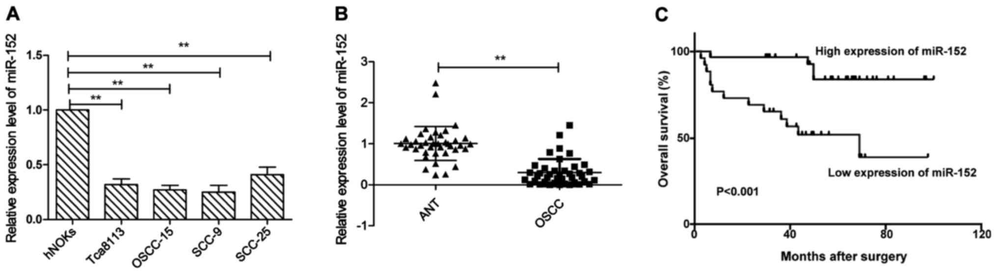
In order to examine miR-152 expression levels, we
first examined miR-152 expression in the human normal oral
keratinocytes (hNOKs) and four human OSCC cell lines Tca8113,
OSCC-15, SCC-9, and SCC-25. As shown in Fig. 1A, miR-152 expression was aberrantly
downregulated in all OSCC cell lines, as compared to normal oral
keratinocytes (hNOKs) (P<0.05). Next, we examined miR-152
expression in 40 pairs of OSCC tissues and matched adjacent normal
tissues. We found that miR-152 expression level in OSCC tissues was
significantly lower than those of their matched adjacent normal
tissues (P<0.01). To further investigate the clinicopathological
significance of miR-152 level in OSCC patients, based on the mean
(0.29) of all samples, patients were divided into two subgroups:
low miR-152 group (<0.29, 24 cases) and a high miR-152 group
(>0.29, 16 cases). The results demonstrated that the decreased
miR-152 expression was significantly associated with lymph node
metastasis (P<0.05), but not with age, sex, clinical stage or
differentiation (Table I, all
P>0.05). In addition, Kaplan-Meier analysis revealed that the
OSCC patients with low miR-152 expression had poor survival rate
(Fig. 1C). These results implied
that miR-152 could serve as a prognostic marker and play a key role
in OSCC progression.
miR-152 inhibits the proliferation,
colony formation, migration, and invasion of OSCC cells
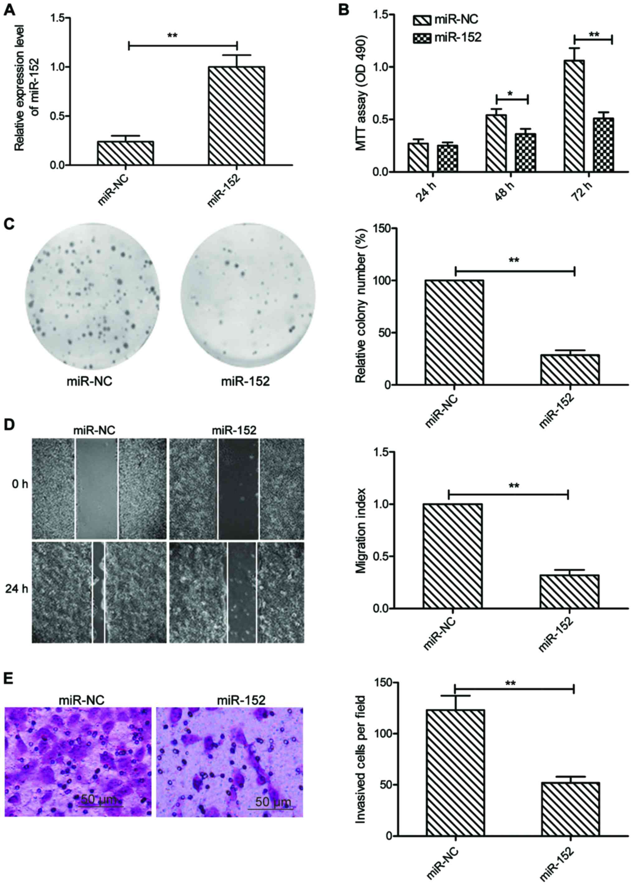
To explore the role of miR-152 in OSCC, we
transfected miR-152 mimic or miR-NC mimic into SCC-9 cells, then
cell proliferation, colony formation, migration and invasion were
determined in the above cells. It was found that SCC-9 cells
transfected with miR-152 mimic could significantly enhance miR-152
expression level compared to cells transfected with miR-NC
(Fig. 2A). MTT assay showed that
cell viability was significantly reduced in miR-152-transfected
cells compared with the miR-NC-transfected cells (Fig. 2B). Consistent with this result,
miR-152 overexpression significantly decreased the colony formation
of SCC-9 cells (Fig. 2C).
Subsequently, we evaluated whether miR-152 was able to suppress the
migration and invasion of OSCC cells by wound healing and Transwell
invasion assays. Our results showed that overexpression of miR-152
caused a significantly suppression of cell migration (Fig. 2D), and invasion (Fig. 2E) capability in SCC-9 cells. These
data implied that miR-152 might play a suppressive role in OSCC
development.
c-MET is a direct target of miR-152 in
OSCC cells
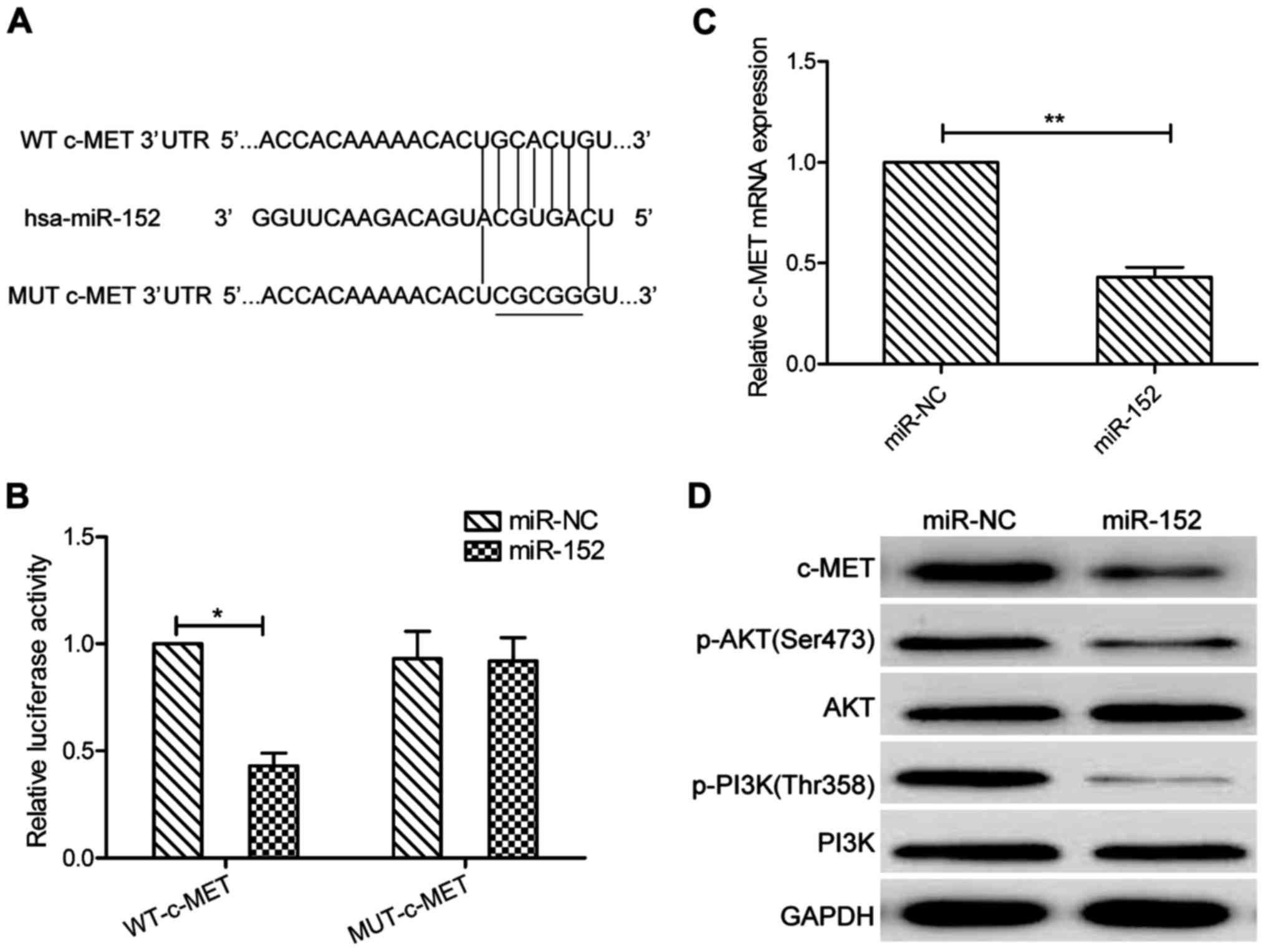
miR-152 targets were predicted using the three
algorithms: TargetScan, PicTar, and miRBase. A complementary
sequence of miR-152 to the 3′UTR of c-MET mRNA is shown in
Fig. 3A. The dual-luciferase
reporter assay showed that that ectopic expression of miR-152
significantly decreased the luciferase activity of the WT-c-MET but
not that of the MUT-c-MET-3′UTR in SCC-9 cells (P>0.05, Fig. 3B). More importantly, c-MET
expression on mRNA and protein levels were both downregulated in
SCC-9 cells transfected with miR-152 mimic compared those of cells
transfected with miR-NC (Fig. 3C and
D). In addition, our results also demonstrated that miR-152
significantly decreased p-PI3K and p-AKT protein expression, a
downstream pathway of c-MET (Fig.
3D). These findings implied that miR-152 directly targeted
c-MET in OSCC cells.
Inverse correlation between c-MET and
miR-152 expression in OSCC tissues
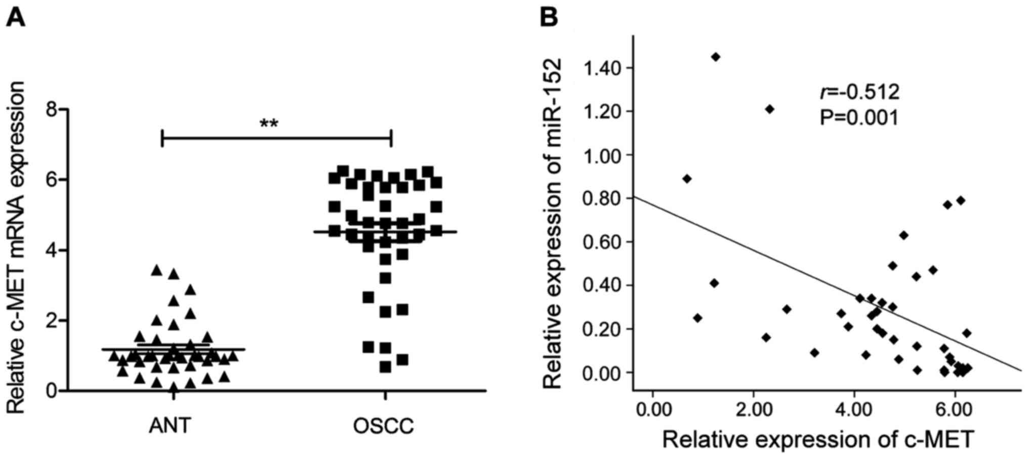
We next applied qRT-PCR to examine the c-MET
mRNA expression in 40 pairs of OSCC tissues and the corresponding
adjacent normal tissues. As shown in Fig. 4A, c-MET mRNA expression level
was significantly upregulated in OSCC tissues compared to the
adjacent normal tissues. Moreover, through Spearman's correlation
analysis, we found that the expression of miR-152 was inversely
correlated with c-MET mRNA expression in the 40 patients
with OSCC (r=−0.512; P=0.001; Fig.
4B).
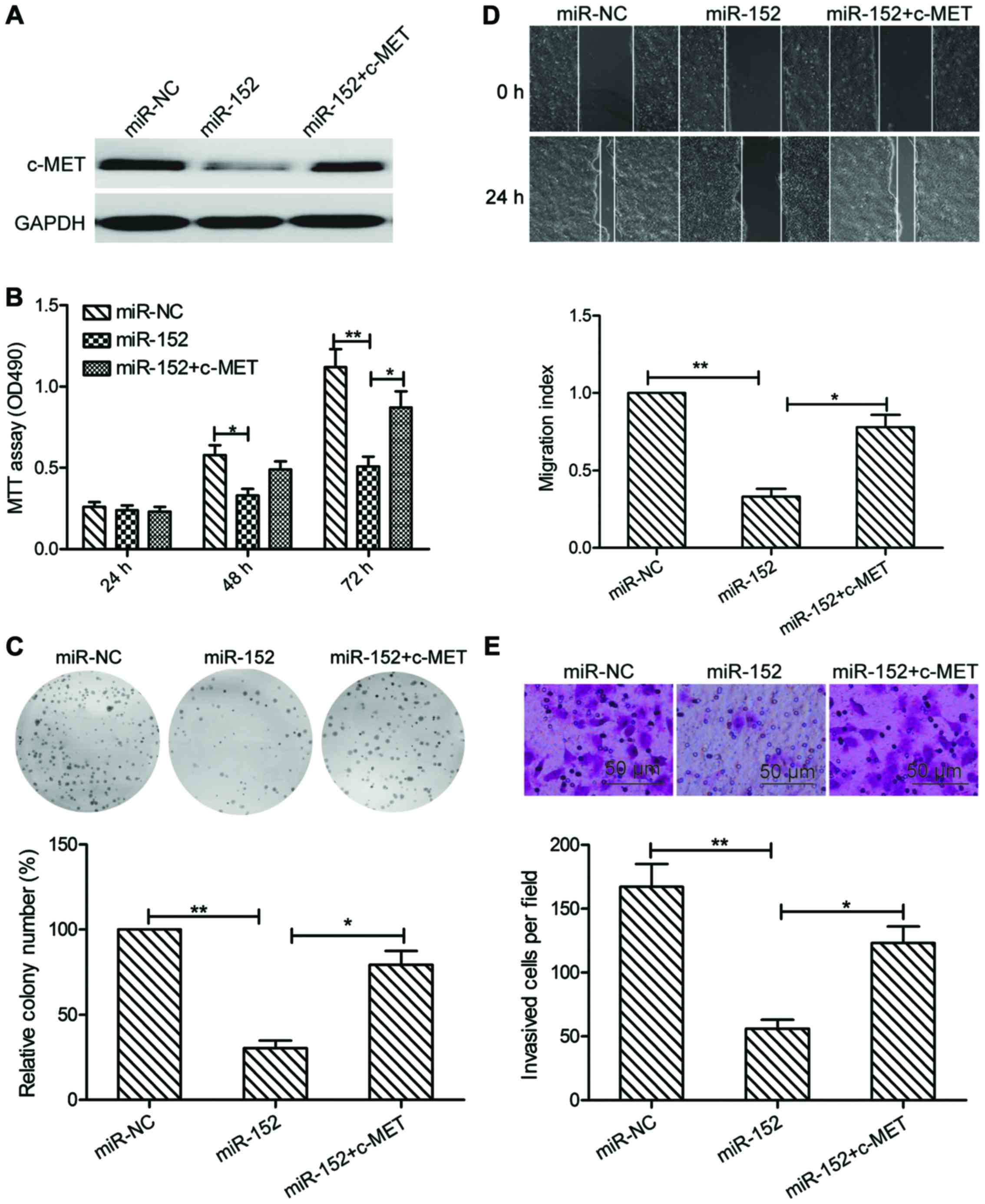
Overexpression of c-MET reverses the
tumor suppressive effect of miR-152 in OSCC
To evaluate if c-MET is responsible for the
functional effects of miR-152 in OSCC cells, SCC9 cells were
transfected with miR-NC, miR-152 mimic, or cotransfected with
miR-152 mimic and c-MET overexpression plasmid, respectively.
Subsequently, cell proliferation, colony formation, migration and
invasion were determined in the above cells. As indicated in
Fig. 5A, the protein expression of
c-MET was increased in the group of cells cotransfected with
miR-152 mimic and c-MET plasmid, compared with that transfected
only with miR-152 mimic. In addition, c-MET overexpression could
partially counteract the decrease of cell proliferation, colony
formation, migration and invasion and migration of SCC-9 cells by
miR-152 induction (Fig. 5B-E).
These results indicated that the suppressive effect of miR-152 on
OSCC cells may be via inhibition of c-MET.
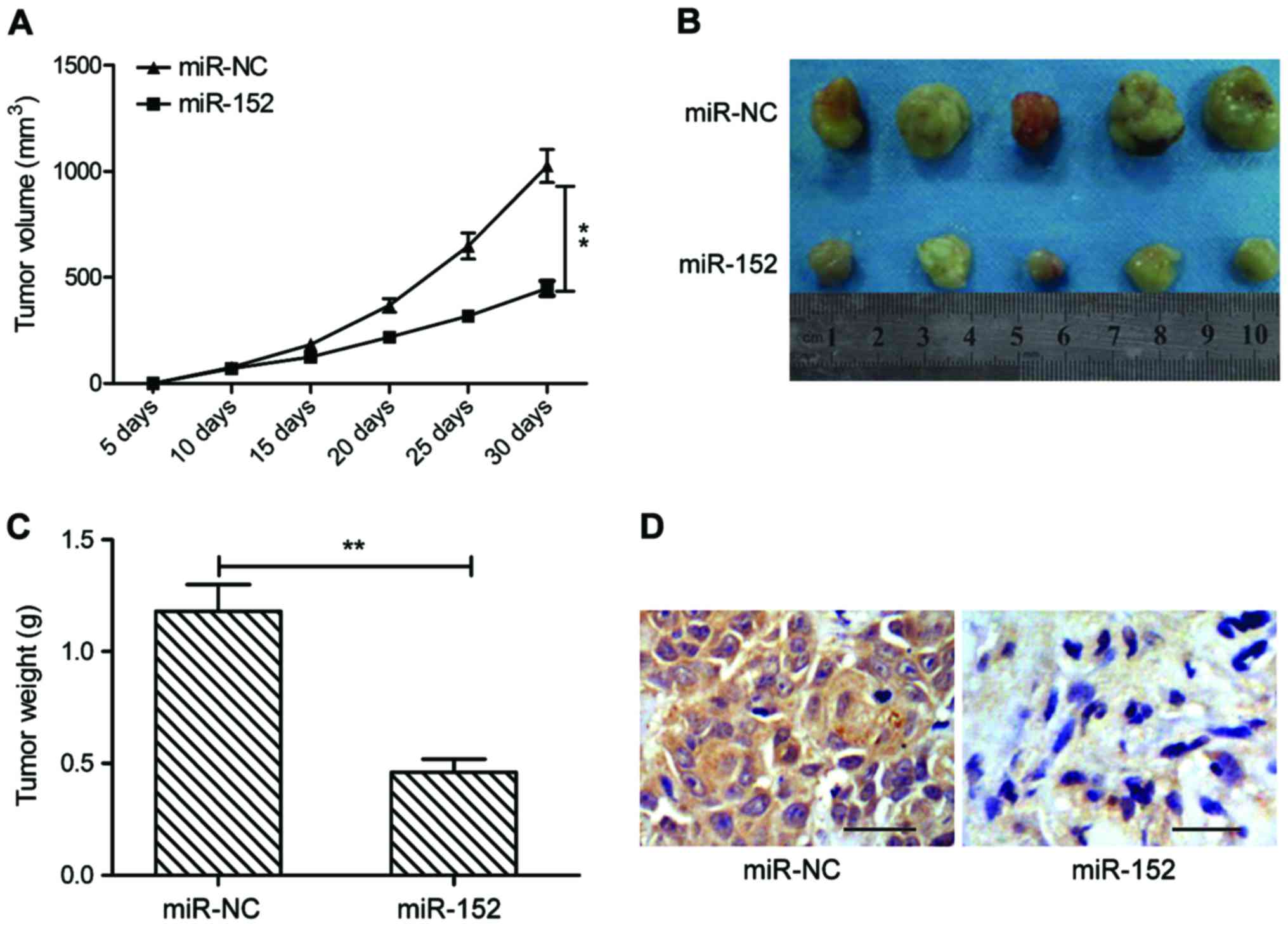
miR-152 suppresses tumorigenesis of
OSCC in vivo
To further test the effect of miR-152 on
tumorigenesis of OSCC in vivo, a xenograft mouse model was
established by subcutaneous injection of SCC-9 cells transfected
with miR-NC or miR-152 mimic, and tumor growth was measured. The
growth curve revealed that miR-152 overexpression group exhibited
significantly reduced tumor growth compared with miR-NC group
(Fig. 6A). At day 30
post-injection, the mice were sacrificed, and tumor tissues were
removed and weighed. miR-152 overexpression group had smaller size
(Fig. 6B) and weight (Fig. 6C) than that of miR-NC group. In
addition, we also found that c-MET expression was weaker in tumor
tissues of miR-152 compared to miR-NC group by
immunohistochemistry. These results implied that miR-152 suppressed
tumor growth of OSCC in vivo by repressing c-MET.
Discussion
It was well known that the development and
progression of OSCC is a very complicated process that involves the
aberrant expression of number of genes and alterations of multiple
signaling pathways (19,20). miRNAs have been reported to play
crucial roles in the development and progression of OSCC through
regulating various target genes (9,10),
thus, exploring novel miRNAs in OSCC is crucially important for
developing diagnosis marker and therapy agent. In this study, we
were interested in the role of miR-152 in OSCC progression.
miR-152, a member of miR-148/152 family, has been
reported to be downregulated, and to function as a tumor
suppressive miRNA in majority types of cancer (11–16).
In contrast, in neuroblastoma and nasopharyngeal carcinoma, miR-152
expression was upregulated, suggesting that miR-152 acts as an
oncogene miRNA (21,22). These results suggested that miR-152
could serve as either oncogene or tumor suppressor in different
types of cancer. Here, we investigated the expression of miR-152 in
OSCC tissues and cell lines, and found that miR-152 expression was
downregulated in OSCC cell lines and tissues, and decreased miR-152
was associated with lymph node metastasis and overall disease-free
survival of patients. Function assay demonstrated that miR-152
overexpression suppressed OSCC cell proliferation, colony
formation, migration and invasion in vitro, as well as
inhibited OSCC growth in nude mouse model. These results implied
that miR-152 function as a tumor suppressor miRNA in OSCC.
It has been shown that miR-152 inhibits the
proliferation and metastases of tumors via repressing multiple
genes, such as B7-H1 (23), PIK3CA
(24), WNT-1 (15), ALCAM (13), RTKN (25), neuropilin-1 (14) and PIK3R3 (12), all of which are known as important
mediators in tumorigenesis or metastasis in enhancing cell
proliferation, survival, migration, invasion capacity or disturbing
normal cell generation cycle, as well as inducing cell apoptosis.
Here, cellular-mesenchymal to epithelial transition factor (c-MET)
is indentified to be another important target of miR-152 by
luciferase reporter assay, qRT-PCR and western blotting. c-MET, a
receptor for hepatocyte growth factor (HGF), has been reported to
promote tumorigenicity in a variety of cancers by increasing
cellular proliferation, vascular invasion, epithelial-mesenchymal
transition, and migration or invasion capacity as well as
decreasing apoptosis (25–27). It has been showed that c-MET exerts
its biological role by activation of downstream signal transducer
molecules, such as protein kinase B, ERK, and signal transducer and
activator of transcription 3 (STAT3), PI3K/AKT, and Wnt signaling
pathway (28–30). These studies indicated that
inhibition of c-MET and its downstream signaling could be the
potential strategy for treatment of cancers. In OSCC, c-MET has
been shown to be upregulated in OSCC tissues, and its expression
was associated with tumor stage (31). In addition, downregulating of c-MET
could inhibit OSCC cell proliferation and invasion (32). Thus, in our study, we selected c-MET
as a direct target of miR-152, and found that overexpression
miR-152 significantly inhibited c-MET and the downstream signaling
pathway, c-MET expression was upregulated in OSCC tissues, and
inversely correlated with miR-152 expression. Of note, we found
that restoring c-MET expression attenuated miR-152 induced
inhibitory effects in OSCC cells. In vivo study confirmed
that restoration of miR-152 suppressed tumor growth in xenograft
nude mice by repressing c-MET. Collectively, these findings
demonstrated that miR-152 exerts suppressive role in OSCC, at least
in part, by repressing c-MET.
Some limitations in this study should be considered.
First, the function of miR-152 was evaluated through overexpression
of miR-152 in a gain-of-function model. Loss-of-function studies
via downregulation of miR-152 in OSCC cell lines are needed to
verify our findings. Second, using two or more cell lines are
needed to validate the role of miR-152 in tumor development and
progression. Third, the miR-152 has multiple target genes which
could influence the progression and metastases of OSCC, it is
anticipated that more molecular mechanisms would be uncovered in
our future study.
Taken together, this study showed that miR-152
expression was downregulated in OSCC cell lines and tissues, and
low expression is associated with lymph node metastasis and the
overall disease-free survival of patients. Further investigation
revealed that overexpression of miR-152 inhibited cell
proliferation and colony formation, migration and invasion, as well
as suppressed tumor growth in vivo by repressing c-MET.
These results indicate that miR-152 may serve as a useful
therapeutic strategy for OSCC.
References
|
1
|
Siegel R, Ma J, Zou Z and Jemal A: Cancer
statistics, 2014. CA Cancer J Clin. 64:9–29. 2014. View Article : Google Scholar : PubMed/NCBI
|
|
2
|
Liao CT, Hsueh C, Lee LY, Lin CY, Fan KH,
Wang HM, Huang SF, Chen IH, Kang CJ, Ng SH, et al: Neck dissection
field and lymph node density predict prognosis in patients with
oral cavity cancer and pathological node metastases treated with
adjuvant therapy. Oral Oncol. 48:329–336. 2012. View Article : Google Scholar : PubMed/NCBI
|
|
3
|
Jerjes W, Upile T, Petrie A, Riskalla A,
Hamdoon Z, Vourvachis M, Karavidas K, Jay A, Sandison A, Thomas GJ,
et al: Clinicopathological parameters, recurrence, locoregional and
distant metastasis in 115 T1-T2 oral squamous cell carcinoma
patients. Head Neck Oncol. 2:92010. View Article : Google Scholar : PubMed/NCBI
|
|
4
|
Bartel DP: MicroRNAs: Genomics,
biogenesis, mechanism, and function. Cell. 116:281–297. 2004.
View Article : Google Scholar : PubMed/NCBI
|
|
5
|
Malan-Müller S, Hemmings SM and Seedat S:
Big effects of small RNAs: A review of microRNAs in anxiety. Mol
Neurobiol. 47:726–739. 2013. View Article : Google Scholar : PubMed/NCBI
|
|
6
|
McManus MT: MicroRNAs and cancer. Semin
Cancer Biol. 13:253–258. 2003. View Article : Google Scholar : PubMed/NCBI
|
|
7
|
Farazi TA, Spitzer JI, Morozov P and
Tuschl T: miRNAs in human cancer. J Pathol. 223:102–115. 2011.
View Article : Google Scholar : PubMed/NCBI
|
|
8
|
Garzon R and Marcucci G: Potential of
microRNAs for cancer diagnostics, prognostication and therapy. Curr
Opin Oncol. 24:655–659. 2012. View Article : Google Scholar : PubMed/NCBI
|
|
9
|
Prasad G, Seers C, Reynolds E and
McCullough MJ: A panel of microRNAs can be used to determine oral
squamous cell carcinoma. J Oral Pathol Med. 46:940–948.
2017.PubMed/NCBI
|
|
10
|
Ries J, Baran C, Wehrhan F, Weber M,
Neukam FW, Krautheim-Zenk A and Nkenke E: Prognostic significance
of altered miRNA expression in whole blood of OSCC patients. Oncol
Rep. 37:3467–3474. 2017. View Article : Google Scholar : PubMed/NCBI
|
|
11
|
Xie G, Li W, Li R, Wu K, Zhao E, Zhang Y,
Zhang P, Shi L, Wang D, Yin Y, et al: Helicobacter pylori promote
B7-H1 expression by suppressing miR-152 and miR-200b in gastric
cancer cells. PLoS One. 12:e01688222017. View Article : Google Scholar : PubMed/NCBI
|
|
12
|
Li B, Xie Z and Li B: miR-152 functions as
a tumor suppressor in colorectal cancer by targeting PIK3R3. Tumour
Biol. 37:10075–10084. 2016. View Article : Google Scholar : PubMed/NCBI
|
|
13
|
Chen MJ, Cheng YM, Chen CC, Chen YC and
Shen CJ: miR-148a and miR-152 reduce tamoxifen resistance in
ER+ breast cancer via downregulating ALCAM. Biochem
Biophys Res Commun. 483:840–846. 2017. View Article : Google Scholar : PubMed/NCBI
|
|
14
|
Zhang YJ, Liu XC, Du J and Zhang YJ:
miR-152 regulates metastases of non-small cell lung cancer cells by
targeting neuropilin-1. Int J Clin Exp Pathol. 8:14235–14240.
2015.PubMed/NCBI
|
|
15
|
Tang XL, Lin L, Song LN and Tang XH:
Hypoxia-inducible miR-152 suppresses the expression of WNT1 and
ERBB3, and inhibits the proliferation of cervical cancer cells. Exp
Biol Med (Maywood). 241:1429–1437. 2016. View Article : Google Scholar : PubMed/NCBI
|
|
16
|
Zhu C, Li J, Ding Q, Cheng G, Zhou H, Tao
L, Cai H, Li P, Cao Q, Ju X, et al: miR-152 controls migration and
invasive potential by targeting TGFα in prostate cancer cell lines.
Prostate. 73:1082–1089. 2013. View Article : Google Scholar : PubMed/NCBI
|
|
17
|
Li B, Yang XX, Wang D and Ji HK:
MicroRNA-138 inhibits proliferation of cervical cancer cells by
targeting c-Met. Eur Rev Med Pharmacol Sci. 20:1109–1114.
2016.PubMed/NCBI
|
|
18
|
Chau L, Jabara JT, Lai W, Svider PF,
Warner BM, Lin HS, Raza SN and Fribley AM: Topical agents for oral
cancer chemoprevention: A systematic review of the literature. Oral
Oncol. 67:153–159. 2017. View Article : Google Scholar : PubMed/NCBI
|
|
19
|
Gharat SA, Momin M and Bhavsar C: Oral
squamous cell carcinoma: Current treatment strategies and
nanotechnology-based approaches for prevention and therapy. Crit
Rev Ther Drug Carrier Syst. 33:363–400. 2016. View Article : Google Scholar : PubMed/NCBI
|
|
20
|
Jasinski-Bergner S, Stoehr C, Bukur J,
Massa C, Braun J, Hüttelmaier S, Spath V, Wartenberg R, Legal W,
Taubert H, et al: Clinical relevance of miR-mediated HLA-G
regulation and the associated immune cell infiltration in renal
cell carcinoma. OncoImmunology. 4:e10088052015. View Article : Google Scholar : PubMed/NCBI
|
|
21
|
Liu DZ, Ander BP, Tian Y, Stamova B,
Jickling GC, Davis RR and Sharp FR: Integrated analysis of mRNA and
microRNA expression in mature neurons, neural progenitor cells and
neuroblastoma cells. Gene. 495:120–127. 2012. View Article : Google Scholar : PubMed/NCBI
|
|
22
|
Huang S, Li X and Zhu H: MicroRNA-152
Targets phosphatase and tensin homolog to inhibit apoptosis and
promote cell migration of nasopharyngeal carcinoma cells. Med Sci
Monit. 22:4330–4337. 2016. View Article : Google Scholar : PubMed/NCBI
|
|
23
|
Wang Y, Wang D, Xie G, Yin Y, Zhao E, Tao
K and Li R: MicroRNA-152 regulates immune response via targeting
B7-H1 in gastric carcinoma. Oncotarget. 8:28125–28134.
2017.PubMed/NCBI
|
|
24
|
Ge S, Wang D, Kong Q, Gao W and Sun J:
Function of miR-152 as a tumor suppressor in human breast cancer by
targeting PIK3CA. Oncol Res. 25:1363–1371. 2017. View Article : Google Scholar : PubMed/NCBI
|
|
25
|
Zhou J, Zhang Y, Qi Y, Yu D, Shao Q and
Liang J: MicroRNA-152 inhibits tumor cell growth by directly
targeting RTKN in hepatocellular carcinoma. Oncol Rep.
37:1227–1234. 2017. View Article : Google Scholar : PubMed/NCBI
|
|
26
|
Skead G and Govender D: Gene of the month:
MET. J Clin Pathol. 68:405–409. 2015. View Article : Google Scholar : PubMed/NCBI
|
|
27
|
Marano L, Chiari R, Fabozzi A, De Vita F,
Boccardi V, Roviello G, Petrioli R, Marrelli D, Roviello F and
Patriti A: c-Met targeting in advanced gastric cancer: An open
challenge. Cancer Lett. 365:30–36. 2015. View Article : Google Scholar : PubMed/NCBI
|
|
28
|
Boccaccio C, Luraghi P and Comoglio PM:
MET-mediated resistance to EGFR inhibitors: An old liaison rooted
in colorectal cancer stem cells. Cancer Res. 74:3647–3651. 2014.
View Article : Google Scholar : PubMed/NCBI
|
|
29
|
Ho-Yen CM, Jones JL and Kermorgant S: The
clinical and functional significance of c-Met in breast cancer: A
review. Breast Cancer Res. 17:522015. View Article : Google Scholar : PubMed/NCBI
|
|
30
|
Mazzone M and Comoglio PM: The Met
pathway: Master switch and drug target in cancer progression. FASEB
J. 20:1611–1621. 2006. View Article : Google Scholar : PubMed/NCBI
|
|
31
|
Freudlsperger C, Alexander D, Reinert S
and Hoffmann J: Prognostic value of c-Met expression in oral
squamous cell carcinoma. Exp Ther Med. 1:69–72. 2010.PubMed/NCBI
|
|
32
|
Yasui H, Ohnishi Y, Nakajima M and Nozaki
M: Migration of oral squamous cell carcinoma cells are induced by
HGF/c-Met signalling via lamellipodia and filopodia formation.
Oncol Rep. 37:3674–3680. 2017. View Article : Google Scholar : PubMed/NCBI
|