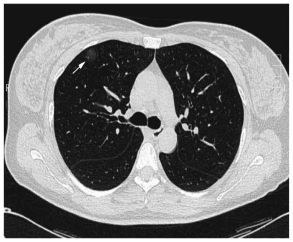
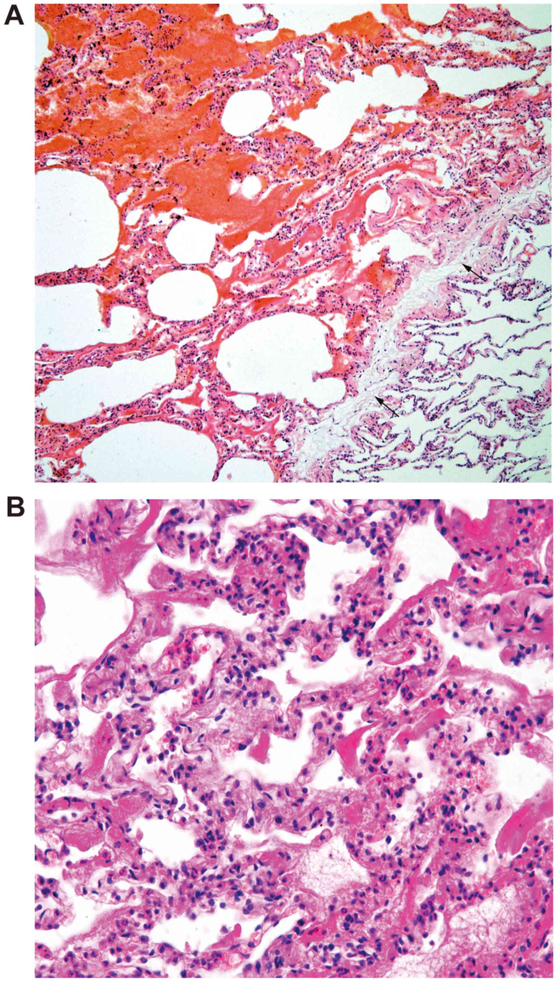
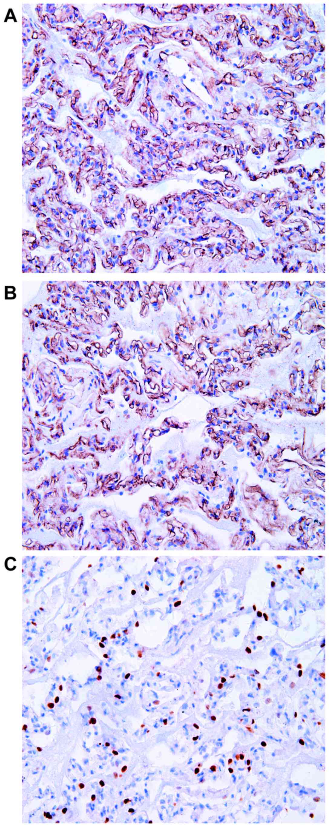
|
1
|
Fugo K, Matsuno Y, Okamoto K, Kusumoto M,
Maeshima A, Kaji M, Takabatake H, Kondo H and Moriyama N: Solitary
capillary hemangioma of the lung: Report of 2 resected cases
detected by high-resolution CT. Am J Surg Pathol. 30:750–753. 2006.
View Article : Google Scholar : PubMed/NCBI
|
|
2
|
Kato H, Oizumi H, Kanauchi N and Sadahiro
M: A case of pulmonary capillary hemangioma diagnosed by
thoraco-scopic segmentectomy. J Jpn Assoc Chest Surg. 23:932–935.
2009. View Article : Google Scholar
|
|
3
|
Hakiri S, Agatsuma H and Yoshioka H: A
resected case of capillary hemangioma of the lung suspected to be
lung cancer on chest computed tomography. Haigan. 50:841–845. 2010.
View Article : Google Scholar
|
|
4
|
Sakaguchi Y, Isowa N, Tokuyasu H and Miura
H: A resected case of solitary pulmonary capillary hemangioma
showing pure ground glass opacity. Ann Thorac Cardiovasc Surg.
20:578–581. 2014. View Article : Google Scholar : PubMed/NCBI
|
|
5
|
Taniguchi D, Taniguchi H, Sano I, Tamura
K, Shindou H, Shimizu K, Hamasaki K, Nakazaki T, Shigematsu K and
Takahara O: Solitary capillary hemangioma in the lung: Report of a
case. Kyobu Geka. 63:423–425. 2010.(In Chinese). PubMed/NCBI
|
|
6
|
Yanagawa N, Kato H and Kanauchi N: Two
cases of solitary peripheral small lung tumor needed to
differentiate from small lung adenocarcinoma. Jpn J Diagn Pathol.
24:426–429. 2007.
|
|
7
|
Uegami S, Hirai S, Mitsui N, Matsuura Y
and Hamanaka Y: A case of solitary capillary hemangioma of the
lung. J Jpn Assoc Chest Surg. 22:641–644. 2008. View Article : Google Scholar
|
|
8
|
Shimada Y, Murakawa T, Sano A, Fukami T,
Yoshida Y, Inoue Y, Morita S, Fukayama M and Nakajima J: Capillary
hemangiomas of the lung presenting as ground glass opacities by
high resolution computed tomography. Kyobu Geka. 65:1038–1043.
2012.(In Chinese). PubMed/NCBI
|
|
9
|
Matsushita M, Kawakami S, Matsushita T,
Sugiyama Y, Endo M, Shimojo H, Toishi M and Kadoya M: Changes in CT
density of solitary capillary hemangioma of the lung upon varying
patient position. Jpn J Radiol. 30:772–776. 2012. View Article : Google Scholar : PubMed/NCBI
|
|
10
|
Isaka T, Yokose T, Ito H, Washimi K,
Imamura N, Watanabe M, Imai K, Nishii T, Yamada K, Nakayama H, et
al: Case of solitary pulmonary capillary hemangioma: Pathological
features based on frozen section analysis. Pathol Int. 63:615–618.
2013. View Article : Google Scholar : PubMed/NCBI
|
|
11
|
Travis WD, Brambilla E, Nicholson AG,
Yatabe Y, Austin JHM, Beasley MB, Chirieac LR, Dacic S, Duhig E,
Flieder DB, et al: WHO Panel: The 2015 World Health Organization
Classification of Lung Tumors: Impact of Genetic, Clinical and
Radiologic Advances Since the 2004 Classification. J Thorac Oncol.
10:1243–1260. 2015. View Article : Google Scholar : PubMed/NCBI
|
|
12
|
Chen XL and Chen XY: The significance of
serum tumor markers in lung cancer diagnosis. J Clin Pulm Med.
9:590–592. 2004.
|
|
13
|
Li X, Jin ML, Wei P, Dai HP, Cui A, Zhang
YG, Diao XL and Zhao HY: Pulmonary capillary hemangiomatosis: A
clinicopathologic analysis of 2 cases with review of literature.
Zhonghua Bing Li Xue Za Zhi. 41:16–19. 2012.(In Chinese).
PubMed/NCBI
|
|
14
|
Frazier AA, Franks TJ, Mohammed TL,
Ozbudak IH and Galvin JR: From the archives of the AFIP: Pulmonary
veno-occlusive disease and pulmonary capillary hemangiomatosis.
Radiographics. 27:867–882. 2007. View Article : Google Scholar : PubMed/NCBI
|
|
15
|
Lv YG, Bao JH, Xu DU, Yan QH, Li YJ, Yuan
DL and Ma JH: Characteristic analysis of pulmonary ground-glass
lesions with the help of 64-slice CT technology. Eur Rev Med
Pharmacol Sci. 21:3212–3217. 2017.PubMed/NCBI
|