Introduction
Endometriosis is an estrogen-dependent condition
affecting 10% of women in their reproductive life (1). It is characterized by the growth of
endometrial-like tissues at sites outside of the uterus and is
associated with pelvic pain and infertility. The pathogenesis of
endometriosis is still unclear, but a number of factors are known,
including retrograde menstruation, coelomic metaplasia, induction
theory and hormonal, immunologic determinant, stem cell and
genetic/epigenetic factors (2).
According to Sampson's theory, endometriotic lesions are originated
from the shed endometrial tissues of the uterus through retrograde
menstrual dissemination, move and attach to the ectopic sites and
then form endometriotic lesions (3). The retrograde endometrium
encounters a number of challenges in the peritoneal cavity,
including mechanical damage, abundant free iron and high oxidative
levels, lack of oxygen, or immune attack (4). Periodic hemorrhage from ectopic
endometriotic lesions is a hallmark of endometriosis and causes
persistent iron overload (5).
Despite a number of difficulties, endometriotic tissues may
overcome these hurdles in the microenvironment by using mechanisms
to escape from cell death. Retrograde menstruation occurs in as a
≥90% of menstruating women, but the incidence rate for
endometriosis is only 10% (6).
Thus, only a fraction of endometriotic cells may escape cell death
in the harsh environment of intraperitoneal iron overload and
hypoxia. This suggests that there must be other genetic and
environmental factors that determine the onset and progression of
this disease, such as mechanisms that prevent cell death (4).
Regulated cell death (RCD) is characterized by
specific signaling cascades orchestrated by diverse biomolecules
(7). RCD is further classified
into apoptotic and non-apoptotic subcategories [i.e., autophagy
(mitophagy), ferroptosis, necroptosis, pyroptosis, anoikis and
cuproptosis] (7). Autophagy,
defined as self-degradation, is a process related to nonapoptotic
cell death induced by a large number of intracellular/extracellular
stimuli (8). Damaged proteins
and organelles are removed and recycled by autophagy (9). This process plays a pivotal role in
maintaining quality control and cellular homeostasis. On the other
hand, ferroptosis is defined as a reactive oxygen species
(ROS)-dependent cell death related to iron accumulation and lipid
peroxidation, which is different from other forms of cell death
(10). We recently reported that
autophagy is dynamically regulated by various intrinsic [e.g.,
phosphatidylinositol 3-kinase (PI3K)/protein kinase B (AKT)/mTOR)
and extrinsic (e.g., hypoxia and oxidative stress)] pathways,
effectively attenuating the induction of apoptosis and promoting
the survival of endometriotic cells (11). The periodic and repeated bleeding
in endometriotic lesions is thought to trigger iron-dependent cell
death known as ferroptosis. However, endometriotic cells may have
acquired a potential mechanism to escape cell death in the harsh
environment of intraperitoneal iron overload, hypoxia and nutrient
deprivation by regulating autophagy and ferroptosis. The present
review summarized the current understanding of the mechanisms
underlying autophagy and ferroptosis in endometriosis and discussed
spatiotemporal orchestration of ferroptosis-mediated cell death
regulation.
Methods
Search strategy and selection
criteria
The present review conducted a narrative review of
the targeted literature that focused on autophagy and ferroptosis
in endometriosis. These mechanisms are well studied in cancer cells
and the present review first drew information from these studies.
It then summarized the roles of molecules regulating autophagy and
ferroptosis that have been reported so far in endometriosis.
Electronic databases including PubMed (https://pubmed.ncbi.nlm.nih.gov/) and Google Scholar
(https://scholar.google.jp/) were
searched for literature published up to the October 31, 2023,
combining the following keywords: 'Autophagy', 'ferroptosis',
'regulated cell death', 'survival', and 'endometriosis'. The search
terms were combined using the Boolean operators And OR (Table I). Additionally, a manual
reference search of published articles was conducted. Included
studies comprised original research publications in English and
reference lists from review articles. Duplicated studies,
literature irrelevant to the research topic and non-English
publications were excluded.
 | Table IKeyword and search term
combinations. |
Table I
Keyword and search term
combinations.
| Search mode | Keyword and search
term combinations |
|---|
| Search term 1 | Endometriosis OR
Ovarian endometriosis OR Endometriotic cyst |
| Search term 2 | Autophagy OR
Autophagic |
| Search term 3 | Ferroptosis OR
Ferroptotic |
| Search term 4 | Regulated cell
death |
| Search term 5 | Survival |
| Search | Search term 1 AND
Search term 2 |
| Search term 1 AND
Search term 3 |
| Search term 1 AND
Search term 2 AND Search term 3 |
| Search term 1 AND
Search term 2 AND Search term 4 OR Search term 5 |
| Search term 1 AND
Search term 3 AND Search term 4 OR Search term 5 |
| Search term 2 AND
Search term 3 AND Search term 4 OR Search term 5 |
The flowchart depicted in Fig. 1 outlines the study selection
process, detailing both inclusion and exclusion criteria. The
initial phase involves identifying records through electronic
database searches, manual searches and the reference lists of
relevant articles and reviews. Titles and abstracts underwent a
preliminary screening. After duplicates were removed, these titles
and abstracts were reviewed to discard non-relevant studies. The
final phase of eligibility involved analyzing the full-text
articles, excluding any from which detailed data could not be
obtained. The authors independently evaluated the articles to
determine their suitability for inclusion or exclusion before
reviewing the full texts. The properties of the identified
molecules were searched in the National Library of Medicine
database (https://www.ncbi.nlm.nih.gov/).
Molecular and regulatory network mechanisms
controlling autophagy
This section briefly summarizes the current
understanding of the development and progression of endometriosis,
focusing on molecules that influence autophagy-mediated quality
control. Autophagy is an evolutionarily conserved self-degrading
process mediated by autophagosomes that eliminates various toxic
substrates and maintains cellular homeostasis and survival
(12). Autophagosomes,
characterized by the formation of lipid-associated double membrane
structures, fuse with lysosomes for cargo degradation (12,13). Mechanistically, autophagy is
controlled by various autophagy-related gene (ATG) proteins
(14). Among the ATG proteins,
microtubule associated protein 1 light chain 3 (MAP1LC3, also known
as LC3) and beclin1 (BECN1) have traditionally been evaluated as
autophagy-related biomarkers (12,13). Beclin1 promotes autophagy and LC3
protein levels reflects autophagy induction (15). For more details, see our recent
review article on the molecular mechanisms of autophagy and
apoptosis in endometriosis (16). Autophagy can determine the
cellular fate (14). For
example, autophagy inhibits tumor initiation by eliminating
potential oncogenic proteins and impaired organelles that cause
genome instability (11,14). On the other hand, autophagy can
maintain energy homeostasis and promote cancer cell growth by
generating the necessary nutrients and energy during stress
(11,14). Autophagy has the dichotomous role
as both a tumor suppressor and tumor promoter. The present review
summarized the role of autophagy in endometriosis, dividing it into
pro-autophagy and anti-autophagy. Autophagy in endometriosis can be
triggered by a number of factors such as oxidative stress, hypoxia,
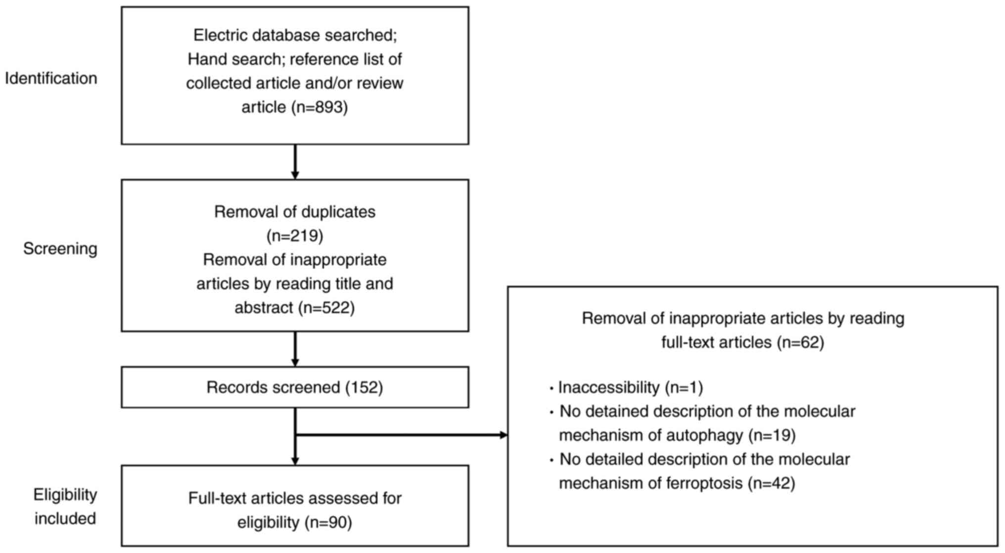
female hormones and specific signaling pathways (Fig. 2) (8).
Anti-autophagy Estrogen
Estrogen promotes the proliferation of human
endometrial stromal cells during the secretory phase via
upregulation of C-X-C motif chemokine ligand 12 (CXCL12)/C-X-C
motif chemokine receptor 4 (CXCR4) expression (17). CXCL12 has been reported to
downregulate autophagy (17). It
has been shown that levels of both CXCR4 and CXCL12 in
endometriotic epithelial cells are higher than in normal
endometrium (17,18). Therefore, estrogen dominance and
progesterone resistance in endometriosis decrease autophagy levels
through the activation of the CXCL12/CXCR4 axis (17,18). Estrogen-induced downregulation of
autophagy is thought to promote the progression of
endometriosis.
mTOR
mTOR expression level markedly increases in the
order of normal endometrium, eutopic endometrium and ectopic
endometrium (19,20). This suggests that the activation
of the PI3K/AKT/mTOR pathway can reduce autophagy induction in
endometriotic tissues. Notably, rapamycin, an mTOR inhibitor,
confers therapeutic efficacy against endometriosis in animal models
for endometriosis through upregulation of autophagy-related protein
expression (13). Rapamycin also
decreases cell viability and increases iron content and ROS
generation (13). Additionally,
phosphatase and tensin homolog (PTEN) and p53 are known to be genes
involved in the regulation of autophagy in endometriosis.
Activating mutations of PTEN gene are able to confer
growth-promoting properties in endometriotic cells through
regulation of autophagy induction (21). The frequency of PTEN gene
mutations is ~21% in ovarian endometriosis (22). Tumor protein p53 (TP53), the most
commonly mutated tumor suppressor gene, initiates cellular
responses, such as cell cycle arrest, in response to stress signals
(23). A reduction in p53
expression activates the mTOR pathway and negatively regulates
autophagy, resulting in endometriosis cell proliferation (23). p53 expression was decreased in
endometriotic tissues compared with normal endometria (24). Furthermore, there was a
significant reduction in p53 expression in women with severe/late
stage disease (25). Therefore,
mTOR is considered a key gene that negatively regulates autophagy
activity not only in cancer but also in endometriosis.
Pro-autophagy ROS
Iron-induced ROS induce autophagy through
suppressing the mTOR signaling pathway (26). Moreover, hemoglobin, heme and
iron caused by periodic hemorrhage induce HO-1 expression via ROS
(27). HO-1 catalyzes heme
breakdown to generate biliverdin, carbon monoxide and iron
(27). HO-1 may further promote
autophagy via iron overload (24).
Hypoxia
Additionally, hypoxia has been reported to induce
enhancements of migration and invasion by activating HIF-1α-induced
autophagy in human endometrial stromal cells (28,29). Indeed, HIF-1 promotes cell
survival and blocks cell death in human ectopic endometriosis
stromal cells (30).
From the aforementioned, both iron-mediated ROS and
hypoxia-induced HIF-1α have been shown to induce autophagy in
endometriotic cells. The generation of iron-mediated ROS that
exceeds endogenous antioxidant capacity leads to oxidative cell
damage. Therefore, well-coordinated quality control mechanisms via
autophagy, involving timely removal of damaged multiple organelles,
are essential for cellular homeostasis and survival. Considering
the microenvironment, which is dominated by iron overload and
hypoxia, effective autophagy could promote the very early stages of
endometriosis development. Meanwhile, estrogen/CXCL12/CXCR4 23
(18) and PI3K/AKT/mTOR pathways
(31) inhibit autophagy
induction in endometriosis. Decreased autophagy may promote the
growth and progression of pre-existing endometriotic lesions. Thus,
both intrinsic and extrinsic factors have emerged as key regulators
influencing autophagy (18,26,30,31). Collectively, autophagy may play a
dual role in endometriosis cell survival, particularly in onset and
progression. Previous reports have shown that autophagy is either
activated or suppressed in endometriosis and some of the
discrepancy between studies may greatly vary depending on
environmental conditions and stage of disease (18,26,30,31).
Molecular and regulatory network mechanisms
controlling iron metabolism and ferritinophagy
The development and progression of endometriosis may
rely not only on the inherent proliferative nature but also on the
ever-changing surrounding microenvironment (31,32). Endometriotic lesion growth can be
promoted via multiple intrinsic pathways, including not only
estrogen and PI3K/AKT/mTOR as aforementioned, but also
mitogen-activated protein kinases (MAPK)/mitogen-activated protein
kinase kinase/extracellular-signal-regulated kinases and nuclear
factor-kappa B (33,34), while it may also be influenced by
extrinsic factors such as iron overload and hypoxia (16,35,36). The present section mainly focused
on the molecular mechanisms of iron, hypoxia-inducible factor 1
(HIF-1) and PI3K/AKT/mTOR pathways that regulate cell survival and
summarized their roles in the development and progression of
endometriosis.
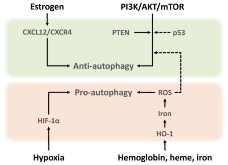
Iron metabolism and its dynamics
Retrograde endometrial tissues encounter several
challenges, including iron overload and hypoxia. Iron, ferritin and
transferrin contents and transferrin saturation are markedly
elevated in the peritoneal fluid of women with endometriosis
compared with the controls (37). Moreover, an increase in
follicular fluid iron and ferritin contents, transferrin saturation
and transferrin receptor (TFRC) expression was demonstrated in the
affected ovaries compared with the unaffected ovaries (38). Additionally, the content of iron
is known to be ~1,000 times higher in the endometriotic cysts
compared with other types of benign cysts (39). Endometriotic cysts contain large
amounts of iron, but absolute concentrations vary by over 10-fold
between patients (244.4±204.9 mg/l; range 65.3-1046.3 mg/l)
(40). Furthermore, Iwabuchi
et al (41) found that
endometriotic cysts contain more ferric (Fe3+) than
ferrous (Fe2+) ions. These findings have drawn much
attention to the potential role of iron uptake, storage,
detoxification, metabolism and iron homeostasis in the development,
progression and pathogenesis of endometriosis (5). Circulating transferrin (TF)
reversibly bound to Fe3+ is recognized by the TFRC and
then transported to the endosome (Fig. 3) (42). After Fe3+ is reduced
to Fe2+ by Steap3 metalloreductase within the endosomes,
Fe2+ is transported out of endosomes via divalent metal
transporter 1 (DMT1; also known as SLC11A2) (43). A portion of the cytosolic
Fe2+ is transported to the mitochondria and the
remaining labile iron is stored by ferritin, made up of ferritin
light chain (FTL) and ferritin heavy chain 1 (FTH1) (42,43). Nuclear receptor coactivator 4
(NCOA4) is required for delivery of ferritin to lysosomes and its
degradation (44). Thus,
downregulation of NCOA4 inhibits ferritin degradation and reduces
free iron level (44).
Ferritinophagy is a specialized form of autophagy that is essential
for transforming intracellular ferritin-bound iron into free iron.
Finally, ferroportin (FPN; also known as SLC40A1) is the cellular
iron exporter, maintaining cellular and systemic iron homeostasis
(45). In addition, the labile
iron pool causes the release of hazardous ROS (7) and oxidative stress (46) via Fenton reaction
(Fe2+ + H2O2 → Fe3+ +
OH− + OH) (46). ROS
include oxygen free radicals, such as superoxide anion, hydrogen
peroxide, hydroxyl radical and free nitrogen radicals (27). On the other hand, endometriotic
cells need increased antioxidant capacity to avoid cell death.
Glutathione (GSH) and glutathione peroxidase (GPX) are known to
play important roles as antioxidant defense systems that protect
cells against oxidative damage in endometriosis (46). However, females with
endometriosis show higher levels of oxidative stress markers [e.g.,
8-hydroxy-2-deoxyguanosine, malondialdehyde (MDA), or lipid
peroxide] and inadequate antioxidant defenses (e.g., superoxide
dismutase, GPX4, HO-1 and catalase, GSH and vitamins A, C and E)
compared with females without endometriosis (46,47). Therefore, endometriosis may avoid
oxidative stress and maintain their viability in ways other than
the antioxidant system. Indeed, an animal model showed that
erythrocyte injection increased the number and size of
endometriotic lesions relative to the untreated control groups
(35). The use of
desferrioxamine, an iron chelator, confirmed that iron was directly
responsible for lesion growth (35). This seems to contradict an
intuitive theory that iron induces cell death through oxidative
damage. Iron may be essential for early lesion development. ROS is
known to play a dual role as both beneficial (e.g., cell survival)
and deleterious (e.g., cell death) effects (46), but the underlying mechanisms are
unknown. The role of iron metabolism in the pathogenesis of
endometriosis is a hot topic.
 | Figure 3Molecular and regulatory network
mechanisms controlling autophagy in endometriosis. Among the
molecules that control autophagy, the upper and lower rows show the
autophagy-inhibiting and autophagy-promoting pathway, respectively.
Solid and dashed arrows represent promotion and inhibition,
respectively. TFRC, transferrin receptor; TF, transferrin; PI3K,
phosphatidylinositol 3-kinase; AKT, protein kinase B; mTOR,
mammalian target of rapamycin; FPN, ferroportin; STEAP3, Steap3
metalloreductase; DMT1, divalent metal transporter 1; NCOA4,
nuclear receptor coactivator 4. |
HIF-1 as a molecule regulating cell
survival
In addition, hypoxia, a hallmark of endometriosis,
plays a key role in the mitochondrial function and metabolic
conversion between glycolysis and oxidative phosphorylation and
metabolic remodeling is known to contribute to cell survival and
disease progression (36). A
hypoxic environment stimulates the proliferation of endometriotic
cells through the upregulation of HIF-1α expression (48). Furthermore, HIF-1α regulates the
expression of estrogen receptors, promotes inflammatory cytokine
levels and accelerates angiogenesis (4). However, oxidative stress is also
caused by hypoxia (48).
Endometriotic cells being exposed to hypoxia-induced oxidative
stress may acquire protective mechanisms to maintain their
survival, avoiding cell cycle arrest or cell death from oxidative
damage (48). Therefore,
endometriotic lesion growth may be due to potential multiple
factors that might affect the signaling mechanisms that control
cell survival and death in response to iron overload and
hypoxia.
Role of PI3K/AKT/mTOR pathway in
regulating cell survival
The PI3K/AKT/mTOR axis is an important cell
signaling pathway that is activated by steroid hormones and growth
factors leading to cellular events (e.g., antioxidant, cell
proliferation, differentiation, migration and survival) (33,49). mTOR regulates metabolism and
energy homeostasis in response to nutrient signals, energy levels
and stress stimuli with growth factors (31). Activation of the mTOR pathway
upregulates the expression of TFRC and FPN, which reduces the net
accumulation of intracellular iron, lowers the release of labile
iron and regulates iron homeostasis (50). This pathway is known to be
aberrantly activated in endometriosis (Fig. 2) (33,51). Therefore, activation of mTOR in
endometriosis may suppress iron-mediated oxidative stress (50). These data suggest that in
addition to the antioxidant defense systems, endometriotic cells
may have evolved cellular mechanisms that control cell death to
adapt to harsh environmental conditions such as iron overload and
hypoxia.
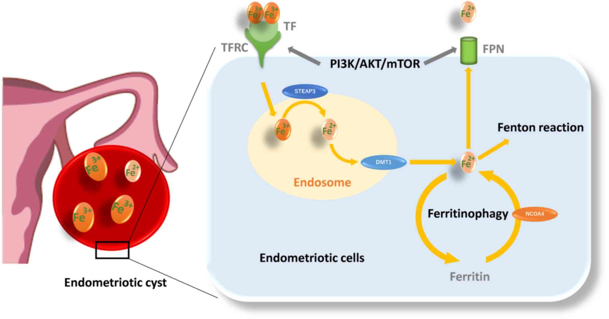
Molecular and regulatory network mechanisms
controlling ferroptosis
Ferroptosis is an iron-dependent non-apoptotic form
of cell death through the accumulation of lipid peroxidation caused
by ROS from the Fenton reaction (52). Ferroptosis can be triggered by
iron overload and dysregulation of the transporter-dependent
pathway [e.g., in a cystine/glutamate antiporter (xCT)-dependent
manner] and/or the enzymic control mechanisms (e.g., in a
GPX4-dependent manner) (53).
The present section summarized the interrelationships of molecules
and signaling pathways involved in ferroptosis. The essential role
and pathways of each molecule are shown in Fig. 4.
 | Figure 4Molecular and regulatory network
mechanisms controlling autophagy and ferroptosis in endometriosis.
The left quarter of the figure shows the autophagy pathway (the
simplified version of Figure 3)
and the right three quarters shows the ferroptosis pathway. An
orange box indicates molecular and regulatory network mechanisms
controlled by iron overload; a green box indicates molecular and
regulatory network mechanisms controlled by existing antioxidant
systems; and a yellow box indicates modulation of ferroptosis by
molecules that control autophagy. The orange and green color
letters represent molecules that may undergo dynamic changes in
early stage and late stage endometriotic lesions, respectively.
HIF-1, hypoxia-inducible factor 1; TF, transferrin; TFRC,
transferrin receptor; xCT, cystine/glutamate antiporter; SLC7A11, a
light-chain subunit of xCT; SLC3A2, a heavy-chain subunit of xCT;
KEAP1, kelch like ECH associated protein 1; HO-1, heme oxygenase-1;
NCOA4, nuclear receptor coactivator 4; DMT1, divalent metal
transporter 1; GPX, glutathione peroxidase; VDAC, voltage-dependent
anion channel; AMPK, AMP-activated protein kinase; PI3K,
phosphatidylinositol 3-kinase; AKT, protein kinase B; mTOR,
mammalian target of rapamycin. |
Molecular and regulatory network
mechanisms controlled by iron overload (Fig. 4, orange box)
Differential gene expression
analyses
Researchers have assessed the differences in
ferroptosis-related genes in eutopic and ectopic endometrial
samples from women with endometriosis and normal endometrium from
women without endometriosis (54). Differential gene expression
analyses based on the datasets in the Gene Expression Omnibus
revealed that resistance to ferroptosis is a feature of eutopic and
ectopic endometrial cells and changes of ferroptosis-related genes
increased progressively from the eutopic endometrium to the ectopic
endometrium (54). Increased
SLC3A2 (a heavy-chain subunit of xCT) and TF expression and
decreased NCOA4 and voltage-dependent anion channel 2/3 (VDAC2/3)
expression were detected in eutopic endometrium compared with
normal endometrium (54).
TF
Transferrin is an iron carrier protein that induces
ferroptosis (55). Women with
endometriosis have increased serum transferrin saturation levels as
compared with controls (56).
Furthermore, transferrin levels are elevated in the circulating and
peritoneal fluid of females with endometriosis (57).
NCOA4
NCOA4-mediated ferritinophagy facilitates ferritin
degradation in autolysosome, increases the cellular labile iron
pool, triggers peroxidation of polyunsaturated fatty
acid-containing phospholipids and activates ferroptosis induction
(10,53,58). Accelerating the ferritinophagy
system and downregulating ferroportin expression further increase
susceptibility to ferroptosis (45). Decreased NCOA4 expression in
endometriosis contributes to suppression of ferroptosis (54).
DMT1
Fe2+ is released into cytoplasm from the
endosome via DMT1 (59). Iron is
released into the cytoplasm by DMT1 overexpressed in the ectopic
endometrium, induces oxidative stress via the Fenton reaction,
leading to lipid peroxidation and ferroptosis (59).
Molecular and regulatory network
mechanisms controlled by existing antioxidant systems (Fig. 4, green box)
xCT-GPX4
The xCT/GSH/GPX4 pathway is an important component
for ferroptosis (60). A feature
of ferroptosis is the reduction of antioxidant activity (e.g.,
intracellular GSH depletion and decreased activity of GPX4)
(7). Excess iron decreases the
expression of solute carrier family 7 member 11 (SLC7A11; a
light-chain subunit of xCT) and GPX4, which efficiently increases
susceptibility to ferroptosis (61). Blood injection results in GPX4
protein depletion in murine models with endometriosis iron overload
(62). In fact, it has been
reported that GPX4 expression in the early secretory phase is
markedly lower in the eutopic endometrium of women with
endometriosis compared with those with normal endometrium (63). These data suggest that
endometriosis is more susceptible to iron-mediated ferroptosis.
Nuclear factor-E2-related factor 2
(Nrf2)
Nrf2, a key transcription factor that regulates
cellular antioxidant response, increases the expression of genes
involved in the anti-oxidative defense system, such as GPX4
(64). Nrf2 inhibits lipid
peroxidation and ferroptosis via enhancing the antioxidant defense
system. Protein expression of Nrf2 and its downstream molecules
[e.g., NAD(P)H quinone dehydrogenase 1 (NQO1) and HO-1] in
endometriotic lesions in a rat model showed an increased level
compared with controls (65).
Nrf2 has been known to be activated during autophagy (66). An adaptor protein Keap1 (kelch
like ECH associated protein 1) binds Nrf2 and promotes
ubiquitin-proteasome-mediated degradation of Nrf2 (65,67). Under oxidative stress conditions,
phosphorylation and upregulation of p62, a selective autophagic
cargo receptor, increases its binding affinity for Keap1, leading
to Nrf2 dissociation from Keap1 (65-67). Free Nrf2 is transferred to the
nucleus to induce the expression of various antioxidant genes and
facilitate restoration of redox homeostasis (67). This suggests that Nrf2 is
involved in ferroptosis resistance via activation of the autophagy
process (66). Conversely, there
is conflicting evidence that Nrf2 levels are lower in ectopic
endometrium of endometriosis patients and in rat models of
endometriosis compared with controls (20,68). Given that only activated Nrf2
that can translocate to the nucleus promotes ferroptosis resistance
(67), intracellular total Nrf2
levels may be unrelated to ferroptosis. Therefore, quantification
of Nrf2 mRNA and protein levels alone cannot determine the net
effect of ferroptosis activation or inhibition.
HO-1
HO-1, a Nrf2-induced antioxidant enzyme, immediately
removes toxic heme, catabolizes heme to biliverdin, carbon monoxide
and iron and effectively protects cells from oxidative damage
(41,69). For example, activation of the
Keap1/Nrf2/HO-1 pathway may inhibit oxidative stress-mediated
ferroptosis induced by lipopolysaccharide (LPS) (70,71). LPS is often used to induce
oxidative stress and inflammation in animal models. On the other
hand, HO-1 overexpression causes the accumulation of intracellular
iron, promotes lipid peroxidation and triggers ferroptotic cell
death (61,72,73). Therefore, HO-1 may exhibit a dual
role as a suppressor or promoter of ferroptosis in response to
oxidative stress, possibly depending on the degree of
ferritinophagy and Nrf2 levels. HO-1 protein expression has been
shown to be increased in ovarian endometriotic lesions (24), but its association with
ferroptosis is still unclear.
Modulation of ferroptosis by molecules
that control autophagy (Fig. 4,
yellow box)
PI3K/Akt/mTOR
Certain oncogene mutations may play an important
role in determining the susceptibility or resistance to ferroptosis
(10). For example, aberrant
constitutive activation of the PI3K/AKT/mTOR pathway markedly
contributes to vital cellular functions, such as carcinogenesis,
proliferation and survival and can acquire resistance to
ferroptosis (10). Specific
oncogenic driver mutations such as Kirsten rat sarcoma viral
oncogene homologue and phosphatidylinositol-4,5-bisphosphate
3-kinase catalytic subunit α promoted ferroptosis resistance in
lung cancer cells and breast cancer cells, respectively (10). Indeed, as illustrated in Fig. 3, the PI3K/Akt/mTOR pathway is
known to influence iron metabolism and ferritinophagy.
AMPK
AMP-activated protein kinase (AMPK), a key cellular
energy sensor, has been reported to either positively or negatively
regulate ferroptosis (53). AMPK
activated in response to energy stress and oxidative damage can
inhibit ferroptosis via phosphorylation of downstream targets,
including acetyl-CoA carboxylase α, the rate-limiting step in fatty
acid synthesis (74) and Nrf2
(75). Meanwhile, the
AMPK/Beclin1 signaling pathway promotes ferroptosis through
inhibition of SLC7A11 activity in cancer cells (76). Beclin1 regulates not only
autophagy but also ferroptosis (14). However, it remains unclear what
role AMPK plays in ferroptosis, as there is conflicting evidence
that expression of Beclin1 is increased (13,77) or decreased (78) in endometriosis.
VDAC
VDAC regulates flow of metabolites, ions and
nucleotides across the outer membrane of the mitochondrion. A study
suggests that VDAC is involved in autophagy, mitophagy, apoptosis
and ferroptosis (79). VDAC
ubiquitination orchestrated by a pathway involving the PTEN induced
kinase 1 kinase and the parkin RBR E3 ubiquitin protein ligase
(PARKIN) ubiquitin ligase eliminates damaged mitochondria by
autophagy (80). Furthermore,
dysregulation of mitochondrial function and overproduction of ROS
by altering the permeability of VDAC promotes ferroptosis in lung
cancer (81). Decreased VDAC2/3
expression in endometriosis may suppress ferroptosis (54).
HIF-1
Hypoxia is known to modulate ferroptosis primarily
by upregulating HIF-1 expression (61). Hypoxia increases HIF-1-mediated
iron uptake through upregulation of TFRC and DMT1 expression,
promotes the accumulation of ROS and affects cell susceptibility to
ferroptosis (61,82). In addition, the RNA-binding
protein ELAV like RNA binding protein 1 interacts with HIF-1 mRNA,
promotes its translation and triggers ferroptosis via activation of
the ferritinophagy-mediated autophagy (61,83). Additionally, treatment of
trophoblast cells with hypoxia reduces cell invasion and induces
ferroptosis by downregulating SCL7A11, GPX4 and FPN1 expression
(84). Furthermore,
hypoxia/reoxygenation induces ferroptosis of cardiomyocytes
(85). Meanwhile, hypoxia
alleviates ferroptosis through decreasing NCOA4 levels, inhibiting
ferritin degradation and increasing FTL and FTH1 storage in HT1080
fibrosarcome cells (86). Also,
hypoxia reduces ferroptosis by inhibiting ferritinophagy in
osteoclasts (87). Additionally,
hypoxia/reoxygenation-induced oxidative stress may reduce
ferroptosis through stearoyl-CoA desaturase 1-mediated fatty acid
desaturation in cancer cells (88). Indeed, HIF-1 has been reported to
inhibit ferroptosis in acute kidney injury (89). Hypoxia is also a central event in
endometriosis. HIF-1α is known to promote eutopic endometrial
stromal cell invasion by upregulating vascular endothelial growth
factor and matrix metalloproteinases in endometriosis (29,90,91). Although HIF-1α has been reported
to upregulate autophagy in endometriosis (29), it is unclear whether HIF-1α is
involved in regulating ferroptosis. However, emerging evidence,
such as decreased NCOA4 expression and increased Nrf2 and GPX4
expression in endometriosis, suggests that HIF-1α may regulate
ferroptosis.
P53
p53 is known to have a dual role as a suppressor or
promoter of ferroptosis (53,92). TP53 inhibits SLC7A11 expression
to suppress tumor growth in human osteosarcoma, breast, or lung
cancer cells through induction of GSH depletion and increased
susceptibility to ferroptosis (93). TP53 triggers ferroptosis through
enhanced arachidonate lipoxygenase expression and glutaminolysis by
inducing the expression of spermidine/spermine N1-acetyltransferase
1 (94) and glutaminase 2
(55,92). Conversely, p53 inhibits
ferroptosis through the upregulation of cyclin dependent kinase
inhibitor 1 A/p21 (cyclin dependent kinase inhibitor 1 A)
expression in cancer cells (95). Downregulation of p53 expression
in endometriosis has been reported to suppress ferroptosis through
activating the SLC7A11 pathway, facilitating disease progression
(96).
The ferroptosis-related pathway in
endometriosis
Based on these data, the present study summarized
ferroptosis-related pathways in endometriosis (Fig. 4). First, it is well known that
the SLC3A2/SLC7A11/Nrf2/GPX4 pathway controls ferroptosis (60). It has been reported that iron
administration reduced GPX4 levels in rat models (early stages of
endometriosis) (62). This
result suggests the importance of ferroptosis induction for cell
survival during the early stages of endometriosis development.
Second, the gene expression levels of GPX4 in eutopic endometrium
are higher than in the normal endometrium through upregulation of
SLC3A2 expression (54).
Upregulation of SLC3A2 (54) and
SLC7A11 (96) expression
suppresses ferroptosis and promotes disease progression.
Furthermore, immunohistochemical studies showed that the positive
rate of p53 in ectopic endometrium was lower than that in normal
endometrium (97). Given that
perturbations of the p53 locus occur frequently in severe/late
stage endometriosis (25),
downregulation of p53 expression may lead to the activation of the
xCT-GPX4 pathway, promoting cell protection and survival by
suppressing ferroptosis. Third, overexpression of TF enhances iron
uptake in eutopic endometrium, but decreased NCOA4 expression
inhibits ferritinophagy, promotes iron accumulation and suppresses
Fe2+ release. In addition, increased expression of HO-1
and ferritin and decreased expression of DMT1 were detected in
ectopic endometrium compared with eutopic endometrium. These data
suggest that inhibition of ferritinophagy suppresses the
accumulation of labile iron and ferroptosis (10,53,58,98,99). Finally, VDAC, which forms part of
the mitochondrial permeability transition pore, is known to be
involved in apoptotic cell death by promoting the release of
cytochrome c (79). It has been
reported that VDAC is closely involved in promoting ferroptosis in
cancer cells (81,100). Similar to cancer cells, VDAC2/3
is markedly downregulated in the eutopic endometrium of patients
with endometriosis (54). This
indicates that decreased VDAC levels suppresses ferroptosis.
Alterations in autophagy and ferroptosis
throughout the development of endometriotic cysts
Autophagy and ferroptosis have been shown to be
induced or conversely suppressed in humans and preclinical animal
models, but the reasons for such discrepancies and their mechanisms
are still not fully understood. The present review discussed the
role of autophagy and ferroptosis by dividing the development of
endometriosis into four stages: Lesion initiation, induction of
cell death, escape from cell death and formation of established
lesions. Assuming that endometriosis evolves from retrograde
endometrium, it was estimated how autophagy and ferroptosis change
as endometriosis progresses (Fig.
5). Endometrial tissues originating from retrograde
menstruation encounter environmental challenges, e.g., iron
overload, oxidative stress and hypoxia caused by periodic bleeding
(Fig. 5 ①) (4,46). Several lines of evidence indicate
that iron has a crucial role in the proliferation of endometriotic
cells. Ding et al (62)
found that in comparison with control mice, endometriotic lesions
in the iron-overload model mice were larger and more numerous.
Under iron overload or hypoxic conditions, autophagy plays a
positive role in scavenging ROS, preserving mitochondrial
integrity, counteracting metabolic insults, avoiding apoptosis and
protecting cells (10).
Endometrial cells derived from retrograde menstruation may be able
to survive even under harsh conditions, possibly through activation
of autophagy-dependent quality control mechanisms (101). In endometriosis, ferroptosis
can occur through two pathways, the activation of the autophagy
pathway controlled by iron and HIF-1 (28,29) and the expression of
ferritinophagy-related proteins such as DMT1 (59). Indeed, the levels of both iron
and MDA, a secondary product of free radical lipid peroxidation,
are increased in the peritoneal fluid and ovaries, whereas
expression of anti-ferroptosis-related proteins (e.g., GPX4 and
GSH) are decreased, highlighting the importance of ferroptosis
(62,102). Moreover, increased ferroptosis
is found to enhance the expression of pro-angiogenic factors
vascular endothelial growth factor and IL-8 (103,104). Ferroptosis has been shown to
contribute to development and maintenance of subsequent
endometriotic lesions through modulating the interaction between
inflammation and angiogenesis (101,103). Additionally, it has been
reported that ferric ammonium citrate-induced ferroptosis may
promote fibrosis process in endometriosis (102). Autophagy is essential for
establishing early lesions by counteracting the
ferroptosis-mediated deleterious effects. Only a rare fraction of
cells are likely able to survive in harsh environments mediated by
iron/hypoxia-dependent oxidative stress. It is easy to understand
that an excessive ferroptosis induces the regression of
endometriotic lesions via lipid peroxidation-driven membrane
destruction (Fig. 5 ②). Indeed,
erastin, a ferroptosis inducer, inhibits endometriotic lesion
growth through iron accumulation and decreased FPN expression in a
mouse model of endometriosis (45). Perhaps, excessive ROS induces
autophagic cell death, accelerates iron-dependent ferroptosis and
amplifies the process of lipid peroxidation and the extent of
membrane rupture (105).
Autophagy also plays a role in eliminating damaged proteins and
subcellular organelles to sustain cell viability, while
unrecoverable damage can trigger cell death (106). By contrast, autophagy removes
damaged intracellular components, such as cellular proteins,
lipids, nucleic acids and mitochondria and can inhibit ferroptosis
(Fig. 5 ③). Nrf2-dependent
autophagy activation may inhibit ferroptosis via enhancing the
defense system against oxidative stress (65). Only a fraction of endometrial
cells may receive survival signals and acquire a
ferroptosis-resistant phenotype. On the other hand, intrinsic
modulators (e.g., estrogen, PI3K/AKT/mTOR, AMPK, p53 and Beclin1)
can inhibit autophagy through the expression of various ATG
proteins (Fig. 5 ④). mTOR
inhibitors have been reported to confer therapeutic efficacy
against endometriosis through activation of autophagy (34). Furthermore, downregulation of the
expression of NCOA4 and VDAC2/3 and upregulation of the expression
of xCT/GPX4 induces the suppression of ferroptosis and causes
further progression of endometriotic lesions.
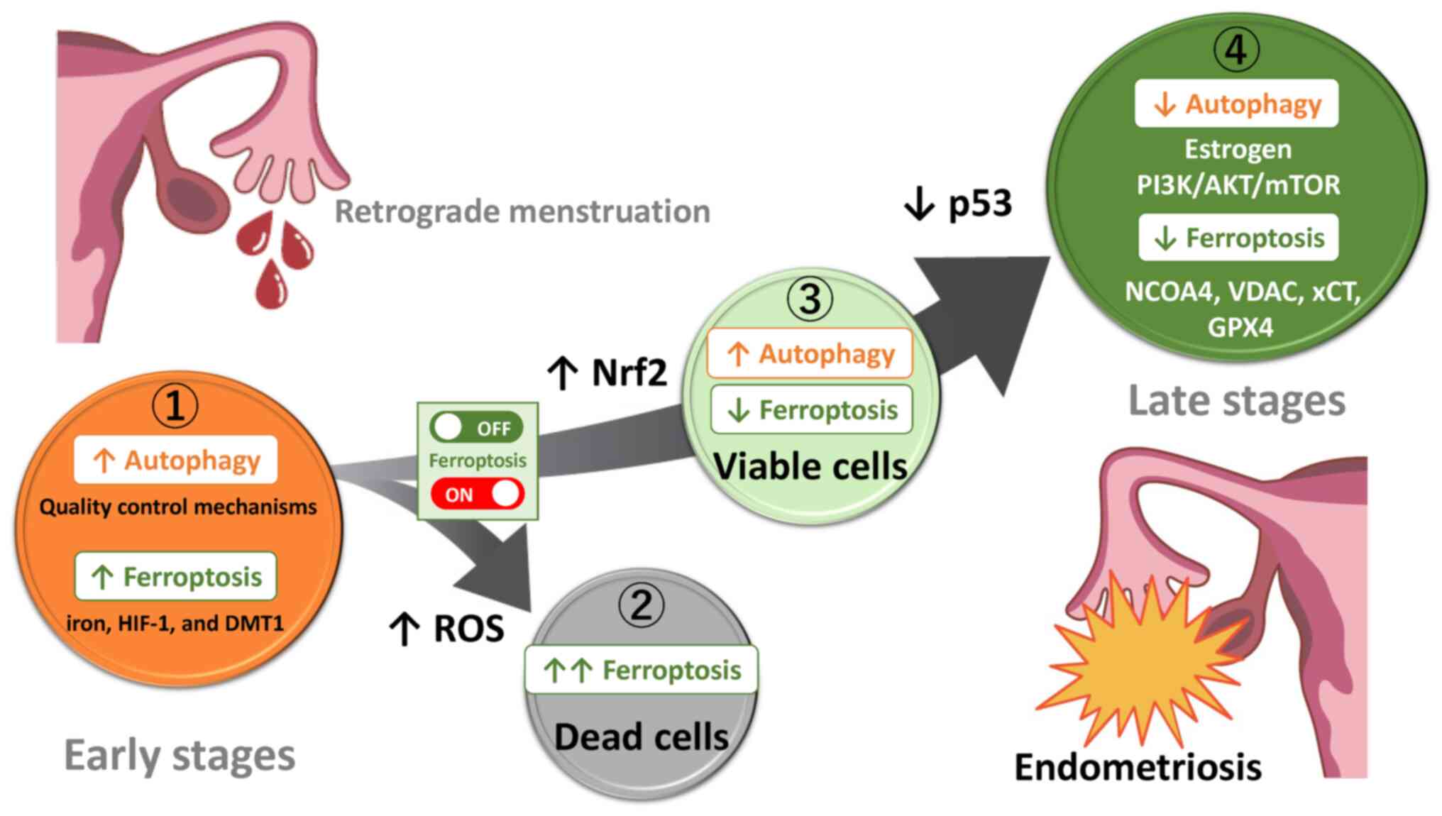 | Figure 5Temporal transition of cell death
regulation during disease progression from retrograde endometrium
to early stage lesions to late stage lesions. The expression levels
of ROS, Nrf2 and p53 may be important molecular actors that can
switch ON/OFF the ferroptosis. ➀, ➁, ➂ and ④ represent initial
lesions, dead cells, viable cells that escape from death and
established lesions, respectively. ROS, reactive oxygen species;
Nrf2, Nuclear factor-E2-related factor 2; HIF-1, hypoxia-inducible
factor 1; DMT1, divalent metal transporter 1. |
Conclusion
Induction of autophagy and ferroptosis may
contribute to the survival and proliferation of the retrograde
tissues originating from the endometrium, whereas suppression of
autophagy and ferroptosis may play an important role in the
progression of endometriotic lesions. The key molecular actors
(e.g., estrogen, PI3K/AKT/mTOR, NCOA4, VDAC, xCT, GPX4, Nrf2, or
p53) may switch ferroptosis on and off. Hence, in light of our
prior report indicating that endometriotic cells may possess a
potential anti-apoptotic capability to survive in changing
environments (11), a timely
fine-tuning of autophagy and ferroptosis levels could regulate the
development and progression of endometriosis. In conclusion,
autophagy and ferroptosis play a dual role in the initiation and
progression of endometriosis through multiple mechanisms regulated
by intrinsic factors (e.g., estrogen and the PI3K/AKT/mTOR pathway)
or extrinsic stressors (e.g., iron overload, oxidative stress and
hypoxia).
Availability of data and materials
Not applicable.
Authors' contributions
HK was responsible for conception and design of the
present study. SM, CY and SI were responsible for acquisition of
the data and HS was responsible for analysis and interpretation of
data. HK drafted the manuscript and SM, CY, HS and SI performed a
critical revision of the manuscript for important intellectual
content. HS performed statistical analysis. H.K. provided
administrative, technical or material support. SI supervised the
present study. All authors read and approved the final manuscript.
Data authentication is not applicable.
Ethics approval and consent to
participate
Not applicable.
Patient consent for publication
Not applicable.
Competing interests
The authors declare that they have no competing
interests.
Authors' information
Dr Hiroshi Kobayashi: ORCID:
0000-0002-8124-6269.
Abbreviations:
|
AKT
|
protein kinase B
|
|
AMPK
|
AMP-activated protein kinase
|
|
ATG
|
autophagy-related gene
|
|
BECN1
|
beclin1
|
|
CXCL12
|
C-X-C motif chemokine ligand 12
|
|
CXCR4
|
C-X-C motif chemokine receptor 4
|
|
DMT1
|
divalent metal transporter 1
|
|
FPN
|
ferroportin
|
|
FTL
|
ferritin light chain
|
|
FTH1
|
ferritin heavy chain 1
|
|
GPX
|
glutathione peroxidase
|
|
GSH
|
glutathione
|
|
HIF-1
|
hypoxia-inducible factor 1
|
|
HO-1
|
heme oxygenase-1
|
|
KEAP1
|
kelch like ECH associated protein
1
|
|
LC3
|
also known as MAP1LC3, microtubule
associated protein 1 light chain 3
|
|
MAPK
|
mitogen-activated protein kinases
|
|
MDA
|
malondialdehyde
|
|
mTOR
|
mammalian target of rapamycin
|
|
NCOA4
|
nuclear receptor coactivator 4
|
|
Nrf2
|
Nuclear factor-E2-related factor
2
|
|
PI3K
|
phosphatidylinositol 3-kinase
|
|
PTEN
|
phosphatase and tensin homolog
|
|
RCD
|
Regulated cell death
|
|
ROS
|
reactive oxygen species
|
|
TF
|
transferrin
|
|
TFRC
|
transferrin receptor
|
|
TP53
|
tumor protein p53
|
|
VDAC
|
voltage-dependent anion channel
|
|
xCT
|
cystine/glutamate antiporter
|
Acknowledgements
Figures were created by Mrs. Toyomi Kobayashi
(Ms.Clinic MayOne, Nara, Japan; https://www.mscl-mayone.com/; accessed on 9 January
31, 2024).
Funding
No funding was received.
References
|
1
|
Giudice LC: Clinical practice.
Endometriosis. N Engl J Med. 362:2389–2398. 2010. View Article : Google Scholar : PubMed/NCBI
|
|
2
|
Vinatier D, Orazi G, Cosson M and Dufour
P: Theories of endometriosis. Eur J Obstet Gynecol Reprod Biol.
96:21–34. 2001. View Article : Google Scholar : PubMed/NCBI
|
|
3
|
Sampson JA: Peritoneal endometriosis due
to the menstrual dissemination of endometrial tissue into the
peritoneal cavity. Am J Obstet Gynecol. 14:422–469. 1927.
View Article : Google Scholar
|
|
4
|
Li WN, Wu MH and Tsai SJ: Hypoxia and
reproductive health: The role of hypoxia in the development and
progression of endometriosis. Reproduction. 161:F19–F31. 2021.
View Article : Google Scholar
|
|
5
|
Scutiero G, Iannone P, Bernardi G,
Bonaccorsi G, Spadaro S, Volta CA, Greco P and Nappi L: Oxidative
stress and endometriosis: A systematic review of the literature.
Oxid Med Cell Longev. 2017:72652382017. View Article : Google Scholar : PubMed/NCBI
|
|
6
|
Viganò P, Parazzini F, Somigliana E and
Vercellini P: Endometriosis: Epidemiology and aetiological factors.
Best Pract Res Clin Obstet Gynaecol. 18:177–200. 2004. View Article : Google Scholar : PubMed/NCBI
|
|
7
|
Huang E, Wang X and Chen L: Regulated cell
death in endometriosis. Biomolecules. 14:1422024. View Article : Google Scholar : PubMed/NCBI
|
|
8
|
Yang HL, Mei J, Chang KK, Zhou WJ, Huang
LQ and Li MQ: Autophagy in endometriosis. Am J Transl Res.
9:4707–4725. 2017.PubMed/NCBI
|
|
9
|
Feng Y, He D, Yao Z and Klionsky DJ: The
machinery of macroautophagy. Cell Res. 24:24–41. 2014. View Article : Google Scholar :
|
|
10
|
Lee S, Hwang N, Seok BG, Lee S, Lee SJ and
Chung SW: Autophagy mediates an amplification loop during
ferroptosis. Cell Death Dis. 14:4642023. View Article : Google Scholar : PubMed/NCBI
|
|
11
|
Li X, He S and Ma B: Autophagy and
autophagy-related proteins in cancer. Mol Cancer. 19:122020.
View Article : Google Scholar : PubMed/NCBI
|
|
12
|
Glick D, Barth S and Macleod KF:
Autophagy: Cellular and molecular mechanisms. J Pathol. 221:3–12.
2010. View Article : Google Scholar : PubMed/NCBI
|
|
13
|
Li H, Yang H, Lu S, Wang X, Shi X and Mao
P: Autophagy-dependent ferroptosis is involved in the development
of endometriosis. Gynecol Endocrinol. 39:22429622023. View Article : Google Scholar : PubMed/NCBI
|
|
14
|
Liu J, Kuang F, Kroemer G, Klionsky DJ,
Kang R and Tang D: Autophagy-dependent ferroptosis: Machinery and
regulation. Cell Chem Biol. 27:420–435. 2020. View Article : Google Scholar : PubMed/NCBI
|
|
15
|
Kong Z and Yao T: Role for
autophagy-related markers beclin-1 and LC3 in endometriosis. BMC
Womens Health. 22:2642022. View Article : Google Scholar : PubMed/NCBI
|
|
16
|
Kobayashi H, Imanaka S, Yoshimoto C,
Matsubara S and Shigetomi H: Molecular mechanism of autophagy and
apoptosis in endometriosis: Current understanding and future
research directions. Reprod Med Biol. 23:e125772024. View Article : Google Scholar : PubMed/NCBI
|
|
17
|
Mei J, Zhu XY, Jin LP, Duan ZL, Li DJ and
Li MQ: Estrogen promotes the survival of human secretory phase
endometrial stromal cells via CXCL12/CXCR4 up-regulation-mediated
autophagy inhibition. Hum Reprod. 30:1677–1689. 2015. View Article : Google Scholar : PubMed/NCBI
|
|
18
|
Shen HH, Zhang T, Yang HL, Lai ZZ, Zhou
WJ, Mei J, Shi JW, Zhu R, Xu FY, Li DJ, et al: Ovarian
hormones-autophagy-immunity axis in menstruation and endometriosis.
Theranostics. 11:3512–3526. 2021. View Article : Google Scholar : PubMed/NCBI
|
|
19
|
Guo J, Gao J, Yu X, Luo H, Xiong X and
Huang O: Expression of DJ-1 and mTOR in eutopic and ectopic
endometria of patients with endometriosis and adenomyosis. Gynecol
Obstet Invest. 79:195–200. 2015. View Article : Google Scholar : PubMed/NCBI
|
|
20
|
Jamali N, Zal F, Mostafavi-Pour Z,
Samare-Najaf M, Poordast T and Dehghanian A: Ameliorative effects
of quercetin and metformin and their combination against
experimental endometriosis in rats. Reprod Sci. 28:683–692. 2021.
View Article : Google Scholar
|
|
21
|
Choi J, Jo M, Lee E, Hwang S and Choi D:
Aberrant PTEN expression in response to progesterone reduces
endometriotic stromal cell apoptosis. Reproduction. 153:11–21.
2017.PubMed/NCBI
|
|
22
|
Sato N, Tsunoda H, Nishida M, Morishita Y,
Takimoto Y, Kubo T and Noguchi M: Loss of heterozygosity on 10q23.3
and mutation of the tumor suppressor gene PTEN in benign
endometrial cyst of the ovary: Possible sequence progression from
benign endometrial cyst to endometrioid carcinoma and clear cell
carcinoma of the ovary. Cancer Res. 60:7052–7056. 2000.
|
|
23
|
Cui D, Qu R, Liu D, Xiong X, Liang T and
Zhao Y: The cross talk between p53 and mTOR pathways in response to
physiological and genotoxic stresses. Front Cell Dev Biol.
9:7755072021.PubMed/NCBI
|
|
24
|
Allavena G, Carrarelli P, Del Bello B,
Luisi S, Petraglia F and Maellaro E: Autophagy is upregulated in
ovarian endometriosis: A possible interplay with p53 and heme
oxygenase-1. Fertil Steril. 103:1244–1251.e1. 2015.PubMed/NCBI
|
|
25
|
Bischoff FZ, Heard M and Simpson JL:
Somatic DNA alterations in endometriosis: High frequency of
chromosome 17 and p53 loss in late-stage endometriosis. J Reprod
Immunol. 55:49–64. 2002.PubMed/NCBI
|
|
26
|
Poillet-Perez L, Despouy G,
Delage-Mourroux R and Boyer-Guittaut M: Interplay between ROS and
autophagy in cancer cells, from tumor initiation to cancer therapy.
Redox Biol. 4:184–192. 2015.PubMed/NCBI
|
|
27
|
Iwabuchi T, Yoshimoto C, Shigetomi H and
Kobayashi H: Oxidative stress and antioxidant defense in
endometriosis and its malignant transformation. Oxid Med Cell
Longev. 2015:8485952015.PubMed/NCBI
|
|
28
|
Xu TX, Zhao SZ, Dong M and Yu XR: Hypoxia
responsive miR-210 promotes cell survival and autophagy of
endometriotic cells in hypoxia. Eur Rev Med Pharmacol Sci.
20:399–406. 2016.PubMed/NCBI
|
|
29
|
Liu H, Zhang Z, Xiong W, Zhang L, Xiong Y,
Li N, He H, Du Y and Liu Y: Hypoxia-inducible factor-1alpha
promotes endometrial stromal cells migration and invasion by
upregulating autophagy in endometriosis. Reproduction. 153:809–820.
2017.PubMed/NCBI
|
|
30
|
Tsuzuki T, Okada H, Shindoh H, Shimoi K,
Nishigaki A and Kanzaki H: Effects of the hypoxia-inducible
factor-1 inhibitor echinomycin on vascular endothelial growth
factor production and apoptosis in human ectopic endometriotic
stromal cells. Gynecol Endocrinol. 32:323–328. 2016.
|
|
31
|
McKinnon BD, Kocbek V, Nirgianakis K,
Bersinger NA and Mueller MD: Kinase signalling pathways in
endometriosis: Potential targets for non-hormonal therapeutics. Hum
Reprod Update. 22:382–403. 2016. View Article : Google Scholar : PubMed/NCBI
|
|
32
|
Wyatt J, Fernando SM, Powell SG, Hill CJ,
Arshad I, Probert C, Ahmed S and Hapangama DK: The role of iron in
the pathogenesis of endometriosis: A systematic review. Hum Reprod
Open. 2023:hoad0332023. View Article : Google Scholar : PubMed/NCBI
|
|
33
|
Hung SW, Zhang R, Tan Z, Chung JPW, Zhang
T and Wang CC: Pharmaceuticals targeting signaling pathways of
endometriosis as potential new medical treatment: A review. Med Res
Rev. 41:2489–2564. 2021. View Article : Google Scholar : PubMed/NCBI
|
|
34
|
Zhang M, Xu T, Tong D, Li S, Yu X, Liu B,
Jiang L and Liu K: Research advances in endometriosis-related
signaling pathways: A review. Biomed Pharmacother. 164:1149092023.
View Article : Google Scholar : PubMed/NCBI
|
|
35
|
Defrère S, Van Langendonckt A, Vaesen S,
Jouret M, González Ramos R, Gonzalez D and Donnez J: Iron overload
enhances epithelial cell proliferation in endometriotic lesions
induced in a murine model. Hum Reprod. 21:2810–2816. 2006.
View Article : Google Scholar : PubMed/NCBI
|
|
36
|
Kobayashi H, Shigetomi H and Imanaka S:
Nonhormonal therapy for endometriosis based on energy metabolism
regulation. Reprod Fertil. 2:C42–C57. 2021. View Article : Google Scholar
|
|
37
|
Defrère S, Lousse JC, González-Ramos R,
Colette S, Donnez J and Van Langendonckt A: Potential involvement
of iron in the pathogenesis of peritoneal endometriosis. Mol Hum
Reprod. 14:377–385. 2008. View Article : Google Scholar : PubMed/NCBI
|
|
38
|
Sanchez AM, Papaleo E, Corti L,
Santambrogio P, Levi S, Viganò P, Candiani M and Panina-Bordignon
P: Iron availability is increased in individual human ovarian
follicles in close proximity to an endometrioma compared with
distal ones. Hum Reprod. 29:577–583. 2014. View Article : Google Scholar : PubMed/NCBI
|
|
39
|
Yamaguchi K, Mandai M, Toyokuni S,
Hamanishi J, Higuchi T, Takakura K and Sujii S: Contents of
endometriotic cysts, especially the high concentration of free
iron, are a possible cause of carcinogenesis in the cysts through
the iron-induced persistent oxidative stress. Clin Cancer Res.
14:32–40. 2008. View Article : Google Scholar : PubMed/NCBI
|
|
40
|
Yoshimoto C, Iwabuchi T, Shigetomi H and
Kobayashi H: Cyst fluid iron-related compounds as useful markers to
distinguish malignant transformation from benign endometriotic
cysts. Cancer Biomark. 15:493–499. 2015. View Article : Google Scholar : PubMed/NCBI
|
|
41
|
Iwabuchi T, Yoshimoto C, Shigetomi H and
Kobayashi H: Cyst fluid hemoglobin species in endometriosis and its
malignant transformation: The role of metallobiology. Oncol Lett.
11:3384–3388. 2016. View Article : Google Scholar : PubMed/NCBI
|
|
42
|
Gao G, Li J, Zhang Y and Chang YZ:
Cellular iron metabolism and regulation. Adv Exp Med Biol.
1173:21–32. 2019. View Article : Google Scholar : PubMed/NCBI
|
|
43
|
Sendamarai AK, Ohgami RS, Fleming MD and
Lawrence CM: Structure of the membrane proximal oxidoreductase
domain of human Steap3, the dominant ferrireductase of the
erythroid transferrin cycle. Proc Natl Acad Sci USA. 105:7410–7415.
2008. View Article : Google Scholar : PubMed/NCBI
|
|
44
|
Hou W, Xie Y, Song X, Sun X, Lotze MT, Zeh
HJ III, Kang R and Tang D: Autophagy promotes ferroptosis by
degradation of ferritin. Autophagy. 12:1425–1428. 2016. View Article : Google Scholar : PubMed/NCBI
|
|
45
|
Li Y, Zeng X, Lu D, Yin M, Shan M and Gao
Y: Erastin induces ferroptosis via ferroportin-mediated iron
accumulation in endometriosis. Hum Reprod. 36:951–964. 2021.
View Article : Google Scholar
|
|
46
|
Kobayashi H, Yoshimoto C, Matsubara S,
Shigetomi H and Imanaka S: Current understanding of and future
directions for endometriosis-related infertility research with a
focus on ferroptosis. Diagnostics (Basel). 13:19262023. View Article : Google Scholar : PubMed/NCBI
|
|
47
|
Matsuzaki S and Schubert B: Oxidative
stress status in normal ovarian cortex surrounding ovarian
endometriosis. Fertil Steril. 93:2431–2432. 2010. View Article : Google Scholar
|
|
48
|
Dai Y, Lin X, Xu W, Lin X, Huang Q, Shi L,
Pan Y, Zhang Y, Zhu Y, Li C, et al: MiR-210-3p protects
endometriotic cells from oxidative stress-induced cell cycle arrest
by targeting BARD1. Cell Death Dis. 10:1442019. View Article : Google Scholar : PubMed/NCBI
|
|
49
|
Makker A, Goel MM, Das V and Agarwal A:
PI3K-Akt-mTOR and MAPK signaling pathways in polycystic ovarian
syndrome, uterine leiomyomas and endometriosis: An update. Gynecol
Endocrinol. 28:175–181. 2012. View Article : Google Scholar
|
|
50
|
Bayeva M, Khechaduri A, Puig S, Chang HC,
Patial S, Blackshear PJ and Ardehali H: mTOR regulates cellular
iron homeostasis through tristetraprolin. Cell Metab. 16:645–657.
2012. View Article : Google Scholar : PubMed/NCBI
|
|
51
|
Driva TS, Schatz C and Haybaeck J:
Endometriosis-associated ovarian carcinomas: How PI3K/AKT/mTOR
pathway affects their pathogenesis. Biomolecules. 13:12532023.
View Article : Google Scholar : PubMed/NCBI
|
|
52
|
Dixon SJ, Lemberg KM, Lamprecht MR, Skouta
R, Zaitsev EM, Gleason CE, Patel DN, Bauer AJ, Cantley AM, Yang WS,
et al: Ferroptosis: An iron-dependent form of nonapoptotic cell
death. Cell. 149:1060–1072. 2012. View Article : Google Scholar : PubMed/NCBI
|
|
53
|
Tang D, Chen X, Kang R and Kroemer G:
Ferroptosis: Molecular mechanisms and health implications. Cell
Res. 31:107–125. 2021. View Article : Google Scholar :
|
|
54
|
Li B, Duan H, Wang S and Li Y: Ferroptosis
resistance mechanisms in endometriosis for diagnostic model
establishment. Reprod Biomed Online. 43:127–138. 2021. View Article : Google Scholar : PubMed/NCBI
|
|
55
|
Gao M, Monian P, Quadri N, Ramasamy R and
Jiang X: Glutaminolysis and transferrin regulate ferroptosis. Mol
Cell. 59:298–308. 2015. View Article : Google Scholar : PubMed/NCBI
|
|
56
|
Xu G, Chen L and Li Q: Association of iron
metabolism markers, socioeconomic and lifestyle factors with
endometriosis: A cross-sectional study. J Trace Elem Med Biol.
78:1271752023. View Article : Google Scholar : PubMed/NCBI
|
|
57
|
Mathur SP: Autoimmunity in endometriosis:
Relevance to infertility. Am J Reprod Immunol. 44:89–95. 2000.
View Article : Google Scholar : PubMed/NCBI
|
|
58
|
Santana-Codina N, Gikandi A and Mancias
JD: The role of NCOA4-mediated ferritinophagy in ferroptosis. Adv
Exp Med Biol. 1301:41–57. 2021. View Article : Google Scholar : PubMed/NCBI
|
|
59
|
Alvarado-Díaz CP, Núñez MT, Devoto L and
González-Ramos R: Endometrial expression and in vitro modulation of
the iron transporter divalent metal transporter-1: Implications for
endometriosis. Fertil Steril. 106:393–401. 2016. View Article : Google Scholar
|
|
60
|
Stockwell BR, Friedmann Angeli JP, Bayir
H, Bush AI, Conrad M, Dixon SJ, Fulda S, Gascón S, Hatzios SK,
Kagan VE, et al: Ferroptosis: A regulated cell death nexus linking
metabolism, redox biology, and disease. Cell. 171:273–285. 2017.
View Article : Google Scholar : PubMed/NCBI
|
|
61
|
Gao X, Hu W, Qian D, Bai X, He H, Li L and
Sun S: The mechanisms of ferroptosis under hypoxia. Cell Mol
Neurobiol. 43:3329–3341. 2023. View Article : Google Scholar : PubMed/NCBI
|
|
62
|
Ding J, Zhao Q, Zhou Z, Cheng W, Sun S, Ni
Z and Yu C: Huayu jiedu fang protects ovarian function in mouse
with endometriosis iron overload by inhibiting ferroptosis. Evid
Based Complement Alternat Med. 2022:14068202022. View Article : Google Scholar : PubMed/NCBI
|
|
63
|
Ota H, Igarashi S, Kato N and Tanaka T:
Aberrant expression of glutathione peroxidase in eutopic and
ectopic endometrium in endometriosis and adenomyosis. Fertil
Steril. 74:313–318. 2000. View Article : Google Scholar : PubMed/NCBI
|
|
64
|
Dodson M, Castro-Portuguez R and Zhang DD:
NRF2 plays a critical role in mitigating lipid peroxidation and
ferroptosis. Redox Biol. 23:1011072019. View Article : Google Scholar : PubMed/NCBI
|
|
65
|
Kapoor R, Sirohi VK, Gupta K and Dwivedi
A: Naringenin ameliorates progression of endometriosis by
modulating Nrf2/Keap1/HO1 axis and inducing apoptosis in rats. J
Nutr Biochem. 70:215–226. 2019. View Article : Google Scholar : PubMed/NCBI
|
|
66
|
Ichimura Y, Waguri S, Sou YS, Kageyama S,
Hasegawa J, Ishimura R, Saito T, Yang Y, Kouno T, Fukutomi T, et
al: Phosphorylation of p62 activates the Keap1-Nrf2 pathway during
selective autophagy. Mol Cell. 51:618–631. 2013. View Article : Google Scholar : PubMed/NCBI
|
|
67
|
Liu S, Pi J and Zhang Q: Signal
amplification in the KEAP1-Nrf2-ARE antioxidant response pathway.
Redox Biol. 54:1023892022. View Article : Google Scholar : PubMed/NCBI
|
|
68
|
Marcellin L, Santulli P, Chouzenoux S,
Cerles O, Nicco C, Dousset B, Pallardy M, Kerdine-Römer S, Just PA,
Chapron C and Batteux F: Alteration of Nrf2 and glutamate cysteine
ligase expression contribute to lesions growth and fibrogenesis in
ectopic endometriosis. Free Radic Biol Med. 110:1–10. 2017.
View Article : Google Scholar : PubMed/NCBI
|
|
69
|
Loboda A, Damulewicz M, Pyza E, Jozkowicz
A and Dulak J: Role of Nrf2/HO-1 system in development, oxidative
stress response and diseases: An evolutionarily conserved
mechanism. Cell Mol Life Sci. 73:3221–3247. 2016. View Article : Google Scholar : PubMed/NCBI
|
|
70
|
Li J, Lu K, Sun F, Tan S, Zhang X, Sheng
W, Hao W, Liu M, Lv W and Han W: Panaxydol attenuates ferroptosis
against LPS-induced acute lung injury in mice by Keap1-Nrf2/HO-1
pathway. J Transl Med. 19:962021. View Article : Google Scholar : PubMed/NCBI
|
|
71
|
Luo L, Huang F, Zhong S, Ding R, Su J and
Li X: Astaxanthin attenuates ferroptosis via Keap1-Nrf2/HO-1
signaling pathways in LPS-induced acute lung injury. Life Sci.
311:1210912022. View Article : Google Scholar : PubMed/NCBI
|
|
72
|
Ryter SW: Heme oxgenase-1, a cardinal
modulator of regulated cell death and inflammation. Cells.
10:5152021. View Article : Google Scholar : PubMed/NCBI
|
|
73
|
Machado SE, Spangler D, Stacks DA,
Darley-Usmar V, Benavides GA, Xie M, Balla J and Zarjou A:
Counteraction of myocardial ferritin heavy chain deficiency by heme
oxygenase-1. Int J Mol Sci. 23:83002022. View Article : Google Scholar : PubMed/NCBI
|
|
74
|
Lee H, Zandkarimi F, Zhang Y, Meena JK,
Kim J, Zhuang L, Tyagi S, Ma L, Westbrook TF, Steinberg GR, et al:
Energy-stress-mediated AMPK activation inhibits ferroptosis. Nat
Cell Biol. 22:225–234. 2020. View Article : Google Scholar : PubMed/NCBI
|
|
75
|
Wang Z, Yao M, Jiang L, Wang L, Yang Y,
Wang Q, Qian X, Zhao Y and Qian J: Dexmedetomidine attenuates
myocardial ischemia/reperfusion-induced ferroptosis via
AMPK/GSK-3β/Nrf2 axis. Biomed Pharmacother. 154:1135722022.
View Article : Google Scholar
|
|
76
|
Song X, Zhu S, Chen P, Hou W, Wen Q, Liu
J, Xie Y, Liu J, Klionsky DJ, Kroemer G, et al: AMPK-mediated BECN1
phosphorylation promotes ferroptosis by directly blocking system
Xc-activity. Curr Biol. 28:2388–2399.e5. 2018.
View Article : Google Scholar
|
|
77
|
Huang J, Chen X and Lv Y: HMGB1 mediated
inflammation and autophagy contribute to endometriosis. Front
Endocrinol (Lausanne). 12:6166962021. View Article : Google Scholar : PubMed/NCBI
|
|
78
|
Sui X, Li Y, Sun Y, Li C, Li X and Zhang
G: Expression and significance of autophagy genes LC3, beclin1 and
MMP-2 in endometriosis. Exp Ther Med. 16:1958–1962. 2018.PubMed/NCBI
|
|
79
|
Zhao Y, Li Y, Zhang R, Wang F, Wang T and
Jiao Y: The role of erastin in ferroptosis and its prospects in
cancer therapy. Onco Targets Ther. 13:5429–5441. 2020. View Article : Google Scholar : PubMed/NCBI
|
|
80
|
Geisler S, Holmström KM, Skujat D, Fiesel
FC, Rothfuss OC, Kahle PJ and Springer W: PINK1/Parkin-mediated
mitophagy is dependent on VDAC1 and p62/SQSTM1. Nat Cell Biol.
12:119–131. 2010. View Article : Google Scholar : PubMed/NCBI
|
|
81
|
Vu NT, Kim M, Stephenson DJ, MacKnight HP
and Chalfant CE: Ceramide kinase inhibition drives ferroptosis and
sensitivity to cisplatin in mutant KRAS lung cancer by
dysregulating VDAC-mediated mitochondria function. Mol Cancer Res.
20:1429–1442. 2022. View Article : Google Scholar : PubMed/NCBI
|
|
82
|
Xiong J, Nie M, Fu C, Chai X, Zhang Y, He
L and Sun S: Hypoxia enhances HIF1 α transcription activity by
upregulating KDM4A and mediating H3K9me3, thus inducing ferroptosis
resistance in cervical cancer cells. Stem Cells Int.
2022:16088062022. View Article : Google Scholar
|
|
83
|
Chen HY, Xiao ZZ, Ling X, Xu RN, Zhu P and
Zheng SY: ELAVL1 is transcriptionally activated by FOXC1 and
promotes ferroptosis in myocardial ischemia/reperfusion injury by
regulating autophagy. Mol Med. 27:142021. View Article : Google Scholar : PubMed/NCBI
|
|
84
|
Wang Y, Zhang L and Zhou X: Activation of
Nrf2 signaling protects hypoxia-induced HTR-8/SVneo cells against
ferroptosis. J Obstet Gynaecol Res. 47:3797–3806. 2021. View Article : Google Scholar : PubMed/NCBI
|
|
85
|
Liu XJ, Lv YF, Cui WZ, Li Y, Liu Y, Xue YT
and Dong F: Icariin inhibits hypoxia/reoxygenation-induced
ferroptosis of cardiomyocytes via regulation of the Nrf2/HO-1
signaling pathway. FEBS Open Bio. 11:2966–2976. 2021. View Article : Google Scholar : PubMed/NCBI
|
|
86
|
Fuhrmann DC, Mondorf A, Beifuß J, Jung M
and Brüne B: Hypoxia inhibits ferritinophagy, increases
mitochondrial ferritin, and protects from ferroptosis. Redox Biol.
36:1016702020. View Article : Google Scholar : PubMed/NCBI
|
|
87
|
Ni S, Yuan Y, Qian Z, Zhong Z, Lv T, Kuang
Y and Yu B: Hypoxia inhibits RANKL-induced ferritinophagy and
protects osteoclasts from ferroptosis. Free Radic Biol Med.
169:271–282. 2021. View Article : Google Scholar : PubMed/NCBI
|
|
88
|
Luis G, Godfroid A, Nishiumi S, Cimino J,
Blacher S, Maquoi E, Wery C, Collignon A, Longuespée R,
Montero-Ruiz L, et al: Tumor resistance to ferroptosis driven by
Stearoyl-CoA desaturase-1 (SCD1) in cancer cells and fatty acid
biding protein-4 (FABP4) in tumor microenvironment promote tumor
recurrence. Redox Biol. 43:1020062021. View Article : Google Scholar : PubMed/NCBI
|
|
89
|
Li W, Xiang Z, Xing Y, Li S and Shi S:
Mitochondria bridge HIF signaling and ferroptosis blockage in acute
kidney injury. Cell Death Dis. 13:3082022. View Article : Google Scholar : PubMed/NCBI
|
|
90
|
Zhan L, Wang W, Zhang Y, Song E, Fan Y and
Wei B: Hypoxia-inducible factor-1 alpha: A promising therapeutic
target in endometriosis. Biochimie. 123:130–137. 2016. View Article : Google Scholar : PubMed/NCBI
|
|
91
|
Xiong W, Zhang L, Xiong Y, Liu H and Liu
Y: Hypoxia promotes invasion of endometrial stromal cells via
hypoxia-inducible factor 1α upregulation-mediated β-catenin
activation in endometriosis. Reprod Sci. 23:531–541. 2016.
View Article : Google Scholar
|
|
92
|
Kang R, Kroemer G and Tang D: The tumor
suppressor protein p53 and the ferroptosis network. Free Radic Biol
Med. 133:162–168. 2019. View Article : Google Scholar
|
|
93
|
Jiang L, Kon N, Li T, Wang SJ, Su T,
Hibshoosh H, Baer R and Gu W: Ferroptosis as a p53-mediated
activity during tumour suppression. Nature. 520:57–62. 2015.
View Article : Google Scholar : PubMed/NCBI
|
|
94
|
Ou Y, Wang SJ, Li D, Chu B and Gu W:
Activation of SAT1 engages polyamine metabolism with p53-mediated
ferroptotic responses. Proc Natl Acad Sci USA. 113:E6806–E6812.
2016. View Article : Google Scholar : PubMed/NCBI
|
|
95
|
Tarangelo A, Magtanong L, Bieging-Rolett
KT, Li Y, Ye J, Attardi LD and Dixon SJ: p53 suppresses metabolic
stress-induced ferroptosis in cancer cells. Cell Rep. 22:569–575.
2018. View Article : Google Scholar : PubMed/NCBI
|
|
96
|
Zou W, Wang X, Xia X, Zhang T, Nie M,
Xiong J and Fang X: Resveratrol protected against the development
of endometriosis by promoting ferroptosis through
miR-21-3p/p53/SLC7A11 signaling pathway. Biochem Biophys Res
Commun. 692:1493382024. View Article : Google Scholar
|
|
97
|
Sang L, Fang QJ and Zhao XB: A research on
the protein expression of p53, p16, and MDM2 in endometriosis.
Medicine (Baltimore). 98:e147762019. View Article : Google Scholar : PubMed/NCBI
|
|
98
|
Wang X, Zhou L, Dong Z and Wang G:
Identification of iron metabolism-related predictive markers of
endometriosis and endometriosis-relevant ovarian cancer. Medicine
(Baltimore). 102:e334782023. View Article : Google Scholar : PubMed/NCBI
|
|
99
|
Goodall M and Thorburn A: Identifying
specific receptors for cargo-mediated autophagy. Cell Res.
24:783–784. 2014. View Article : Google Scholar : PubMed/NCBI
|
|
100
|
Oh SJ, Ikeda M, Ide T, Hur KY and Lee MS:
Mitochondrial event as an ultimate step in ferroptosis. Cell Death
Discov. 8:4142022. View Article : Google Scholar : PubMed/NCBI
|
|
101
|
Ng SW, Norwitz SG, Taylor HS and Norwitz
ER: Endometriosis: The role of iron overload and ferroptosis.
Reprod Sci. 27:1383–1390. 2020. View Article : Google Scholar : PubMed/NCBI
|
|
102
|
Zhang Y, Liu X, Deng M, Xu C, Zhang Y, Wu
D, Tang F, Yang R and Miao J: Ferroptosis induced by iron overload
promotes fibrosis in ovarian endometriosis and is related to
subpopulations of endometrial stromal cells. Front Pharmacol.
13:9306142022. View Article : Google Scholar : PubMed/NCBI
|
|
103
|
Li G, Lin Y, Zhang Y, Gu N, Yang B, Shan
S, Liu N, Ouyang J, Yang Y, Sun F and Xu H: Endometrial stromal
cell ferroptosis promotes angiogenesis in endometriosis. Cell Death
Discov. 8:292022. View Article : Google Scholar : PubMed/NCBI
|
|
104
|
Wang X, Wei Y, Wei F and Kuang H:
Regulatory mechanism and research progress of ferroptosis in
obstetrical and gynecological diseases. Front Cell Dev Biol.
11:11469712023. View Article : Google Scholar : PubMed/NCBI
|
|
105
|
Su LJ, Zhang JH, Gomez H, Murugan R, Hong
X, Xu D, Jiang F and Peng ZY: Reactive oxygen species-induced lipid
peroxidation in apoptosis, autophagy, and ferroptosis. Oxid Med
Cell Longev. 2019:50808432019. View Article : Google Scholar : PubMed/NCBI
|
|
106
|
Yadav AK, Yadav PK, Chaudhary GR, Tiwari
M, Gupta A, Sharma A, Pandey AN, Pandey AK and Chaube SK: Autophagy
in hypoxic ovary. Cell Mol Life Sci. 76:3311–3322. 2019. View Article : Google Scholar : PubMed/NCBI
|