Introduction
Undifferentiated carcinoma of the gallbladder
predominantly comprising sarcomatous admixed with carcinomatous
elements has been referred to as ‘sarcomatoid carcinoma’,
‘so-called carcinosarcoma’ and ‘spindle-cell carcinoma’, and is at
times considered to be carcinosarcoma, while the terminology
remains to be unified (1–9). Undifferentiated carcinoma of the
gallbladder is now classified into four types, according to the
World Health Organization (WHO) classification (10), in addition to the diagnostic
classification of carcinosarcoma. Similarly, the American Joint
Committee on Cancer (AJCC) distinguishes between undifferentiated
gallbladder carcinoma, sub-divided into i) the spindle- and
giant-cell type and ii) the small-cell type, and carcinosarcoma
(11). To further determine the
characteristics of this rare carcinoma type, advanced
immunostaining-based and gene analyses are required.
The present study reported on a case of
undifferentiated gallbladder carcinoma of the spindle- and
giant-cell type with endothelial differentiation and performed a
review of the literature.
Case report
A female patient (age, 61 years) was admitted to
Sodegaura Satsukidai Hospital (Sodegaura, Japan) with the chief
complaint of right hypochondralgia with Murphy's sign. The patient
had diabetes mellitus. Laboratory findings revealed mild
inflammation and hyperglycemia; the white blood cell count was
10,600/mcl (normal range, 4,000–9,000 mcl), C-reactive protein
levels were 3.41 mg/dl (normal range, <0.5 mg/dl) and fasting
blood glucose levels were 210 mg/dl (normal range, 70–110 mg/dl).
Examination for tumor markers revealed that carcinoembryonic
antigen and carbohydrate antigen 19-9 were within normal limits.
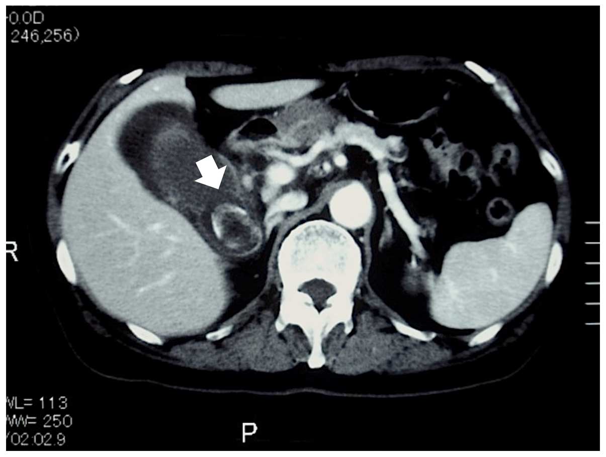
Abdominal computed tomography (CT) revealed acute cholecystitis
with a thickened, edematous wall of the gallbladder (Fig. 1), a single stone with a diameter of
20 mm in the neck of the gallbladder and no demonstrable lymph
nodes. Ultrasonography (US) revealed identical results to CT,
including debris in the gallbladder and a thickened gallbladder
wall. Drip infusion cholangiography CT demonstrated that the cystic
duct was occluded despite improvement of the edematous gallbladder
wall after conservative treatment with antibiotics for 2 weeks at
the Department of Internal Medicine of Sodegaura Satsukidai
Hospital (Sodegaura, Japan). One month after leaving our hospital,
laparoscopic cholecystectomy was performed, and a diagnosis of
chronic cholecystitis with a stone was made.
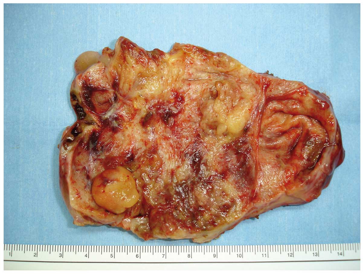
The resected gallbladder was 11×7 cm in length, and
its whitish-yellow mucosa was atrophic with two slight elevations
measuring 2.0×1.5 and 1.5×1.5 cm in the body and fundus,
respectively, as well as a smooth mass measuring 1.5×1.5 cm in the
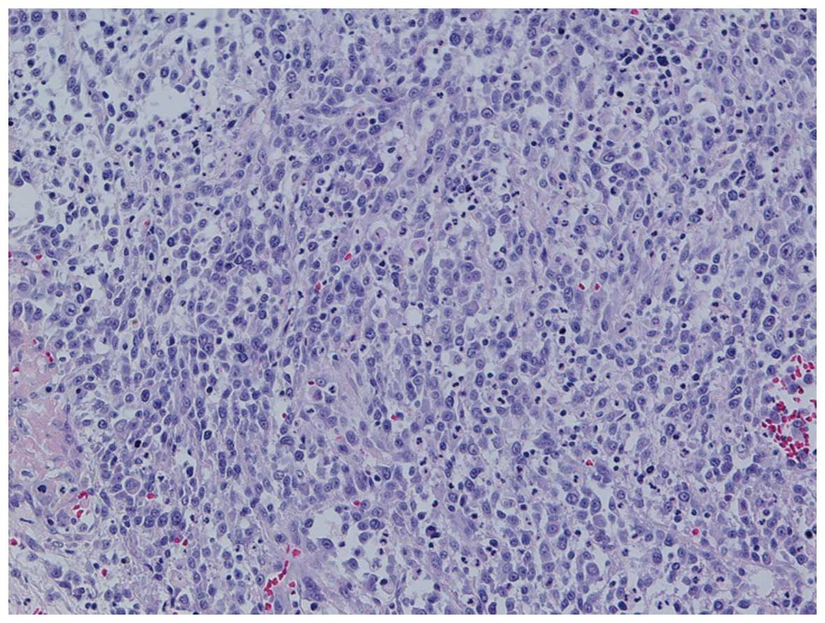
fundus (Fig. 2). Histological
examination revealed undifferentiated carcinoma with spindle- and
polygonal-shaped cells (Fig. 3). No
evidence of cartilaginous, osseous or rhabdomyosarcomatous
differentiation was found. The tumor cells were spread throughout
approximately two-thirds of the gallbladder with invasion of
perimuscular connective tissue; however, no cancer was observed in
the neck of the gallbladder, the liver or sentinel lymph nodes. The
pathological stage was T2 N0 M0, stage II according to the
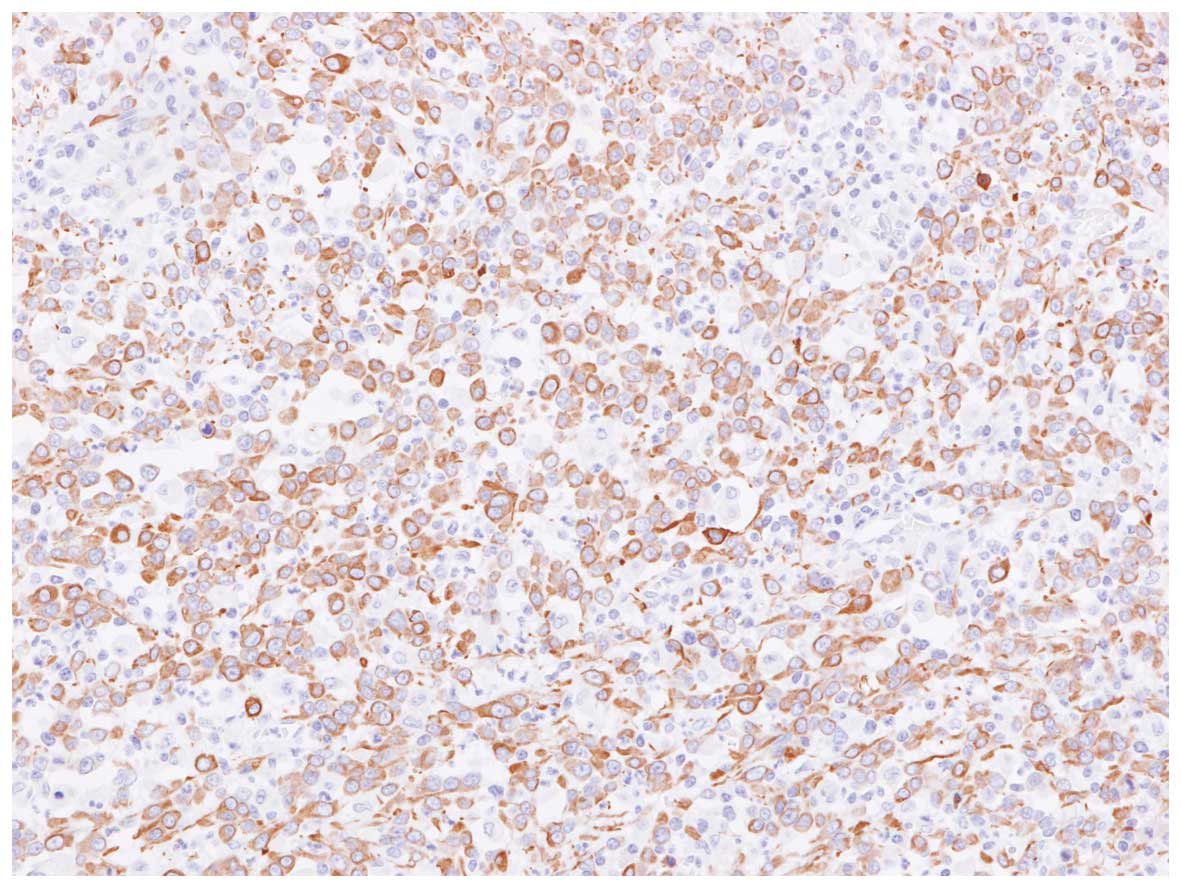
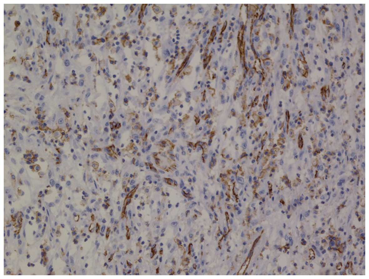
Tumor-Nodes-Metastasis classification. Immunohistochemical staining
revealed that spindle- and polygonal-shaped cells were positive for
cytokeratin (CK) AE1/3 (Fig. 4),
vimentin and vascular endothelial growth factor (VEGF) (data not
shown). Furthermore, numerous spindle-shaped cells were positive
for CD31 (Fig. 5) and CD34, and
certain spindle-shaped cells were positive for Factor VIII (data
not shown).
The post-operative course was uneventful. However,
at two months following surgery, CT demonstrated liver metastasis.
The patient rejected hepatectomy, but consented to chemotherapy.
After the first administration of gemcitabine and
cis-diamminedichloro-platinum (CDDP), the liver tumors had
responded well and the tumor size was considerably reduced; the
maximum tumor size (5 cm diameter was measured by CT) in the liver
decreased by 50%. After two cycles, the regimen was discontinued
due to the appearance of Grade 2 or 3 side effects (vomiting, bone
marrow suppression and renal disfunction), and tegafur, gimeracil
and oteracil potassium (S-1) were administered. The patient
succumbed to multiple liver metastases and peritoneal dissemination
at ~5 months following surgery.
Discussion
In 2000, a new WHO classification of
undifferentiated carcinoma was published (12), according to which undifferentiated
carcinoma of the gallbladder are classified into the following four
types: i) Undifferentiated carcinoma, spindle-and giant-cell type;
ii) undifferentiated carcinoma with osteoclast-like giant cells;
iii) undifferentiated carcinoma, small-cell type; and iv)
undifferentiated carcinoma, nodular or lobular type. In the new 4th
edition of the WHO classification of undifferentiated gallbladder
carcinoma from 2010 (10), the
pathological description is more concise compared with that in the
classification from 2000 (12). Even
after the the 2000 version of WHO classification was published, the
terms ‘sarcomatoid carcinoma’, ‘so-called carcinosarcoma’ and
‘spindle-cell carcinoma’ have been used in the English literature
for the same diagnosis of undifferentiated carcinoma of the
gallbladder. However, the use of the pathological diagnostic name
‘undifferentiated carcinoma’ has gradually increased, while the use
of the other names has decreased, but still occurs. The various
above-mentioned diagnostic names must be unified under the name
‘undifferentiated carcinoma’ immediately, so that the
characteristics, prognosis and novel treatments of this rare tumor
type can be clarified by meta-analysis. The present case was
properly classified as ‘undifferentiated carcinoma, spindle- and
giant-cell type’ according to the WHO classification. To the best
of our knowledge, the present study provided the first case report
of ‘undifferentiated carcinoma with endothelial differentiation’
diagnosed using sub-type analysis. Notably, the final diagnosis was
made not only based on morphological findings, but also using
immunohistochemical staining.
Although the WHO and AJCC differentiate between
undifferentiated carcinoma and carcinosarcoma, certain pathologists
classify them into the same group. The prognosis for
undifferentiated carcinoma as well as carcinosarcoma of the
gallbladder is poor. The present study performed an analysis of
previous studies containing statistically significant, reliable
data, while excluding those containing combined data for
undifferentiated carcinoma and carcinosarcoma. The following groups
were analyzed in order to clarify their clinical characteristics
and differences: A) The undifferentiated carcinoma (2–5,7) and sarcomatoid carcinoma (6) group, and B) the carcinosarcoma group
(13,14). The data from each article were
re-evaluated and re-calculated as required. The results of the
analysis, which are shown in Table
I, showed that the patients' mean age was 66.6 years and that a
high percentage of affected patients were female (female-to-male
ratio, 1.57–7.00). Of note, in group A, the prognostic data in more
recent studies (5–7) tended to be improved compared with those
in earlier studies (2–4). Among the more recent studies, no
significant difference between the prognostic data in groups A
(5–7)
and B (13,14) was present; however, the tumor size in
group B was slightly larger compared with that in group A. The
implementation of diagnostic methods, including CT, US and magnetic
resonance imaging to detect small-sized tumors, the development of
extended radical surgery may have accounted for the abovementioned
findings. The differences in the malignant potential between groups
A and B may be based on the fact that the cells have different
origins: True carcinosarcoma is derived from totipotent stem cells,
which separately differentiate into epithelial and sarcomatous
cells, while undifferentiated carcinoma is a morphological variant
of carcinoma, which is transformed to exhibit sarcomatous features
(4). The prognosis of patients with
undifferentiated carcinoma of the gallbladder may be identical or
slightly worse compared with that of patients with carcinosarcoma;
however, this requires further assessment by future meta-analyses
of data from patients worldwide.
 | Table I.Characteristics of undifferentiated
carcinoma and carcinosarcoma. |
Table I.
Characteristics of undifferentiated
carcinoma and carcinosarcoma.
| A, Undifferentiated
carcinoma |
|---|
|
|---|
| Author, year | No. of cases | Mean age, years | Female-to-male
ratio | Median tumor size, cm
(mean) | Median survival,
months (mean) | 1-year survival rate
(%) | Refs. |
|---|
| Guo et al,
1988 | 21 | 66.3 | 2.50 | ND | 3.0 (10.0) | 18.2 | (2) |
| Nishihara et
al, 1993 | 11 | 66.5 | 2.67 | 5.8 (6.7) | 3.0 (8.1) | 18.2 | (3) |
| Akatsu et al,
2005 | 18 | 67.5 | 2.60 | 5.0 (6.1) | 3.0 (6.9) | 12.5 | (4) |
| Mizusaki et
al, 2007 | 18 | 65.7 | 1.57 | 7.0 (7.2) | 7.0 (11.1) | 38.5 | (5) |
| Hu et al,
2010 | 10 | 67.0 | 4.00 | 5.2 (ND) | 9.0 (ND) | ND | (6) |
| Park et al,
2014 | 8 | 57.2 | 7.00 | 5.0 (5.8) | 7.3 (18.0) | 37.5 | (7) |
|
| B,
Carcinosarcoma |
|
| Author, year | No. of cases | Mean age, years | Female-to-male
ratio | Median tumor size, cm
(mean) | Median survival,
months (mean) | 1-year survival rate
(%) | Refs. |
|
| Okabayashi et
al, 2010 | 28 | 67.4 | 2.62 | 8.5 (8.6) | 5.5 (10.8) | 33.3 | (13) |
| Gao et al,
2015 | 21 | 69.0 | 1.63 | 6.5 (7.0) | 7.0 (13.7) | 50.0 | (14) |
Certain studies have pointed out the difficulty of
treating undifferentiated carcinoma of the gallbladder because of
the large tumor size, liver and lymph node metastasis, and
peritoneal dissemination (1,15,16),
even if it is detected without patient's symptom. Although surgical
resection is the predominant treatment, a study by Doval et
al (17) reported the use of
palliative chemotherapy in a case of sarcomatoid carcinoma;
however, chemotherapy and radiotherapy were generally shown not to
be beneficial by numerous other studies (5,9,13,16,18–21). In
the present case, the secondary liver tumors responded well to
adjuvant gemcitabine and CDDP, and the tumor size decreased
considerably. The regimen was discontinued due to the appearance of
side effects, and tegafur, gimeracil and oteracil potassium (S-1)
were administered. Owing to the rapid growth of the patient's
tumor, she succumbed to it ~5 months following surgery. VEGF
inhibitors, which are not approved for patients with
undifferentiated gallbladder carcinoma by the National Health
Insurance Program of Japan, may have improved the prognosis of the
present case, as the tumor showed endothelial differentiation and
VEGF-positive staining. It is expected that the prognosis of
undifferentiated gallbladder carcinoma will be improved in the
future by using gene analysis and gene therapy.
In conclusion, the present study reported on a case
histopathologically identified as undifferentiated carcinoma of the
gallbladder. However, immunohistochemical staining revealed
characteristics that differed from those of other studies. The
present case was diagnosed as ‘undifferentiated carcinoma with
endothelial differentiation’ due to its immunohistochemical
features.
References
|
1
|
Kubota K, Kakuta Y, Kawamura S, Abe Y,
Inamori M, Kawamura H, Kirikoshi H, Kobayashi N, Saito S and
Nakajima A: Undifferentiated spindle-cell carcinoma of the
gallbladder: An immunohistochemical study. J Hepatobiliary Pancreat
Surg. 13:468–471. 2006. View Article : Google Scholar : PubMed/NCBI
|
|
2
|
Guo KJ, Yamaguchi K and Enjoji M:
Undifferentiated carcinoma of the gallbladder. A clinicopathologic,
histochemical, and immunohistochemical study of 21 patients with a
poor prognosis. Cancer. 61:1872–1879. 1988. View Article : Google Scholar : PubMed/NCBI
|
|
3
|
Nishihara K and Tsuneyoshi M:
Undifferentiated spindle cell carcinoma of the gallbladder: A
clinicopathologic, immunohistochemical and flow cytometric study of
11 cases. Hum Pathol. 24:1298–1305. 1993. View Article : Google Scholar : PubMed/NCBI
|
|
4
|
Akatsu T, Ueda M, Shimazu M, Wakabayashi
G, Aiura K, Tanabe M, Kawachi S, Kameyama K and Kitajima M: Primary
undifferentiated spindle-cell carcinoma of the gallbladder
presenting as a liver tumor. J Gastroenterol. 40:993–998. 2005.
View Article : Google Scholar : PubMed/NCBI
|
|
5
|
Mizusaki K, Saito E and Kobayashi H: A
case of undifferentiated carcinoma of the gallbladder with rapidly
progress after operation. J Jpn Biliary Assoc. 21:567–573.
2007.(Japanese).
|
|
6
|
Hu ZH, Li ZW, Shen L, Zhang M and Zheng
SS: Surgical therapy and prognosis of sarcomatoid carcinoma of the
gallbladder. Hepatobiliary Pancreat Dis Int. 9:175–179.
2010.PubMed/NCBI
|
|
7
|
Park HJ, Jang KT, Choi DW, Heo JS and Choi
SH: Clinicopathologic analysis of undifferentiated carcinoma of the
gallbladder. J Hepatobiliary Pancreat Sci. 21:58–63. 2014.
View Article : Google Scholar : PubMed/NCBI
|
|
8
|
Zhang L, Chen Z, Fukuma M, Lee LY and Wu
M: Prognostic significance of race and tumor size in carcinosarcoma
of gallbladder: A meta-analysis of 68 cases. Int Clin Exp Pathol.
1:75–83. 2008.
|
|
9
|
Hotta T, Tanimura H, Yokoyama S, Ura K and
Yamaue H: So-called carcinosarcoma of the gallbladder; spindle cell
carcinoma of the gallbladder: Report of a case. Surg Today.
32:462–467. 2002. View Article : Google Scholar : PubMed/NCBI
|
|
10
|
Albores-Saavedra J, Adsay NV, Crawford JM,
Klimstra DS, Kloppel G, Sripa B, Tsui WMS and Paradis V: Carcinoma
of the gallbladder and extrahepatic bile ductsWHO Classification of
Tumors of the Digestive System. 4th. Bosman FT, Carneiro F, Hruban
RH and Theise ND: IARC Press; Lyon: pp. pp266–pp273. 2010
|
|
11
|
Edge SB and Compton CC: The american joint
committee on cancer: The 7th edition of the AJCC cancer staging
manual and the future of TNM. Ann Surg Oncol. 17:1471–1474. 2010.
View Article : Google Scholar : PubMed/NCBI
|
|
12
|
Albores-Saavedra J, Scoazec JC, Wittekind
C, Sripa B, Menck HR, Soehendra N and Sriram PVJ: Carcinoma of the
gallbladder and extrahepatic bile ductsWorld Health Organization
Classification of Tumours. Pathology and Genetics of Tumours of the
Digestive System. IARC Press; Lyon: pp. pp206–pp214. 2000
|
|
13
|
Okabayashi T, Sun ZL, Montgomey RA and
Hanazaki K: Surgical outcome of carcinosarcoma of the gall bladder:
A review. World J Gastroenterol. 15:4877–4882. 2009. View Article : Google Scholar : PubMed/NCBI
|
|
14
|
Gao S, Huang L, Dai S, Chen D, Dai R and
Shan Y: Carcinosarcoma of the gallbladder: A case report and review
of the literature. Int J Clin Exp Pathol. 8:7464–7469.
2015.PubMed/NCBI
|
|
15
|
Badmos KB, Seada L Salah, Al Rashid F
Fahad and Oreiby HA: Undifferentiated spindle-cell carcinoma of the
gallbladder: A report of a case, an immunohistochemistry profile,
and a review of the literature. Case Rep Pathol.
2013:2671942013.PubMed/NCBI
|
|
16
|
Nagata J, Kijima H, Watanabe N and Mine T:
Undifferentiated carcinoma of the gallbladder. Nihon Rinsho.
3:(Suppl 73). S769–S773. 2015.(In Japanese).
|
|
17
|
Doval DC, Azam S, Mehta A, Pruthi A, Batra
U, Choudhury KD and Kumar K: A report of sarcomatoid carcinoma of
the gallbladder treated with palliative deocetaxel and gemcitabine
chemotherapy. J Gastrointest Cancer. 45:270–274. 2014. View Article : Google Scholar : PubMed/NCBI
|
|
18
|
Ajiki T, Nakamura T, Fujino Y, Suzuki Y,
Takeyama Y, Ku Y, Kuroda Y and Ohbayashi C: Carcinosarcoma of the
gallbladder with chondroid differentiation. J Gastroenterol.
37:966–971. 2002. View Article : Google Scholar : PubMed/NCBI
|
|
19
|
Kataria K, Yadav R and Seenu V:
Sarcomatoid carcinoma of the gall bladder. J Surg Case Rep.
2012:52012. View Article : Google Scholar : PubMed/NCBI
|
|
20
|
Huguet KL, Hughes CB and Hewitt WR:
Gallbladder carcinocsarcoma: A case report and literature review. J
Gastrointest Surg. 9:818–821. 2005. View Article : Google Scholar : PubMed/NCBI
|
|
21
|
Liu KH, Yeh TS, Hwang TL, Jan YY and Chen
MF: Surgical management of gallbladder sarcomatoid carcinoma. World
J Gastroenterol. 15:1876–1879. 2009. View Article : Google Scholar : PubMed/NCBI
|