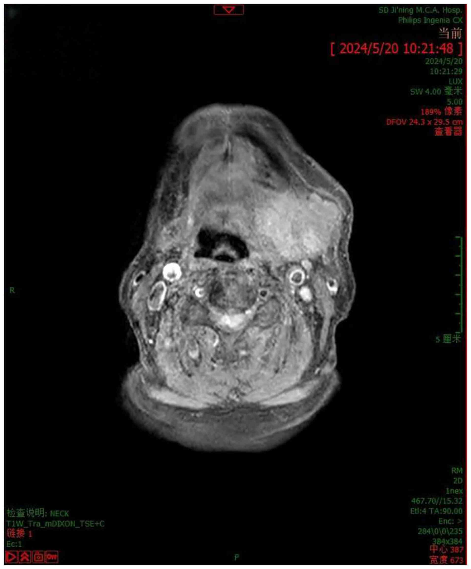
|
1
|
Kim SH, Park SE, Bae HG, Song DH, Oh HH,
Cho KR, Kim HJ and Sohn BS: Epithelial-myoepithelial carcinoma of
the nasopharynx: A case report and review of the literature. Oncol
Lett Aug. 10:927–930. 2015. View Article : Google Scholar : PubMed/NCBI
|
|
2
|
Shah AA, Mulla AF and Mayank M:
Pathophysiology of myoepithelial cells in salivary glands. J Oral
Maxillofac Pathol. 20:480–490. 2016. View Article : Google Scholar : PubMed/NCBI
|
|
3
|
Lavareze L, Scarini JF, de Lima-Souza RA,
Kimura TC, Gondak RO, Egal ESA, Altemani A and Mariano FV:
Clinicopathological and survival profile of patients with salivary
gland myoepithelial carcinoma: A systematic review. J Oral Pathol
Med. 52:101–108. 2023. View Article : Google Scholar : PubMed/NCBI
|
|
4
|
Xu B and Katabi N: Myoepithelial
carcinoma. Surg Pathol Clin Mar. 14:67–73. 2021. View Article : Google Scholar : PubMed/NCBI
|
|
5
|
Rosen LE, Singh RI, Vercillo M and Gattuso
P: Myoepithelial carcinoma of the lung: A review. Appl
Immunohistochem Mol Morphol. 23:397–401. 2015. View Article : Google Scholar : PubMed/NCBI
|
|
6
|
Henning A, Pennington G, Deeken A and
Srivastava S: Myoepithelial carcinoma of the digit. J Cutan Pathol.
49:111–115. 2022. View Article : Google Scholar : PubMed/NCBI
|
|
7
|
Kanesv SV and Bagwanin IN:
Myoepithelialcarcinoma of the salivary glands: A clinicopathologic
study of 51 cases in atertiary cancer center. Arch Otolaryngol Head
Neck Surg. 136:702–712. 2010. View Article : Google Scholar : PubMed/NCBI
|
|
8
|
Jo VY, Antonescu CR, Hornick JL and Patel
RM: Myoepithelioma, myoepithelial carcinoma, and mixed tumor. In:
WHO Classification of Tumours Editorial Boardeditor. WHO
classification of tumours, soft tissue and bone tumours. 5th
edition. Geneva, Switzerland: World Health Organization; pp.
277–279. 2020
|
|
9
|
Seethala RR, Barnes EL and Hunt JL:
Epithelial-myoepithelial carcinoma: A review of the
clinicopathologic spectrum and immunophenotypic characteristics in
61 tumors of the salivary glands and upper aerodigestive tract. Am
J Surg Pathol. 31:44–57. 2007. View Article : Google Scholar : PubMed/NCBI
|
|
10
|
Peilong CA, Shaoqiang Z and Jiyuan ZH:
Clinicopathological analysis of 12 cases of myoepithelial carcinoma
of salivary gland. J Clin Exp Pathol. 33:174–177. 2017.
|
|
11
|
Nagao T, Sugano I and Ishida Y: Salivary
gland malig-nant myoepithelioma: A clinicopathologic and
immunohisto-chemical study of ten cases. Cancer. 83:1292–1299.
1998. View Article : Google Scholar : PubMed/NCBI
|
|
12
|
Guo-Liang PI, Peng XU and Guang H:
Research progress of myoepithelial carcinoma. China Cancer Clinics.
44:253–257. 2017.(In Chinese).
|
|
13
|
Aiba H, Errani C, Ciani G, Gambarotti M,
Righi A, Maioli M, Spinnato P, Frega G, Ibrahim T and Longhi A:
Myoepithelial carcinoma of soft tissues and bone. Eur J Cancer.
194:1133532023. View Article : Google Scholar : PubMed/NCBI
|
|
14
|
Dinker D, Rajan K, Sharma S and Kumar NA:
Myoepithelial carcinoma of sinonasal cavity: Peculiar diagnosis,
conventional treatment. Indian J Otolaryngol Head Neck Surg.
75:3929–3935. 2023. View Article : Google Scholar : PubMed/NCBI
|
|
15
|
Vilar-González S, Bradley K, Rico-Pérez J,
Vogiatzis P, Golka D, Nigam A, Sivaramalingam M and Kazmi S:
Epithelial-myoepithelial Carcinoma-review of clinicopathological
and immunohistochemical features. Clin Transl Oncol. 17:847–855.
2015. View Article : Google Scholar : PubMed/NCBI
|
|
16
|
Masuya D, Haba R, Huang CL and Yokomise H:
Myoepithelial carcinoma of the lung. Eur J Cardiothorac Surg.
28:775–777. 2005. View Article : Google Scholar : PubMed/NCBI
|
|
17
|
Nicholas RG, Hanson JA and Meiklejohn DA:
Myoepithelial cell carcinoma of the oral tongue: Case report and
review of the literature. Clin Med Insights Oncol.
13:11795549198382542019. View Article : Google Scholar : PubMed/NCBI
|
|
18
|
Wu H, Zhang F, Peng J, Wu Z, Zhang X and
Wu X: Epithelial-myoepithelial carcinoma of the esophagus: A Case
Report. Front Surg. 9:9420192022. View Article : Google Scholar : PubMed/NCBI
|
|
19
|
Shaoyan L, Song NI, Yiming Z and Jian W:
Analysis of diagnosis and treatment of myoepithelial carcinoma of
the submandibular gland. Chin Med J. 50:28–29. 2015.
|
|
20
|
Waitzman J, Waitzman A, Powers J and
Deraniyagala R: Epithelial-myoepithelial carcinoma of the nasal
cavity: A case report. Ear Nose Throat J. 31:14556132311899622023.
View Article : Google Scholar : PubMed/NCBI
|
|
21
|
Zhao F'Zhu H and Huang Y: Myoepithelial
carcinoma inside of maxilla bone: A case report. Mol Clin Oncol.
1:315–317. 2013. View Article : Google Scholar : PubMed/NCBI
|
|
22
|
Passador-Santos F, Grönroos M, Irish J,
Gilbert R, Gullane P, Perez-Ordonez B, Mäkitie A and Leivo I:
Clinicopathological characteristics and cell cycle proteins as
potential prognostic factors in myoepithelial carcinoma of salivary
glands. Virchows Arch. 468:305–312. 2016. View Article : Google Scholar : PubMed/NCBI
|
|
23
|
Kaur R, Melgandi W, Rathi AK, Singh K,
Rahman F and Mandal S: Epithelial myoepithelial carcinoma: A case
report. J Cancer Res Ther. Jan 22–2024.(Epub ahead of print).
|
|
24
|
Ru K, Srivastava A and Tischler AS:
Primary myoepithelial carcinoma of palate. Arch Pathol Lab Med.
128:92–94. 2004. View Article : Google Scholar : PubMed/NCBI
|
|
25
|
Thompson LDR and Xu B: Top ten
differentials to mull over for head and neck myoepithelial
neoplasms. Head Neck Pathol. 17:1–15. 2023. View Article : Google Scholar : PubMed/NCBI
|
|
26
|
Yokose C, Asai J, Kan S, Nomiyama T,
Takenaka H, Konishi E, Goto K, Ansai SI and Katoh N: Myoepithelial
carcinoma on the right shoulder: A case report with published work
review. J Dermatol. 43:1083–1087. 2016. View Article : Google Scholar : PubMed/NCBI
|
|
27
|
Ren J, Liu Z, Liu X, Li Y, Zhang X, Li Z,
Yang Y, Yang Y, Chen Y and Jiang S: Primary myoepithelial carcinoma
of palate. World J Surg Oncol. 9:1042011. View Article : Google Scholar : PubMed/NCBI
|
|
28
|
Gore MR: Epithelial-myoepithelial
carcinoma: A population-based survival analysis. BMC Ear Nose
Throat Disord. 18:152017. View Article : Google Scholar : PubMed/NCBI
|